2. LEARNING OBJECTIVES
To define
To enumerate the risk factors
To discuss the pathophysiology and
site of occurrence
To describe the clinical features and
management of ectopic preg
3. Ectopic (Ektopos) out of place
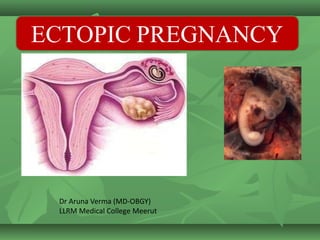
Definition -Ectopic pregnancy: fertilized
embryo implanted outside the uterine cavity.
Incidence are 0.25-2%
Mechanism of occurrence is either anatomic
and physiological abnormalities of tube or some
chemotactic factors which predispose for EP.
4.
5.
6. Mechanical factors
Congenital: long narrow tube, diverticulae and
accessory ostia.
Traumatic: operation on the tube as salpingoplasty and
tubal reversal following ligation.
Inflammatory: Chronic salpingitis
Neoplastic: Narrowing of the tube by a fibroid or a broad
ligament tumor.
Functional: As tubal spasm or antiperistaltic
contractions.
Endometriosis in the tube. encourages embedding of
the fertilized ovum.
7. RISK FACTORS
Hz of tubal surgery
Hx of STD’s (such as chlamydia)
Hx of ART
Hx of ectopic (esp if conservatively
managed without surgery)
Smoking
IUD in place at time of conception
18. Symptoms & Signs:
In a woman of child bearing age with
pelvi-abdominal pain and/ or vaginal
bleeding …… ALWAYS….think
19. Amenorrhoea
A dull aching pain is usually present in
one iliac fossa. It is due to distension of the
tube and stretching of its peritoneal coat.
Classic signs – adnexal or
cervical motion tenderness.
ClinicalFinding: Undistrubedectopic
20. Signs:
■ Abdominal examination: Tenderness in one
iliac fossa.
■ Vaginal examination:
(cervical motion tenderness or jumping sign)
The cervix is soft and severe pain occurs
when itis moved from side to side
■ A mass may be felt to one side of the uterus.
It is very tender, soft and may be pulsating.
21. Subacute type:Symptoms:
■ Short period of amenorrhea in (25%) no history
of conceptional bleeding that mistakenas a true
menstrual period
■ Pain: It is felt in one iliac fossa. It may be dull
aching or sharp stabbing or colicky
■ Fainting attacks or even shock
■ Vaginal bleeding occurs after pain
22. With ruptured ectopic pregnancy
abdominal guarding and rigidity,
■ shoulder pain
■ fainting attacks
■ and shock.
23. Investigations
a. UPT
b. USG
c. Hemogram and blood grouping
d. Culdocentesis
e. Correlation of beta hCG and
USG
f. Color doppler
24. When a woman presents with an
early pregnancy…
AsAskkyyoouursrselfelft
w
t
w
o
oqqueuesstiontionss……
W
W
h
e
h
e
r
r
e
eiisstthihiss p
p
r
r
e
e
g
n
a
g
n
a
n
n
c
c
y
y
?
?
Ask yourself two questions…
Where is this pregnancy?
Is it viable?
25. Where is this pregnancy?
In a woman with an early pregnancy you
must determine if the pregnancy is
intrauterine or an ectopic, because her
life could depend on it!
26. How to you determine location of
the pregnancy?
First determine dating by LMP
Then perform ultrasound
If you can see location of the pregnancy, you
are done!
If you cannot…it becomes more
complicated…
27. β-hCG discriminatory value (or zone)
It is the lower limit of hCG at which an
examiner can reliably visualize pregnancy
on ultrasound. It is 1000-2000 IU/L with
vaginal ultrasound and 5000-6000 IU/L
with abdominal ultrasound.
28. If β-hCG levels above the
discriminatory value
The absence of uterine pregnancy
signifies an abnormal pregnancy; ectopic,
incomplete abortion
If β-hCG levels are still below the
discriminatory value, serial β-hCG and
ultrasound should be done.
29. Doubling sign:
In normal pregnancy a 66% or greater increase
in serum β-hCG levels should be observed
every 48 hours (nearly doubles).
Inappropriately rising serum β-hCG levels
suggest (but do not diagnose) an abnormal
pregnancy including ectopic, however, they do
not identify its location.
32. Early pregnancy with unknown
location
Check a serum BHCG
If it is above the discriminatory zone (DZ)an
intrauterine pregnancy should be seen
Then do an ultrasound to see if you see the
pregnancy
34. Treatment of tubal pregnancy
If the patient is shocked: antishock measures.
If the patient is Rh negative and not sensitized
anti-D serum is given.
Medical therapy:
methotrexate (a folic acid antagonist).
IM methotrexate given as a single dose.
35. The best candidate is the woman who is
asymptomatic, compliant with follow-up, with
an initial serum value <5000 IU/L.
Contraindications:
Breastfeeding
Immunodeficiency / active infection
Chronic liver disease
Active pulmonary disease
Active peptic ulcer or colitis
Blood disorder
Hepatic, Renal or Haematological
dysfunction
36.
37.
38. Significantly worsening abdominal pain,
Haemodynamic instability
Level of HCG do not decline by at least 15%
between Day 4 & 7 post treatment
or plateauing HCG level after first week of
treatment
Signs and Treatment failure and tubal
rupture:
39. Follow-Up:
If the β-hCG level does not decline (plateau or
increase), the patient may require either a
second dose of methotrexate or surgery.
Surgical management:
Laparoscopy approach – salpingostomy
Laprotomy – salpingostomy salpingectomy
40. Salpingostomy / Salpingotomy is only indicated
when:
1. The patient desires to conserve her fertility
2. Patient is haemodinmically stable
3. Tubal pregnancy is accessible
4. Unruptured and < 4Cm. In size
5. Contralateral tube is absent
or damaged
43. laparatomy (if the mass is greater than 4 cm in
diametar, internal bleeding, cardiovascular colapse)
44. Algorithm for the diagnosis of unruptured ectopic pregnancy
without laparoscopy.
45. 1- Positive pregnancy test
Lowe abdominal pain +
Minimal Vaginal bleeding
Asymptomatic with factors
for ectopic pregnancy
2. History + clinical examination
Management of ectopic pregnancy
46. If sure of date of LMP and /or
Regular cycle, i.e.
>6 wks. gestation,
Arrange TV ultrasound
If unsure of date of LMP
and /or irregular cycle,
Measure serum hCG
If hCG <1000
(?early Intrauterine/
? Ectopic pregnancy
If Hcg >1000, use
protocol for
suspected
Ectopic pregnancy
3. Empty uterus + free fluid in POD + adnexal + serum hCG > 1000
Meet criteria for
Methorexate treatment
Does not meet criteria
for methotrexate treatment
Use methotrexate
protocol
Laproscopic /salpingotomy/
Salpingectomy ?Proceed to
laparotomy OR Laparotomy if
haemodynamically unstable
47. Time for small quiz
T/F
i. The most common site of ectopic pregnancy is
ovary?
ii. Laparoscopy is the ideal mode of treatment in
ruptured ectopic with hemodynamic unstability?
iii. Mx can be administered in pts with ruptured
ectopic?
iv. Abdominal USG is the gold standard to
diagnose ectopic?
v. IUCD decrease the chances of ectopic?
48. Questions for SDL
I. Ring of fire sign?
II. Aria stella reaction?
III. Studdiford criteria?
IV. Spiegelberg criteria?
V. Heterotrpic pregnancy?