Maternal hypertension, induced through nitric oxide inhibition (L-NAME model) or spontaneous hypertension (SHR model), caused structural changes in the hearts of offspring rats. Specifically, the study found increased aorta wall area and wall-to-lumen ratio in hypertensive offspring at all ages examined (fetal, 2 days, and 15 days). Microvessel wall area in the heart was also greater in hypertensive offspring at 15 days. Additionally, collagen content in the heart was higher for SHR offspring at 2 and 15 days and L-NAME offspring at 15 days compared to controls. The results suggest that maternal hypertension is a risk factor for cardiovascular changes in offspring.
![www.symbiosisonline.org
www.symbiosisonlinepublishing.com
Symbiosis
Research Article
Journal of Clinical Trials in Cardiology
Open Access
The Heart is a Target Organ in Offspring Rats Due to
Maternal Hypertension
Sonia Regina Jurado1*, Roberto Jorge da Silva Franco2, Antonia Dalla Pria
Bankoff3, Andrea Sanchez3
2
1
Federal University of Mato Grosso do Sul, Brazil
Universidade Estadual Paulista Julio de Mesquita Filho, Brazil
3
Federal University of Mato Grosso do Sul, Brazil
Received: 29.10.2013, Accepted: 10.12.2013, Published: 16.12.2013
Corresponding author: Sonia Regina Jurado, Federal University of Mato Grosso do Sul, Tres Lagoas, Mato Grosso do Sul, Avenue Ranulpho Marques
Leal, Brazil, Tel: +55 67-3509-3714, E-mail: srjurado@bol.com.br
*
Abstract
Purpose: The aim of this study was to analyze the effect of
hereditary hypertension or induced by chronic nitric oxide inhibition
during pregnancy on the structural changes in rats fetal and neonate
coronary microvessels, aorta and myocardial collagen content.
Methods: Total nine sub-groups allocated from three main groups
of fetuses (20th d) and newborns (2nd and 15th d) offspring’s from
normotensive mothers (C, control), SHR (spontaneously hypertensive
rats) and L-NAME (Nω-Nitro-L-Arginine Methyl Ester) were studied.
Heart and aorta sections were stained with hematoxylin and eosin,
Mallory’s thichrome, Picrosirius red and periodic acid-Schiff reagent.
Pro-Plus image analysis system was used to assess the thickness of
aorta and myocardial vessels. The tunica media inner and outer
border was traced in each microvessel (external diameter < 50 µm)
image at x100 and, in aorta at x400 magnification, and areas encircled
by tracings were calculated.
Results: Aorta wall area and wall-to-lumen ratio increased in
hypertensive animals in all ages. The number of elastic lamellae
was increased in hypertension (L-NAME: 7.10±0.18, p<0.05 and
SHR: 7.45±0.17, p<0.05) at 2nd d compared to C (7.60±0.17). The
microvessels wall area was greater in hypertensive offspring at 15th
d. Lumen area and external perimeter decreased in both hypertension
models at 15th d. Wall-to-lumen ratio increased in hypertensive
at 2nd and 15th d. Collagen content (%) was higher in SHR at 2nd d
(0.74±0.02, p<0.05) and 15th d (2.53±0.04, p<0.05) and in L-NAME at
15th (2.25±0.04, p<0.05) versus C (0.61±0.02 at 2nd d and 2.12±0.05
at 15th d).
Conclusion: Maternal hypertension, as in the model L-NAME and
SHR, caused microvasculature remodeling in heart the neonate at 2
and 15 days.
Keywords: Nitric oxide; Microvasculature; Aorta; Myocardium;
Hypertension; Pregnancy; Morphometry; Fetus; Newborn; Rat
Abbreviations: ACE: Angiotension I Converting Enzyme; C20:
Control group at 20 post conception days; C2: Control group at 2
post-natal days; C15: Control group at 15 post-natal days; g: grams;
Kg: Kilograms; L20: L-NAME group at 20 post conception days;
L2: L-NAME group at 2 post-natal days; L15: L-NAME group at 15
Symbiosis Group
post-natal days; L-NAME: Nω-Nitro-L-Arginine Methyl Ester; mg:
milligrams; NO: Nitric Oxide; NOS: Nitric Oxide Synthase; S20:
SHR group at 20 post conception days; S2: SHR group at 2 post-natal
days; S15: SHR group at 15 post-natal days; SHR: Spontaneously
Hypertensive Rats; µm: micrometers
Introduction
Nitric oxide (NO) is synthesized from L-arginine by the
constitutive NO synthase in vascular endothelial cells, and
plays an important role in the regulation of blood pressure
and coronary vasomotion [1]. Normal pregnancy is associated
with major adaptations in maternal cardiovascular function,
which help the woman to accommodate the growing fetus.
Although there is an increase of maternal blood pressure
volume and cardiac output as well, the systemic blood pressure
actually declines during pregnancy [2]. Furthermore, pressure
responsiveness or vascular reactivity to several vasoconstrictors
is attenuated. This is probably due to the potential contribution
of nitric oxide to the vasodilator phenomena of pregnancy [3].
Preeclampsia constitutes the syndrome of vasoconstriction with
elevated arterial blood pressure, edema, proteinuria, kidney and
liver dysfunction, and intrauterine growth retardation [4,5]. The
vascular endothelium is stimulated during pregnancy to release
increased amounts of NO and the abnormality in the L-arginineNO pathway may play a role in the etiology of preeclampsia [3].
It is known that the administration of L-NAME (Nω-NitroL-Arginine Methyl Ester) in adult rats promotes arterial
hypertension [6,7], cardiac hypertrophy, myocytic necrosis,
perivascular and interstitial fibrosis and microvascular lumen
occlusion [8,9] and cardiomyocyte apoptosis [10]. However, low
dose of L-NAME administrated in rats under prolonged period
caused arterial hypertension accompanied by a significant
reduction in cardiac weight and cardiomyocyte size [11]. In
pregnant rats, this nitric oxide synthesis inhibitor causes fetal
growth restriction by a reduction in cellular proliferation due
*Corresponding author email: parsons@SDRMI.org, parsons@xcelthera.com](https://image.slidesharecdn.com/cardiology01-140108102816-phpapp01/75/The-Heart-is-a-Target-Organ-in-Offspring-Rats-Due-to-Maternal-Hypertension-1-2048.jpg)
![The Heart is a Target Organ in Offspring Rats Due to Maternal Hypertension
to induction of apoptosis [12], reducing the body weight and
causing hemorrhagic necrosis of neonate’s hind limbs [13, 14].
The role of nitric oxide on fetal cardiovascular development
is only partially known. Mice with endothelial nitric oxide
synthase deficiency had bicuspid aortic valves [15], increased
cardiomyocyte apoptosis, congenital septal defects [16], fetal
growth restriction and reduced survival [17].
The NO-generation is required for cardiomyogenesis since
NO synthase inhibitors prevent the maturation of terminally
differentiated cardiomyocytes in vitro and the number of
differentiated cardiomyocytes is significantly reduced [18].
The purpose of this work was to determine whether the fetus
and neonate myocardium microvasculature and aorta as well as
myocardial collagen content were affected due to oxide nitric
synthase inhibition during pregnancy in rats.
Material and Methods
Thirty females 14-16-week-old Wistar and fifteen
spontaneously hypertensive rats (SHR) with a body weight of
200-250 g were mated with male rats. Day 1 of pregnancy was
determined by the presence of spermatozoa in vaginal smear.
The animals were housed under controlled lighting (12:12h
light-dark cycle) and temperature (21±3ºC). The Wistar dams
were randomly assigned to two groups of 15 rats each: control
and L-NAME (Nω-Nitro-L-Arginine Methyl Ester). The L-NAME
animals received the NO synthase inhibitor (hydrochloride,
L-NAME, Sigma, St Louis, MO, lot 70H7703) in drinking water
(12 mg/day/rat), throughout the pregnancy (21days). The rat
arterial pressure was evaluated by tail cuff plethysmography at
the beginning (on day 1 of gestation) and end of gestation (on day
21 of gestation). The average of three successive measurements
was taken as the mean systolic pressure value. A total of nine subgroups of fetuses and pups were studied from three main groups
(Control, L-NAME and SHR). Fifteen fetuses were separated into
three sub-groups of five each: C20, L20 and S20 (C: control,
L: L-NAME, S: SHR; 20: at 20 post conception days). Thirty
newborns also were allocated in the following sub-groups of five
each: C2, C15, L2, L15, S2, S15 (2 and 15: at 2 and 15 post-natal
days). They were sacrificed under pentobarbital anesthesia. All
handling and procedures observed minimum animal suffering,
and procedures and experimental protocols were approved by
the Ethical Committee of the Botucatu Medical School – UNESP.
The heart and aorta were dissected, fixed for 24 hours by
immersion in buffered formalin 10 % (pH 7.2). The samples were
dehydrated in ethanol, embedded in paraplast and sectioned
5-6 µm thick. Sections were stained with hematoxylin and eosin,
Masson’s trichrome, picrosirius red and periodic acid-Schiff
(PAS) reagent.
Quantitative assessment of myocardial collagen
It was analyzed thirty fields of view from each heart section
(6 µm thick) stained with picrosirius red to provide a range of
Copyright:
© 2013 Jurado SR, et al.
collagen contents (types I and III). Specifically, we examined
tissue from the free wall of the left ventricle, right ventricle,
and intraventricular septum from newborn at 2 and 15 days.
The areas of the myocardium that contain collagen, such the
perivascular space, were excluded. It was not performed the
collagen content in fetal heart because the collagen network
was still in development. The myocardial collagen content was
assessed by digital image analysis using Pro-Plus analysis system
(Media Cybernetics, USA) and polarized light microscopy at level
x40. At polarized light, picrosirius red stained sections are viewed
in brightfield, type I collagen appears red and type III green. This
technique provides a convenient and a quantitative histologic
approach that can also be used to quantify collagen content [19].
Cardiac microvasculature and aorta morphometry
Heart and aorta sections stained by hematoxylin-eosin were
observed through a light microscope (Leica DMLB) and their
images were recorded by a video camera and then loaded into
a Pro-Plus analysis system (Media Cybernetics, USA). All vessels
in the section had their tunica media, lumen and outer border
traced at x400 magnification. Aorta section was studied at x100
magnification. The following variables for each blood vessel were
then calculated by the Pro-Plus analysis system: total vessel area,
vessel wall area, vessel lumen area, wall-to-lumen ratio (the area
of the vessel wall divided by the area of the blood vessel lumen),
minimum and maximal diameters and external perimeter. More
than 150 blood vessels were analyzed in each group. To compare
the myocardial microvasculature tunica media – capillaries and
arterioles, minimum diameter ranging from 5 to 50 µm, only
transverse sectional images (minimum diameter/maximum
diameter > 0.60) were considered.
The aortas of newborns at 2 and 15 days also had the number
of elastic lamellae in each of four quadrants counted using light
microscopy.
Statistics
Data are expressed as mean±SD. Comparisons of the maternal
arterial pressure, fetal and neonatal length, body and cardiac
weights and relative cardiac weight were performed by one-way
ANOVA. Comparisons of the vessel external perimeter, external
area, lumen area, wall area, wall-to-lumen ratio, collagen content
and number of elastic lamellae were performed by two-way
(treatment x age) ANOVA followed by multiple comparisons tests.
A probability of p 0.05 was considered statistically significant.
Results
Maternal arterial pressure
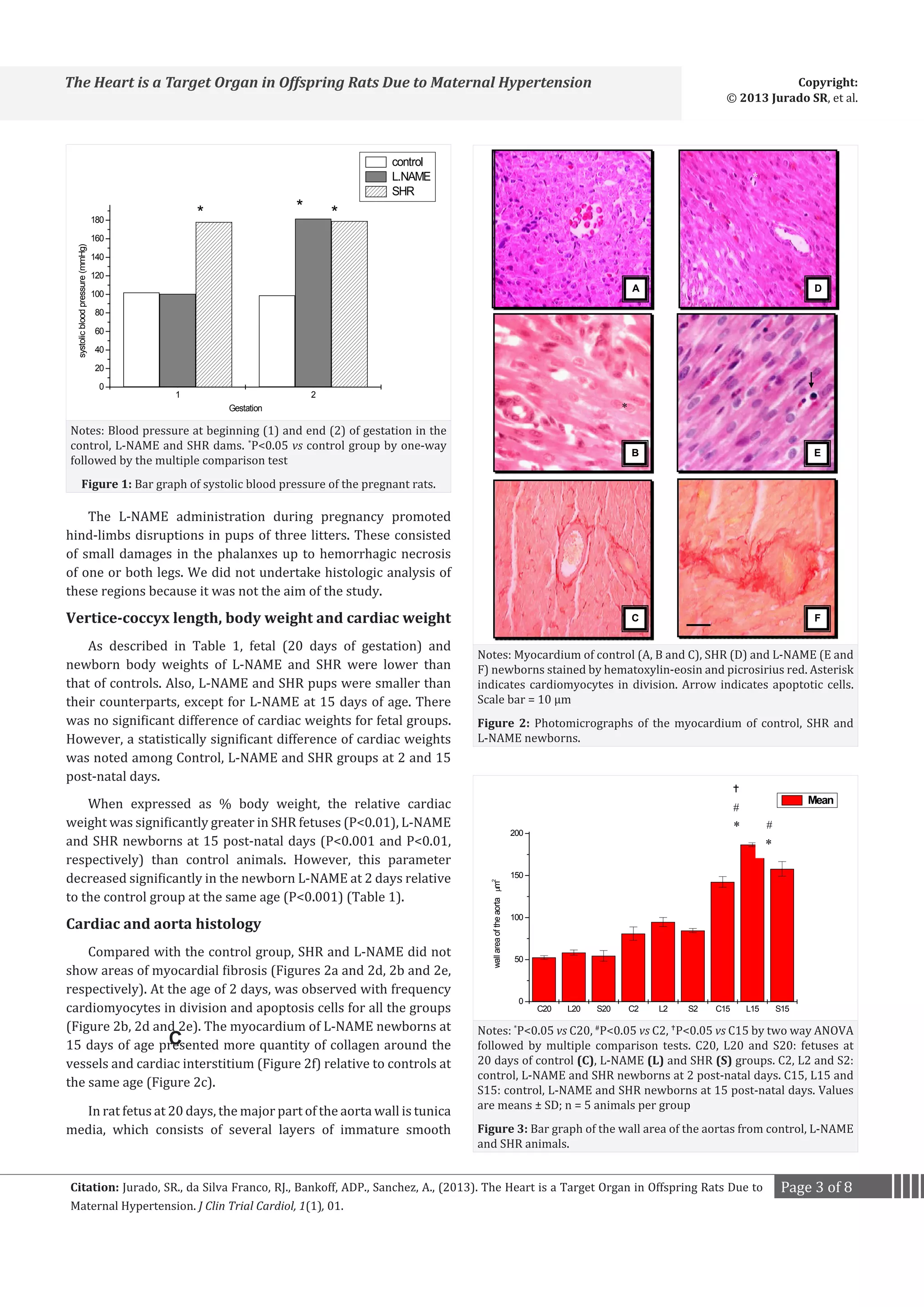
L-NAME and SHR pregnant rats showed higher arterial
pressure relative to dams normotensive (Figure 1). An
insignificant fall in blood pressure was observed in the Wistar
(98.8±4.6 mmHg) and SHR (178±5.2 mmHg) dams at the end of
gestation when compared at the beginning (102.0±5.2 mmHg
and 179.0±6.4 mmHg, respectively).
Citation: Jurado, SR., da Silva Franco, RJ., Bankoff, ADP., Sanchez, A., (2013). The Heart is a Target Organ in Offspring Rats Due to
Maternal Hypertension. J Clin Trial Cardiol, 1(1), 01.
Page 2 of 8](https://image.slidesharecdn.com/cardiology01-140108102816-phpapp01/75/The-Heart-is-a-Target-Organ-in-Offspring-Rats-Due-to-Maternal-Hypertension-2-2048.jpg)
![Copyright:
© 2013 Jurado SR, et al.
The Heart is a Target Organ in Offspring Rats Due to Maternal Hypertension
V-C
(mm)
3.222 ± 0.048
2.972 ± 0.036*
Control
Fetus
2.958 ± 0.086
L-NAME
*
SHR
4.886 ± 0.112
4.370 ± 0.077*
Control
2-day
4.290 ± 0.318
L-NAME
*
SHR
6.898 ± 0.136
7.466 ± 0.192
Control
15-day
L-NAME
6.198 ± 0.050*
2.815 0.248
2.138 ± 0.267*
2.092 ± 0.105
8.114 0.440
*
6.463 ± 0.312*
6.550 ± 0.265
*
29.832 1.315
27.906 ± 2.424
17.192 ± 0.495*
Data are given as means ± SD; n = 5 animals per group
P<0.05 vs controls by one-way ANOVA followed by the multiple comparison test
a
*
*
Total Body
(g)
SHR
Cardiac
(g)
Cardiac/Body
(%)
0.015 ± 0.002
0.602 0.075*
0.017 ± 0.002
0.015 ± 0.003
0.061 ± 0.007
0.034 ± 0.003*
0.048 ± 0.006
*
0.135 ± 0.008
0.163 ± 0.013
*
0.117 ± 0.004*
0.404 ± 0.050
0.370 0.104
0.763 ± 0.057
0.569 ± 0.035*
0.731 ± 0.112
0.570 ± 0.029
0.728 ± 0.067*
0.697 ± 0.041*
Table 1: Details of fetuses and newborns (control, L-NAME and SHR mothers) at 2 and 15 daysa.
muscle cells where the internal elastic membrane is observed.
There were no differences among aortas of control, L-NAME
and SHR fetuses. In newborn at 2 days, there was thickening
of media for all groups, however the elastic lamellae were less
individualized. In newborn at 15 days, the elastic lamellae were
well developed.
Aorta morphometry
The wall area of the aorta was greater in hypertensive groups
(L-NAME and SHR) than in control groups for all ages (Figure
3). The wall-to-lumen ratio increased significantly in L-NAME
fetuses in relative to controls (Figure 4).
There was an increase in the number of elastic lamellae in
L-NAME and SHR groups compared with control animals at 2
days, and stop increasing the number of elastic lamellae at 15
days comparing the same groups (Table 2).
Cardiac microvasculature morphometry
Representative examples of coronary vessels in the groups
studied at 2 and 15 days are shown in Figure 5.
Total blood vessel area (external area) increased with postnatal cardiac growth, but there was a significant difference
between control, L-NAME and SHR neonates only in the age of 15
days (Figure 6).
The wall area in the microvessels was greater in the L-NAME
and SHR groups than in the control group, however significant
difference was noted only at 15 post-natal days (Figure 7). Also,
the lumen area and external perimeter decreased significantly in
L-NAME and SHR newborn at 15 post-natal days when compared
to controls at the same age.
The wall-to-lumen ratio increased significantly in newborns
at 2 and 15 post-natal days of L-NAME and SHR groups relative
to controls at the same ages (Figure 8).
Collagen content
The collagen content is summarized in Table 2. Collagen
content was higher in the heart of SHR newborns at 2 days and
L-NAME and SHR at 15 days.
Discussion
There are few data in literature about the importance of nitric
oxide in the fetus and neonate development and growing. Our
results agree with previous ones showing that inhibition of nitric
oxide by L-NAME during the gestation disturbed the development
of fetus limbs [13,14].
Gardiner and collaborators [20] demonstrated that the
initial hypertensive response after intravenous administration
of L-NAME is accompanied by marked vasoconstriction in the
hindquarter, mesenteric and renal vascular beds. Over time,
both mesenteric and renal vasculature returns to baseline tone,
whereas the hind limb circulation demonstrates a sustained
vasoconstriction. This suggests that L-NAME crosses the placental
barrier and affects the fetal nitric oxide synthesis, leading to cell
death in the limbs because the NO has a role for in limb and digit
development [21].
In this study, apoptotic cells were observed with a frequency
in the myocardium of the all the groups at 2 post-natal days. Thus,
apoptosis or programmed cell death takes place in situations
of heart remodeling during or after the pathological processes
[22,23]. Therefore, it is logical to believe that apoptosis may also
occur in post-natal maturation of the heart and other tissues of
the cardiovascular system, which have to adapt to their new
hemodynamic role.
When expressed as % body weight, the relative cardiac
weight was significantly greater in SHR fetuses and L-NAME
and SHR newborns at 15 post-natal days than control animals.
The increase in relative cardiac weight of these animals may
Citation: Jurado, SR., da Silva Franco, RJ., Bankoff, ADP., Sanchez, A., (2013). The Heart is a Target Organ in Offspring Rats Due to
Maternal Hypertension. J Clin Trial Cardiol, 1(1), 01.
Page 4 of 8](https://image.slidesharecdn.com/cardiology01-140108102816-phpapp01/75/The-Heart-is-a-Target-Organ-in-Offspring-Rats-Due-to-Maternal-Hypertension-4-2048.jpg)
![Copyright:
© 2013 Jurado SR, et al.
The Heart is a Target Organ in Offspring Rats Due to Maternal Hypertension
Mean
3,5
3,0
wall to lumen ratio
2,5
*
#
2,0
1,5
#
*
#
*
A
E
D
E
B
E
C
*
F
1,0
0,5
0,0
Notes: P<0.05 vs C20, P<0.05 vs C2, P<0.05 vs C15 by two way ANOVA
followed by multiple comparison tests. Abbreviations as in Figure 3
*
C20
L20
S20
#
C2
L2
S2
C15
L15
S15
Figure 4: Bar graph of the wall-to-lumen ratio area of the aortas from
control, L-NAME and SHR animals.
The effect of NO inhibition of the rate of collagen synthesis
has been poorly characterized. A study demonstrated that the
NO-generation inhibits the protein synthesis in cultured vascular
smooth muscle cells [24]. Thus, we observed that NO inhibition
is reflected in the rate of synthesis of collagen during neonatal
development of the rat heart, especially in L-NAME neonate at
15 days. Therefore, the nitric oxide inhibition in vivo may lead
to increase collagen synthesis in the myocardium of the L-NAME
newborns.
Reduction in number of elastic lamellae in the aorta of
newborns L-NAME and SHR may impair the contractile properties
of the aorta. As a possible mechanism we speculate that, in fetuses
with growth retardation, such as L-NAME and SHR models, there
is impairment in the synthesis of elastin during a critical period
of blood vessel development. As a result of the elastin deficiency,
the compliance of the conduit arteries is reduced, leading to
higher pulse pressure.
Elastin synthesis in the aorta of SHRs was found to exceed
control levels in the prehypertensive period, decrease during the
development of hypertension and increase again in the period of
the established hypertensive state [25] In this study, the number
of elastic lamellae increases in L-NAME e SHR neonates at 2 days
and latter it stop increasing at 15 days.
The rat offspring born from L-NAME parents, with sustained
NO induced hypertension, had a remarkably higher blood
pressure [26]. In this study we assess not blood pressure of
newborns L-NAME and SHR, and we can not infer at the age of 15
days were hypertensive, however, the synthesis of elastic fibers
Notes: Control (A and D), L-NAME (B and E) and SHR newborns (C
and F) at 2 and 15 post-natal days stained by Mallory’s trichrome and
hematoxylin-eosin, respectively. A, B and C at 2 days and D, E and F at
15 days. L-NAME and SHR groups had increased wall-to-lumen ratio
compared with the control group. Scale bar = 10 μm
Figure 5: Photomicrographs of the coronary arterioles of control, SHR
and L-NAME newborns.
Mean
1200
1000
External area µm
be explained by an increase of collagen content or hypertrophy
cardiomyocyte. However, newborns L-NAME at 2 days had
lower relative cardiac weight probably due to lower number
of cardiomyocytes because apoptotic activity increased in the
hearts of these animals.
*
*
800
*
600
400
200
0
C2
L2
S2
C15
External diameter 50 µm
L15
S15
Notes: *P0.05 vs C15 by two way ANOVA followed by multiple
comparison tests. Abbreviations as Figure 3. Values are means ± SD; n
= 5 animals per group
Figure 6: Bar graph of the external area of coronary arteries with
external diameters 50 µm.
Citation: Jurado, SR., da Silva Franco, RJ., Bankoff, ADP., Sanchez, A., (2013). The Heart is a Target Organ in Offspring Rats Due to
Maternal Hypertension. J Clin Trial Cardiol, 1(1), 01.
Page 5 of 8](https://image.slidesharecdn.com/cardiology01-140108102816-phpapp01/75/The-Heart-is-a-Target-Organ-in-Offspring-Rats-Due-to-Maternal-Hypertension-5-2048.jpg)
![The Heart is a Target Organ in Offspring Rats Due to Maternal Hypertension
Age
Group
0.611 ± 0.022
7.10 ± 0.16*
0.602 ± 0.020
SHR
7.45 ± 0.17*
0.743 ± 0.020*
Control
15-day
Collagen content (%)
6.05 ± 0.18
L-NAME
2-day
Elastic lamellae (n)
Control
7.60 ± 0.17
2.118 ± 0.048
L-NAME
7.00 ± 0.16
2.251 ± 0.040
Data are given as means ± SD; n = 5 animals per group
P0.05 vs controls by two-way ANOVA followed by the multiple
comparison test
SHR
b
7.30 ± 0.19
2.531 ± 0.040*
*
Table 2: Number of elastic lamellae of the aorta and collagen content of
the heart of neonatesb.
Mean
500
*
*
600
*
*
*
wall area µm2
400
300
200
100
0
C2
L2
S2
C15
External diameter 50 µm
L15
S15
Notes: *P0.05 vs the corresponding values of the control group at 15
days by two way ANOVA followed by multiple comparison tests. Values
are means ± SD; n = 5 animals per group
Figure 7: Bar graph of the wall area of intramyocardial arteries with
external diameters 50 µm.
Mean
#
#
4
*
wall to lumen ratio
3
#
#
*
*
*
2
1
0
C2
L2
S2
C15
External diameter 50 µm
L15
S15
Notes: *P0.05 vs C2. #P0.05 vs C15 by two way ANOVA followed by
multiple comparison tests. Values are means ± SD; n = 5 animals per
group
Figure 8: Bar graph of the wall-to-lumen ratio of the myocardial
microvessels with external diameters 50 µm.
Copyright:
© 2013 Jurado SR, et al.
in the aorta in these animals was similar to that of adult animals
before and after the establishment of hypertension.
The modest increase of ratio wall-to-lumen of the aorta
in offspring at 2 days from L-NAME-treated mothers in this
study could be explained by a high level of NO synthesis and/
or its activity in the wall of major arteries even after L-NAME
treatment. This is supported by findings of high endothelial NO
production in the pulmonary artery during the fetal and early
post-natal period [27,28]. NO has been shown to inhibit vascular
smooth muscle cell proliferation [24].
The present study showed that the most significant change on
the L-NAME neonate myocardium involves the microvasculature,
meanly arterioles. The wall-to-lumen ratio of arterioles was
significantly higher in the L-NAME and SHR than in the control
group at 2 and 15 post-natal days. Numaguchi, K., et al. [8] also
found an increase of wall-to-lumen ratio in arterioles and small
coronary arterioles on the myocardium of L-NAME-treated rats
for 8 weeks. They observed an accumulation of collagen in the
media of microvessels and perivascular fibrosis and believed
that chronic L-NAME administration might increase sympathetic
nerve activity, which may contribute to vascular remodeling.
L-NAME-treated rats (50mg/Kg/day) during 8 weeks showed
an increase of wall thickness (tunica intima and tunica media)
of carotid and coronary arteries in relation to control rats [29].
Using the same dosage during the same period, other authors [30]
found a significant increase in the capillary domain in treating
animals, suggesting that there is no capillary proliferation, but
myocyte hypertrophy during this period.
In spontaneous hypertension, not all vascular beds display
the same increase in the ratio between the wall-to-lumen ratio. In
SHR, for instance, the change is less marked in the kidney than in
the mesenteric, cerebral or hindquarter skeletal muscle vascular
bed. Furthermore, not all vessels are affected simultaneously
during development [31]. Korsgaard, N., Mulvany, M.J. [32]
reported a smaller lumen diameter for mesenteric small arteries
of hypertensive rats. Also, in subcutaneous resistance arteries of
untreated hypertensive patients, a significant reduction of lumen
diameter was observed [32].
In established hypertension, peripheral vascular resistance
is elevated, primarily due to structural changes in the arterial
tree, most notably to an elevation of the wall thickness to lumen
diameter ratio of small arteries. According to Poiseuille’s law,
resistance is more markedly affected by arterial diameter than
by the length and number of arteries segments. So, arterial
resistance resides primarily in small “resistance-sized” arteries
(diameter 300µm) and in arterioles with characteristics of
precapillary vessels that present only one layer of smooth muscle
cells [33]. During the establishment of the arterial hypertension,
the cardiac output is normal, but the resistance is elevated
[31,34]. The arterial structural changes observed in hypertension
include an increase in the wall-to-lumen ratio and reduction of
the number of microvessels (rarefaction), leading to a reduction
Citation: Jurado, SR., da Silva Franco, RJ., Bankoff, ADP., Sanchez, A., (2013). The Heart is a Target Organ in Offspring Rats Due to
Maternal Hypertension. J Clin Trial Cardiol, 1(1), 01.
Page 6 of 8](https://image.slidesharecdn.com/cardiology01-140108102816-phpapp01/75/The-Heart-is-a-Target-Organ-in-Offspring-Rats-Due-to-Maternal-Hypertension-6-2048.jpg)
![The Heart is a Target Organ in Offspring Rats Due to Maternal Hypertension
of the average overall lumen diameter of the arterial system
[35]. However, neither arterial hypertrophy nor rarefaction
necessarily led to a reduction of lumen diameter and increase
in the vascular resistance. Vascular resistance depends on the
relationship between wall mass and lumen diameter [36].
Some questions can be rise regarding the increase of
wall-to-lumen ratio in the myocardial arterioles of L-NAME
neonates in this study: What does promote the increase of the
vascular wall thickness: smooth muscle cell hypertrophy and/
or hyperplasia? What factors stimulate the vascular smooth
muscle cell hypertrophy and/or hyperplasia? Only the nitric
oxide inhibition can promote the arterioles changes or other
factors that also contribute to these changes? In general, the
smooth muscle hyperplasia cells in the media of hypertensive
animals are considered a primary change, whereas hypertrophy
is considered a secondary one [37]. In the SHR and Goldblatt
hypertensive rats, the media mass increase in the aorta was due
to cellular hypertrophy and polyploidy, whereas in rats made
hypertensive by aortic coarctation, the median change was due
to hyperplasia of the smooth muscle cells [37].
Probably, the myocardial vascular changes induced by
long-term blockade of nitric oxide synthesis in rats result from
local angiotensin I converting enzyme (ACE) activation. Using
immunohistochemistry methods, Takemoto, M., et al. [38] found
an increase of ACE in the coronary arteries during the first week
of L-NAME administration. These investigators also reported an
increase of wall-to-lumen ratio in the myocardial vasculature as
well as in the aorta of L-NAME rats.
The increase of the wall-to-lumen ratio in L-NAME neonates
at 2 and 15 post-natal days are probably due to the nitric oxide
inhibition during the pregnancy joined to arterial pressure
increased in the newborns. Both effects induce smooth muscle
cell proliferation on myocardial arterioles. It is know that NO
inhibits smooth muscle cell proliferation [39]. A decreased NO
generation induces the synthesis of growth-promoting factors
from the endothelium [40]. The angiotensin-converting enzyme
(ACE) activation would increase the formation of angiotensin
II, which in turn directly induces vascular smooth muscle
proliferation [41] and the release of platelet-derived growth
factor [38], transforming growth factor-40 and endothelins [42].
The findings support the angiotensin II involvement in the
vascular smooth muscle cell hypertrophy/hyperplasia during
hypertension promoted by the administration of L-NAME in rats.
The majority of nitric oxide synthase (NOS) enzyme in the rat
heart is related to vessels and its production by the eighteenth
day of the embryo development [43]. Our results point out that
nitric oxide inhibition during pregnancy induces vascular smooth
muscle cell proliferation and accumulation of collagen in the
neonatal hearts.
Not only the vascular smooth muscle development is affected
by L-NAME, but also the administration of these nitric oxide
inhibitors during pregnancy compromises pyloric musculature,
hypertrophy and hyperplasia, in neonate rats [44].
Copyright:
© 2013 Jurado SR, et al.
Another factor that may contribute to myocardial
microvasculature changes in neonates from L-NAME-treated
mothers is the increase in the sympathetic activity in these
newborns. In L-NAME-treated rats, the increase in the
sympathetic activity leads to vascular remodeling: smooth
muscle cell proliferation and lumen occlusion in the myocardial
arterioles [8].
Conclusion
In conclusion, our results demonstrated that the chronic
inhibition of NO synthesis with L-NAME during the pregnancy
caused microvasculature remodeling in the ages of 2 and 15
days, and increased collagen content in the neonate hearts at
15th post-natal days. These changes were also described in SHR
newborns leading similarity between models of hypertension
during pregnancy.
References
1. Noll, G., Luscher, T. F. (1998). The endothelium in acute coronary
syndromes. European Heart Journal, 19(8 Pt 2), C30–38.
2. King, R. G., Gude, N. M., Di Julio, J. L., Brennecke, S. P. (1995).
Regulation of human placental fetal vessel tone: role of nitric oxide.
Reproduction Fertility and Development, 7(6), 1407–1411.
3. Sladek, S. M., Magness, R. R., Conrad K. P (1997). Nitric oxide and
pregnancy. American Journal of Physiology, 272(2 Pt 2), R441–R463.
4. Salas, S. P., Altermatt, F., Campos, M., Giacaman, A., Rosso, P(1995).
Effects of long–term nitric oxide synthesis inhibition on plasma volume
expansion and fetal growth in the pregnant rat. Hypertension,26(6 Pt
2), 1019–1023.
5. Zhang, Y., Kaufman, S. (2000). Effect of nitric oxide synthase
inhibition on cardiovascular and hormonal regulation during
pregnancy in the rat. Can J Physiol Pharmacol, 78(5), 423–427.
6. Ribeiro, M. O., Antunes, E., de Nucci, G., Lovisolo, S. M., Zatz, R. (1992)
. Chronic inhibition of nitric oxide synthesis. A new model animal of
arterial hypertension. Hypertension, 20(3), 298–303.
7. Arnal, J. F., el Amrani, A.I., Chatellier, G., Menard, J., Michel J.B(1993).
Cardiac weight in hypertension induced by nitric oxide synthase
blockade. Hypertension, 22(3), 380–387.
8. Numaguchi, K., Egashira, K., Takemoto, M., Kadokami, T., Shimokawa,
H., Sueishi, K., Takeshita A.(1995). Chronic inhibition of nitric
oxide synthesis causes coronary microvascular remodeling in rats.
Hypertension, 26(6 Pt 1),957–962.
9. Pessanha, M.G., Mandarin–de–Lacerda, C.A., Hahn, M.D.(1999).
Stereologyandimmunohistochemistryof
the
myocardium
in experimental hypertension: long–term and low–dosage
administration of inhibitor of the nitric oxide synthesis. Pathobiology,
67(1), 26–33.
10. Mandarim–de–Lacerda, C.A., Pereira, L.M.M.( 2000). Numerical
density of cardiomyocytes in chronic nitric oxide synthesis inhibition.
Pathobiology,68(1), 36–42.
11. de Oliveira, C.F., Cintra, K.A., Teixeira, S.A., De Luca I.M.S, Antunes E.,
De Nucci G. (2000). Development of cardiomyocyte hypotrophy in rats
under prolonged treatment with a low dose of a nitric oxide synthesis
inhibitor. European Journal of Pharmacology,391(1-2), 121–126.
12. Miller, M.J.S, Voelker, C. A., Olister, S., Thompson, J. H., Zhang, X.–J.,
Citation: Jurado, SR., da Silva Franco, RJ., Bankoff, ADP., Sanchez, A., (2013). The Heart is a Target Organ in Offspring Rats Due to
Maternal Hypertension. J Clin Trial Cardiol, 1(1), 01.
Page 7 of 8](https://image.slidesharecdn.com/cardiology01-140108102816-phpapp01/75/The-Heart-is-a-Target-Organ-in-Offspring-Rats-Due-to-Maternal-Hypertension-7-2048.jpg)