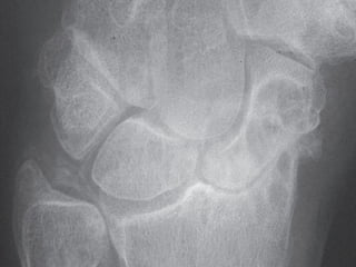
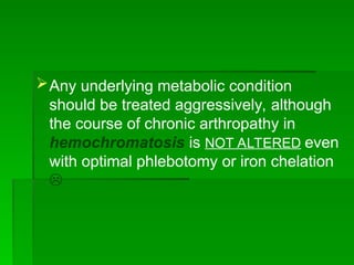
HISTORY
HISTORY
 CPPD crystals were first identified in 1962
CPPD crystals were first identified in 1962
when McCarty and colleagues discovered non-
urate crystals of patients with acute synovitis
and CC.
and CC.
 The clinical similarity to gout prompted the
The clinical similarity to gout prompted the
term
term pseudogout
pseudogout for this new “crystal-induced
for this new “crystal-induced
arthropathy
arthropathy.
EPIDEMIOLOGY
EPIDEMIOLOGY
•
CPPD is a disease of the elderly, mainly > 50
years
years
.
.
•
•
CPPD in patients < 50 years with
chondrocalcinosis (CC) should be evaluated for a
familial form or metabolic diseases associated
with CPPD
with CPPD
.
.
•
•
Chronic CPPD should be considered in any
elderly patient with symptoms suggesting
seronegative RA or PMR
.
CLASSIFICATION
CLASSIFICATION
•
Asymptomatic
•
Acute CPPD
“
Pseudogout
”
•
Chronic CPPD
“
Pseudo-OA
”
“
Pseudo-RA
”
“
Pseudo-neuropathic” (Rare)
Spinal involvement (Rare)
RISK FACTORS
RISK FACTORS
•
Idiopathic (sporadic): advancing age
•
Joint trauma and/or surgery
•
Familial: in young-onset CPPD:Patients tend to
present in their 20s and 30s and are more likely to
have spinal involvement
.
•
Associated Conditions:(‘5 Hs’)
.
HYPERPARATHYROIDISM, Hypophosphatasia,
Hemochromatosis
Hypomagnesemia, Hypothyroidism
?!
CLINICAL PICTURE
CLINICAL PICTURE
Asymptomatic CPPD
•
Identified when CPPD crystals are seen accidentally
in cartilage on radiographs
.
•
This finding is termed chondrocalcinosis(CC),the
most common sites for CC are the knees and
triangular fibrocartilage of the wrists
(>90%) ,Calcifications should be bilateral
.
•
20%
of CPPD can cause arthritis without
being seen as CC
If calcifications found unilaterally may be
basic calcium phosphate (BCP) deposition due
to trauma “Hydroxyapatite (HA) crystals
!!??”
Acute CPPD/Pseudogout
•
More common in men
.
•
Attacks may be precipitated by severe illness,
trauma, or surgery
.
•
It manifests as severe, sudden-onset mono- or
oligoarthritis with erythema, swelling, pain
.
•
Usually self-limiting, and resolve within 1–2 weeks
.
•
Knee most commonly affected, followed by the
wrist, shoulder, ankle, and elbow
.
Situations That May Trigger Acute CPPD
Definite
Direct trauma to a joint
Recurrent illness (e.g., chest infection, UTI, MI)
Surgery (especially parathyroidectomy)
Blood transfusion, parenteral fluid administration
Joint lavage
Possible
Institution of thyroxine replacement therapy
Intraarticular injection of hyaluronan  
Bisphosphonate treatment
Chronic CPPD
•
“
Pseudo-OA
”
-
Occurs in 50% of patients with CPPD
.
-
Pseudo-OA is more common in women
.
There is involvement of joints that are not
typically associated with OA
(
e.g., wrists, MCP, elbows, Glenohumeral, and
ankles
)
-
Isolated or predominant involvement of the
radiocarpal or patellofemoral joint-space
narrowing
.
-
Valgus deformity of the knees is especially
suggestive of underlying CPPD or BCP, as it
affects the lateral compartment
•
“ •
Pseudo-RA”
- Occurs in 5% of patients with CPPD.
- Patients will typically present with
symmetric joint involvement and prominent
systemic complaints (e.g., morning stiffness
and fatigue).
- May have pseudo-RA appearance with
ulnar drift and MCP subluxation
- 10% of patients with CPPD will test
positive for RF (low titer) because of their
age
.
•
Spinal involvement (Rare)
Spinal deposits occur in the ligametum flavum,
resulting in clinical symptoms of cord
compression, in either the cervical or lumbar
spine; lumbar spine involvement may produce
an acute radiculopathy or neurogenic
claudication resulting from spinal stenosis
.
INVESTIGATIONS
INVESTIGATIONS
Synovial Fluid Analysis
Polarized light microscopy: CPPD crystals are
rhomBoid-shaped and Positively Birefringent. They
appear Blue when parallel to the long axis of the
compensator
.
WBCs: typically ranges between 5000 and 50,000
cells/mcL
.
IMAGING
Plain Radiographs
:
Chondrocalcinosis It is classically seen in
-
Fibrocartilage (knee menisci, triangular
ligament of the wrist, symphysis pubis) →
punctate
punctate
-
Hyaline articular cartilage (knee, shoulder,
hip) → linear
linear
-
Tendon insertion sites (Achilles) → linear
-
Bursa
Structural Joint Changes
-
CPPD can be associated with
subchondral sclerosis, subchondral
cyst, and joint space narrowing. Although
the findings are also seen in OA, the
location may help differentiate
.
-
Unlike RA, CPPD does not have typical
bony erosions
.
 Other Imaging
MSKUS
:
•
CPPD can appear as a thin hyperechoic
band within the substance of hyaline
cartilage, and as rounded or amorphous
deposits in fibrocartilage
.
•
MSKUS has high specificity (96%) and
moderate to high sensitivity (86 %)
CT
It is the preferred method of identifying
calcium deposits in upper cervical spine
Laboratories
Screening for metabolic diseases after CPPD diagnosis,
particularly in young patients < 50 years or patients with
severe arthritis
.
•
Calcium (rule out hyperparathyroidism, total
calcium should be ↑↑ > 10.5 mg/dL)
•
Phosphorus (rule out hyperparathyroidism, should be
less than 2.5 mg/dL)
•
Alkaline phosphatase (rule out hypophosphatasia,
should be low)
•
Magnesium (rule out hypomagnesemia, usually
from renal wasting)
•
Ferritin, iron, total iron-binding capacity (rule
out hemochromatosis)
•
Renal function
•
TSH (rule out hypothyroidism)
,
 Starting thyroxin therapy in a hypothyroid
patient who has CPPD may  precipitate a
pseudogout attack
!?
Management
Management
Acute CPPD/Pseudogout
•
•
NSAIDs
NSAIDs
•
•
Aspiration and injection of corticosteroids
•
•
Oral corticosteroids
•
•
Colchicine
Colchicine
•
•
IL-1 inhibitors such as anakinra
!?
!?
Management
Management
Chronic CPPD
Chronic CPPD
•
•
Colchicine (0.6 mg once to three times daily)
may decrease the frequency of attacks
.
.
•
• Low-dose corticosteroids
Low-dose corticosteroids
•
•
MTX and HCQ may be considered in
resistant cases
resistant cases
.
.
•
IL-1 inhibitors such as anakinra
Any underlying metabolic condition
should be treated aggressively, although
the course of chronic arthropathy in
hemochromatosis is NOT ALTERED even
with optimal phlebotomy or iron chelation

Calcium Pyrophosphate Deposition (CPPD) overview.ppt

Calcium Pyrophosphate Deposition (CPPD) overview.ppt

  • 2.
    HISTORY HISTORY  CPPD crystalswere first identified in 1962 CPPD crystals were first identified in 1962 when McCarty and colleagues discovered non- when McCarty and colleagues discovered non- urate crystals of patients with acute synovitis urate crystals of patients with acute synovitis and CC. and CC.  The clinical similarity to gout prompted the The clinical similarity to gout prompted the term term pseudogout pseudogout for this new “crystal-induced for this new “crystal-induced arthropathy arthropathy.
  • 3.
    EPIDEMIOLOGY EPIDEMIOLOGY • CPPD is adisease of the elderly, mainly > 50 CPPD is a disease of the elderly, mainly > 50 years years . . • • CPPD in patients < 50 years with CPPD in patients < 50 years with chondrocalcinosis (CC) should be evaluated for a chondrocalcinosis (CC) should be evaluated for a familial form or metabolic diseases associated familial form or metabolic diseases associated with CPPD with CPPD . . • • Chronic CPPD should be considered in any Chronic CPPD should be considered in any elderly patient with symptoms suggesting elderly patient with symptoms suggesting seronegative RA or PMR seronegative RA or PMR .
  • 4.
  • 5.
    RISK FACTORS RISK FACTORS • Idiopathic(sporadic): advancing age • Joint trauma and/or surgery • Familial: in young-onset CPPD:Patients tend to present in their 20s and 30s and are more likely to have spinal involvement . • Associated Conditions:(‘5 Hs’) . HYPERPARATHYROIDISM, Hypophosphatasia, Hemochromatosis Hypomagnesemia, Hypothyroidism ?!
  • 6.
    CLINICAL PICTURE CLINICAL PICTURE AsymptomaticCPPD • Identified when CPPD crystals are seen accidentally in cartilage on radiographs . • This finding is termed chondrocalcinosis(CC),the most common sites for CC are the knees and triangular fibrocartilage of the wrists (>90%) ,Calcifications should be bilateral .
  • 7.
    • 20% of CPPD cancause arthritis without being seen as CC If calcifications found unilaterally may be basic calcium phosphate (BCP) deposition due to trauma “Hydroxyapatite (HA) crystals !!??”
  • 8.
    Acute CPPD/Pseudogout • More commonin men . • Attacks may be precipitated by severe illness, trauma, or surgery . • It manifests as severe, sudden-onset mono- or oligoarthritis with erythema, swelling, pain . • Usually self-limiting, and resolve within 1–2 weeks . • Knee most commonly affected, followed by the wrist, shoulder, ankle, and elbow .
  • 9.
    Situations That MayTrigger Acute CPPD Definite Direct trauma to a joint Recurrent illness (e.g., chest infection, UTI, MI) Surgery (especially parathyroidectomy) Blood transfusion, parenteral fluid administration Joint lavage Possible Institution of thyroxine replacement therapy Intraarticular injection of hyaluronan   Bisphosphonate treatment
  • 12.
    Chronic CPPD • “ Pseudo-OA ” - Occurs in50% of patients with CPPD . - Pseudo-OA is more common in women . There is involvement of joints that are not typically associated with OA ( e.g., wrists, MCP, elbows, Glenohumeral, and ankles )
  • 13.
    - Isolated or predominantinvolvement of the radiocarpal or patellofemoral joint-space narrowing . - Valgus deformity of the knees is especially suggestive of underlying CPPD or BCP, as it affects the lateral compartment
  • 16.
    • “ • Pseudo-RA” - Occursin 5% of patients with CPPD. - Patients will typically present with symmetric joint involvement and prominent systemic complaints (e.g., morning stiffness and fatigue). - May have pseudo-RA appearance with ulnar drift and MCP subluxation - 10% of patients with CPPD will test positive for RF (low titer) because of their age .
  • 17.
    • Spinal involvement (Rare) Spinaldeposits occur in the ligametum flavum, resulting in clinical symptoms of cord compression, in either the cervical or lumbar spine; lumbar spine involvement may produce an acute radiculopathy or neurogenic claudication resulting from spinal stenosis .
  • 18.
    INVESTIGATIONS INVESTIGATIONS Synovial Fluid Analysis Polarizedlight microscopy: CPPD crystals are rhomBoid-shaped and Positively Birefringent. They appear Blue when parallel to the long axis of the compensator . WBCs: typically ranges between 5000 and 50,000 cells/mcL .
  • 19.
    IMAGING Plain Radiographs : Chondrocalcinosis Itis classically seen in - Fibrocartilage (knee menisci, triangular ligament of the wrist, symphysis pubis) → punctate punctate - Hyaline articular cartilage (knee, shoulder, hip) → linear linear - Tendon insertion sites (Achilles) → linear - Bursa
  • 23.
    Structural Joint Changes - CPPDcan be associated with subchondral sclerosis, subchondral cyst, and joint space narrowing. Although the findings are also seen in OA, the location may help differentiate . - Unlike RA, CPPD does not have typical bony erosions .
  • 24.
     Other Imaging MSKUS : • CPPDcan appear as a thin hyperechoic band within the substance of hyaline cartilage, and as rounded or amorphous deposits in fibrocartilage . • MSKUS has high specificity (96%) and moderate to high sensitivity (86 %) CT It is the preferred method of identifying calcium deposits in upper cervical spine
  • 25.
    Laboratories Screening for metabolicdiseases after CPPD diagnosis, particularly in young patients < 50 years or patients with severe arthritis . • Calcium (rule out hyperparathyroidism, total calcium should be ↑↑ > 10.5 mg/dL) • Phosphorus (rule out hyperparathyroidism, should be less than 2.5 mg/dL) • Alkaline phosphatase (rule out hypophosphatasia, should be low) • Magnesium (rule out hypomagnesemia, usually from renal wasting)
  • 26.
    • Ferritin, iron, totaliron-binding capacity (rule out hemochromatosis) • Renal function • TSH (rule out hypothyroidism) ,  Starting thyroxin therapy in a hypothyroid patient who has CPPD may  precipitate a pseudogout attack !?
  • 27.
    Management Management Acute CPPD/Pseudogout Acute CPPD/Pseudogout • • NSAIDs NSAIDs • • Aspirationand injection of corticosteroids Aspiration and injection of corticosteroids • • Oral corticosteroids Oral corticosteroids • • Colchicine Colchicine • • IL-1 inhibitors such as anakinra IL-1 inhibitors such as anakinra !? !?
  • 28.
    Management Management Chronic CPPD Chronic CPPD • • Colchicine(0.6 mg once to three times daily) Colchicine (0.6 mg once to three times daily) may decrease the frequency of attacks may decrease the frequency of attacks . . • • Low-dose corticosteroids Low-dose corticosteroids • • MTX and HCQ may be considered in MTX and HCQ may be considered in resistant cases resistant cases . . • IL-1 inhibitors such as anakinra IL-1 inhibitors such as anakinra
  • 29.
    Any underlying metaboliccondition should be treated aggressively, although the course of chronic arthropathy in hemochromatosis is NOT ALTERED even with optimal phlebotomy or iron chelation 