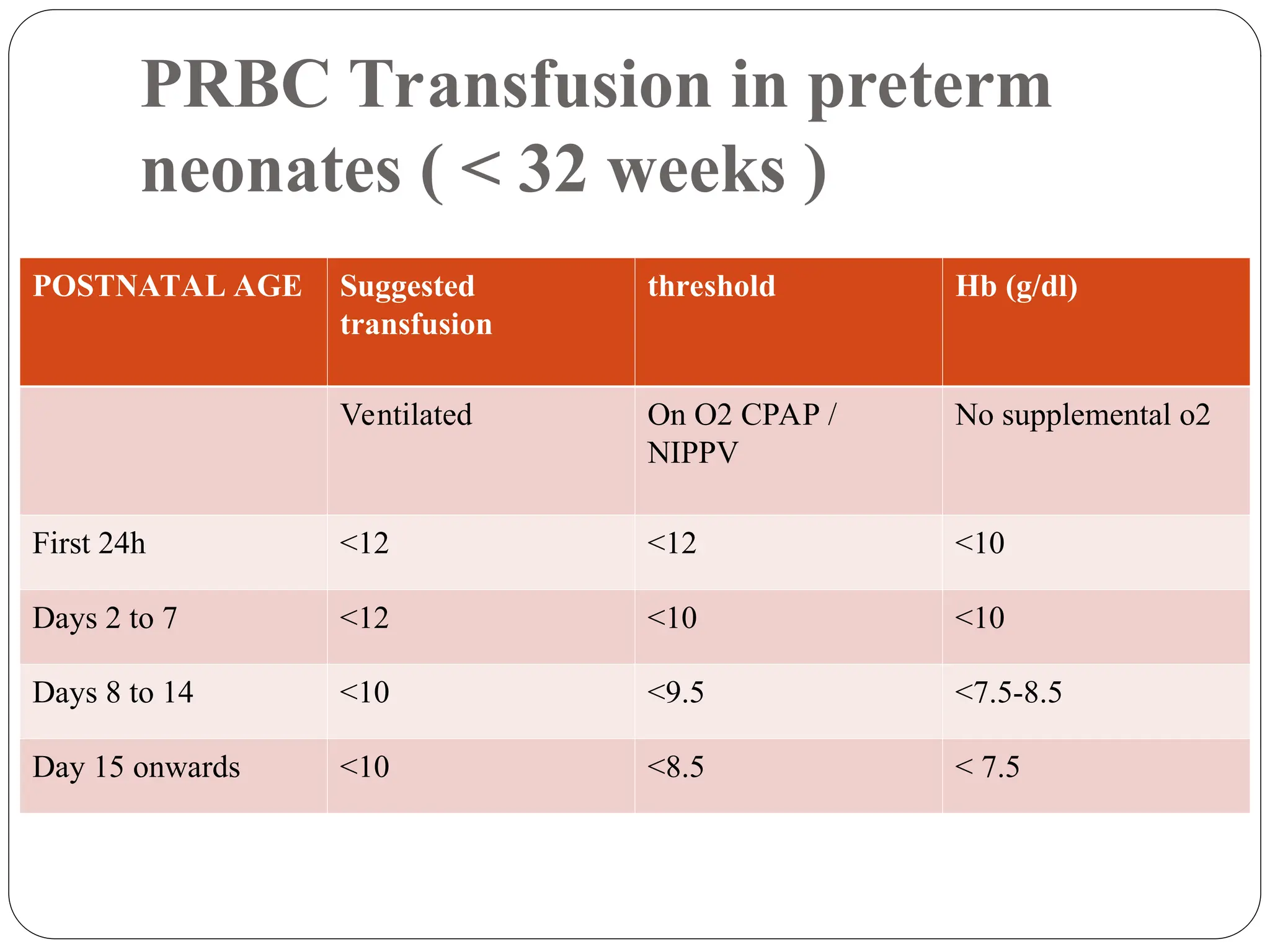
Presented by: Dr. Love Khanna (PG-1)
Moderator : Dr. Santosh Kumar
Associate Professor,AMCH
BLOOD COMPONENTS
TRANSFUSION IN NEONATES
Packed Red Blood Cells
1. PRBC s are prepared from whole blood after centrifugation within 8 hours of
collection. Preservative solutions are used such as :
2. SAGM ( Saline , Adenine , Glucose , Mannitol )
3. Shelf life of 42 days
4. Citrate phosphate dextrose (CPD) shelf life of 21 days.
5. Subsequently Adenine was added to the preservative thus forming CPD-A which
improved the ATP content of the stored blood and thus increased the shelf life to 35
days.
6. Stored at a temp of 2-6 C irrespective of preservative used.
Directed or designated donor blood
 Blood donated by family member or friends for specific patients is commonly
known as directed or designated donor blood.
 Directed donations have a small increase in the rate of infectious disease
transmission.
 Additionally, in a case of hemolytic disease of the newborn or neonatal alloimmune
thrombocytopenia, the neonate’s blood contains maternal antibodies that are
directed against paternally inherited antigens on blood cells. In these cases, paternal
relative’s blood may carry the same antigens rendering their blood incompatible
with the baby.
Directed or designated donor blood
 Finally, directed donor blood from relatives can induce
an immune response against human leukocyte antigen
(HLA) and other antigens against those relatives. This
would complicate future therapy if the relatives were to
be considered as donors of other tissue for the patient
later in life.
Leucocyte Reduction of PRBCs
To reduce the concentration of leucocytes to less than 5x10⁶
leukocytes per unit of RBCs by using special filters.
Preventing :
1. Non-hemolytic febrile transfusion reactions (NHFTR),
2. HLA alloimmunization,
3. Transmission of leukotropic viruses (CMV, EBV,HTLV-1),
4. Transfusion related GVHD,
5. Transfusion related acute lung injury (TRALI).
Gamma irradiation
1. Done to inactivate donor T cells, and the associated risk of transfusion
associated graft versus host disease (TA-GVHD),
2. TA-GVHD which may occur in immuno suppressed.patients, very small
babies, in large volume transfusions and during intrauterine transfusions or
when the donor is related.
3. Irradiation reduces the shelf life of RBCs to 28 days
4. Also causes leakage of potassium out of RBCs.
5. Should be used within 4 hours in neonates to avert the risk of hyperkalemia.
Washed RBCs
1. Washing RBCs with saline is done to remove plasma and
to reduce potassium in the RBCs.
2. Washed RBCs are recommended for intrauterine
transfusions, exchange transfusion and large volume
transfusions (more than 20 mL/kg).
CMV reduced RBCs
CMV reduction can be achieved by either
 Leucoreduction of blood components,
 By pre-selecting donors who are CMV negative.
PLATELETS
 RANDOM DONOR PLATELET (RDP)
 Isolated from the whole blood donations or by
Apheresis.
 From whole blood, platelet can be produced either by
platelet rich plasma (PRP) method or buffy- coat
method.
APHERESIS
1. Blood components are produced from whole blood donations by
selectively collecting one or more components directly from
donors and returning the rest to the circulation.
2. Apheresis can be used to collect platelets, plasma, red cells
or granulocytes from the donor.
3. Main advantage of apheresis collections are that more than one
dose of platelets or red cells can be collected from one donor per
donation, thus reducing patient exposure to multiple donors.
Single donor platelet (SDP)
1. Obtained by platelet Apheresis wherein multiple units of
platelets are collected from single donor and the RBCs and
platelet poor plasma are returned to the donor.
2. The procedure is repeated 4 to 6 times, yielding 4 to 6
units of platelets from one individual.
3. It is especially useful to prevent alloimmunization in
multiply transfused patients.
1. The concentration of platelets is more in SDP than in RDP, with SDP having a
platelet concentration of 3x10¹¹/unit and RDP having a concentration of
0.5x10¹⁰ per unit.
2. In neonatal transfusion practice, RDP is generally adequate to treat thrombocytopenia.
3. SDP is required only if prolonged and severe thrombocytopenia is anticipated,
requiring multiple platelet transfusions.
4. Platelets should be stored at 22-24°C with continuous gentle agitation in platelet
incubator and agitator for 5 days.
FRESH FROZEN PLASMA (FFP)
1. Produced by rapidly freezing the plasma within 8 hours of
collection in order to preserve the Activity of coagulation
factors V and VIII which are relatively labile.
2. Can be stored for up to 24 to 36 months depending on the
storage temperature, which is usually below -30°C.
3. Once thawed, FFP should be used immediately but can be
stored for up to 24 hours at 4°C.
CRYOPRECIPITATE
 Prepared from FFP by thawing at 2 to 4o C.
 Un dissolved cryoprecipitate is collected by centrifugation and
supernatant plasma is aseptically expressed into a satellite bag.
 Can be stored for 12 months at -30° C or lower.
 Thawed Cryoprecipitate can be stored for 6 hours at 2-6° C .
 Pooled cryoprecipitate kept at 2-6°C should be used within 4
hours.
PRBC Transfusion in preterm
neonates ( < 32 weeks )
POSTNATAL AGE Suggested
transfusion
threshold Hb (g/dl)
Ventilated On O2 CPAP /
NIPPV
No supplemental o2
First 24h <12 <12 <10
Days 2 to 7 <12 <10 <10
Days 8 to 14 <10 <9.5 <7.5-8.5
Day 15 onwards <10 <8.5 < 7.5
PRBC Transfusion in term neonates
Condition Hb (g/dl)
Severe pulmonary Disease <12
Moderate pulmonary disease <10
Severe cardiac disease <12
Major surgery <10
Symptomatic anemia <7
PRBC Transfusion
1. Transfuse 10-15ml/kg in non bleeding neonate , at 5ml/kg/hr .
2. Fresh RBCs (less than 7 days old) with high 2, 3-DPG levels ensure higher
tissue oxygen delivery. They also reduce the risk of hyperkalemia.
3. It is preferable to take samples from both, mother and the newborn, for initial
testing prior to transfusion. Mother’s sample should be tested for blood group
and for any atypical red cell antibodies.
4. Each transfusion of 9 mL/kg of body weight should increase hemoglobin level
by 3 g/dL.
PRBC Transfusion
 In exchange transfusions for Rh hemolytic disease of
newborn, blood transfused should be compatible with
mother’s serum.
 Ideally Rh negative blood of the baby’s ABO group has to be
used after cross matching with maternal serum. If compatible
ABO group is not available then group O and Rh negative
blood can be used.
Platelet Transfusion
 Thrombocytopenia is defined as platelet count less than 1.5
lakh/cubic mm.
PLATELET COUNT CONDITION
< 25,000/mm3 Neonates with no bleeding ( including
neonates with NAIT if no bleeding and
no family history of ICH )
<50,0000/mm3 Neonates with bleeding
Evidence of coagulopathy
Before surgery
NAIT if previously affected subling
with ICH
<1,00,000/mm3 Major bleeding eg. Significant IVH
Major surgery
Platelet Transfusion
 Usual recommended dose of platelets for neonates is 1 unit of
platelets per 10 kg body weight, which amounts to 5 mL/kg.
 The predicted rise in platelet count from a 5-mL/kg dose
would be 20 to 60,000/cubic mm.
 Doses of up to 10-20 ml/kg may be used in case of severe
thrombocytopenia
 Rate of platelet transfusion is 10-20 ml/kg/hr.
Fresh frozen plasma
Only valid indications for transfusing FFP in a newborns-
1. Disseminated intravascular coagulopathy
2. Vitamin K deficiency bleeding
3. Inherited deficiencies of coagulation factors
 Transfuse 15ml-20ml/kg , at 5ml/kg/hr .
Cryoprecipitate
 Cryoprecipitate contains about
 80 to 100 U of factor VIII
 Minimum150 mg of fibrinogen
 and varying amounts of factor XIII
 Transfuse 5ml-10ml/kg , at 10-20ml/kg/hr
Transfusion Associated Risks
Blood transfusion reactions may be broadly classified as
1. Infectious
2. Non-infectious
a. Acute
i. Immunologic
ii. Non-immunologic
b. Delayed
Infectious complications
 Mandatory to test every unit of blood collected for hepatitis
B, hepatitis C, HIV/AIDS, syphilis and malaria.
 NAT screening is also available for emerging infections like
Zika,West Nile virus and Babesia Microti.
Non Infectious Complications
 Acute- immune mediated reactions
Immune mediated hemolysis
 Newborns do not form red blood cell (RBC) antibodies; all antibodies present are maternal
in origin.
(1) Newborns must be screened for maternal RBC antibodies, including ABO antibodies if
non-O RBCs are to be given as the first transfusion.
(2) If the initial results are negative, no further testing is needed for the initial 4 postnatal
months.
 Symptoms. include hypotension, fever, tachycardia, infusion site pain, and hematuria.
 Treatment- Administer fluids and furosemide to protect the kidneys. If necessary, treat
hypotension with pressors and use hemostatic agents for bleeding; there may be a need to
transfuse compatible RBCs.
TRALI (Transfusion related acute lung injury )
 Noncardiogenic pulmonary edema complicating transfusion therapy.
 Most common symptoms associated with TRALI are dyspnea, cough, and fever, associated
with hypo- or hypertension.
 It occurs most commonly worsening of respiratory distress (described as increased need of
FiO2 by 10 or mean airway pressure of 2) within the initial 6 hours after transfusion.
 The presence of anti-HLA and/or anti-granulocyte antibodies in the plasma of donors is
implicated in the pathogenesis of TRALI.
 Diagnosis requires a high index of suspicion, and confirmation of donor serum cross-
reacting antibodies against the recipient.
 Treatment is mainly supportive in this self-limiting condition.
Acute- Immune Mediated Reactions
 Febrile nonhemolytic transfusion reactions
(FNHTR) are suspected in the absence of hemolysis
with an increase in body temperature of less than 2°C.
 Allergic reactions Allergic reactions are caused by
presence of preformed immunoglobulin E antibody against
an allergen in the transfused plasma, and are a rare
occurrence in newborns.
Transfusion/Anemia-associated NEC
 There are multiple retrospective studies reporting association between NEC occurring within 24 to 48 hours
of RBC transfusions in VLBW babies.
 Significantly lower NEC in units that used less RBC transfusions.
 NEC was common in lower gestations and birth weight. Patent ductus arteriosus (PDA) was often present.
 There are conflicting reports of RBC transfusion having protective effect.
 Prospective studies showed weak association between transfusion and NEC; in fact, severe anemia (<8 g/dL)
itself, not transfusions, is likely in the causative pathway of NEC (transfusion/anemia associated NEC).
 Several studies have reported decreased incidence of NEC by withholding feeding around the time of
transfusion, but good-quality evidence is lacking.
Acute non immune reactions
 Fluid overload
 Metabolic complications
 Hyperkalemia: In stored blood, potassium levels tend to be high. It has been seen that
after storage for around 42 days, potassium levels may reach 50 mEq/L in a RBC
unit.
 Hypothermia. Cool blood can cause hypothermia. Transfusion through warmers can
prevent this.
 Hypoglycemia: Blood stored in CPD blood has a high content of glucose leading to a
rebound rise in insulin release 1-2 hours after transfusion. This may lead to
hypoglycemia and routine monitoring is necessary, particularly after exchange
transfusion, after 2 and 6 hours, to ensure that this complication does not occur.
 Acid- base derangements: Metabolism of citrate in CPD
leads to late metabolic alkalosis. Metabolic acidosis is an
immediate complication occurring in sick babies who cannot
metabolize citrate.
 Hypocalcemia and Hypomagnesemia are caused by binding of
these ions by citrate present in CPD blood.
Delayed Complications
 Alloimmunization: Alloimmunization is an uncommon occurrence before
the age of 4 months, and is caused by transfusion of blood products with are
mismatched for highly immunogenic antigens like Rh.
 Transfusion associated graft versus host disease (TA-GVHD): Newborns
are at risk for TA-GVHD if they have received intrauterine transfusions,
exchange transfusions, or are very small, or immunocompromised. Unchecked
donor T cell proliferation is the cause of TA-GVHD, and it can be effectively
prevented by leucoreduction of the transfused blood products in at risk patients.

blood components @ neonates.ppt blood transfusion guidelines

  • 1.
    Presented by: Dr.Love Khanna (PG-1) Moderator : Dr. Santosh Kumar Associate Professor,AMCH BLOOD COMPONENTS TRANSFUSION IN NEONATES
  • 2.
    Packed Red BloodCells 1. PRBC s are prepared from whole blood after centrifugation within 8 hours of collection. Preservative solutions are used such as : 2. SAGM ( Saline , Adenine , Glucose , Mannitol ) 3. Shelf life of 42 days 4. Citrate phosphate dextrose (CPD) shelf life of 21 days. 5. Subsequently Adenine was added to the preservative thus forming CPD-A which improved the ATP content of the stored blood and thus increased the shelf life to 35 days. 6. Stored at a temp of 2-6 C irrespective of preservative used.
  • 3.
    Directed or designateddonor blood  Blood donated by family member or friends for specific patients is commonly known as directed or designated donor blood.  Directed donations have a small increase in the rate of infectious disease transmission.  Additionally, in a case of hemolytic disease of the newborn or neonatal alloimmune thrombocytopenia, the neonate’s blood contains maternal antibodies that are directed against paternally inherited antigens on blood cells. In these cases, paternal relative’s blood may carry the same antigens rendering their blood incompatible with the baby.
  • 4.
    Directed or designateddonor blood  Finally, directed donor blood from relatives can induce an immune response against human leukocyte antigen (HLA) and other antigens against those relatives. This would complicate future therapy if the relatives were to be considered as donors of other tissue for the patient later in life.
  • 5.
    Leucocyte Reduction ofPRBCs To reduce the concentration of leucocytes to less than 5x10⁶ leukocytes per unit of RBCs by using special filters. Preventing : 1. Non-hemolytic febrile transfusion reactions (NHFTR), 2. HLA alloimmunization, 3. Transmission of leukotropic viruses (CMV, EBV,HTLV-1), 4. Transfusion related GVHD, 5. Transfusion related acute lung injury (TRALI).
  • 6.
    Gamma irradiation 1. Doneto inactivate donor T cells, and the associated risk of transfusion associated graft versus host disease (TA-GVHD), 2. TA-GVHD which may occur in immuno suppressed.patients, very small babies, in large volume transfusions and during intrauterine transfusions or when the donor is related. 3. Irradiation reduces the shelf life of RBCs to 28 days 4. Also causes leakage of potassium out of RBCs. 5. Should be used within 4 hours in neonates to avert the risk of hyperkalemia.
  • 7.
    Washed RBCs 1. WashingRBCs with saline is done to remove plasma and to reduce potassium in the RBCs. 2. Washed RBCs are recommended for intrauterine transfusions, exchange transfusion and large volume transfusions (more than 20 mL/kg).
  • 8.
    CMV reduced RBCs CMVreduction can be achieved by either  Leucoreduction of blood components,  By pre-selecting donors who are CMV negative.
  • 9.
    PLATELETS  RANDOM DONORPLATELET (RDP)  Isolated from the whole blood donations or by Apheresis.  From whole blood, platelet can be produced either by platelet rich plasma (PRP) method or buffy- coat method.
  • 10.
    APHERESIS 1. Blood componentsare produced from whole blood donations by selectively collecting one or more components directly from donors and returning the rest to the circulation. 2. Apheresis can be used to collect platelets, plasma, red cells or granulocytes from the donor. 3. Main advantage of apheresis collections are that more than one dose of platelets or red cells can be collected from one donor per donation, thus reducing patient exposure to multiple donors.
  • 11.
    Single donor platelet(SDP) 1. Obtained by platelet Apheresis wherein multiple units of platelets are collected from single donor and the RBCs and platelet poor plasma are returned to the donor. 2. The procedure is repeated 4 to 6 times, yielding 4 to 6 units of platelets from one individual. 3. It is especially useful to prevent alloimmunization in multiply transfused patients.
  • 12.
    1. The concentrationof platelets is more in SDP than in RDP, with SDP having a platelet concentration of 3x10¹¹/unit and RDP having a concentration of 0.5x10¹⁰ per unit. 2. In neonatal transfusion practice, RDP is generally adequate to treat thrombocytopenia. 3. SDP is required only if prolonged and severe thrombocytopenia is anticipated, requiring multiple platelet transfusions. 4. Platelets should be stored at 22-24°C with continuous gentle agitation in platelet incubator and agitator for 5 days.
  • 13.
    FRESH FROZEN PLASMA(FFP) 1. Produced by rapidly freezing the plasma within 8 hours of collection in order to preserve the Activity of coagulation factors V and VIII which are relatively labile. 2. Can be stored for up to 24 to 36 months depending on the storage temperature, which is usually below -30°C. 3. Once thawed, FFP should be used immediately but can be stored for up to 24 hours at 4°C.
  • 14.
    CRYOPRECIPITATE  Prepared fromFFP by thawing at 2 to 4o C.  Un dissolved cryoprecipitate is collected by centrifugation and supernatant plasma is aseptically expressed into a satellite bag.  Can be stored for 12 months at -30° C or lower.  Thawed Cryoprecipitate can be stored for 6 hours at 2-6° C .  Pooled cryoprecipitate kept at 2-6°C should be used within 4 hours.
  • 15.
    PRBC Transfusion inpreterm neonates ( < 32 weeks ) POSTNATAL AGE Suggested transfusion threshold Hb (g/dl) Ventilated On O2 CPAP / NIPPV No supplemental o2 First 24h <12 <12 <10 Days 2 to 7 <12 <10 <10 Days 8 to 14 <10 <9.5 <7.5-8.5 Day 15 onwards <10 <8.5 < 7.5
  • 16.
    PRBC Transfusion interm neonates Condition Hb (g/dl) Severe pulmonary Disease <12 Moderate pulmonary disease <10 Severe cardiac disease <12 Major surgery <10 Symptomatic anemia <7
  • 17.
    PRBC Transfusion 1. Transfuse10-15ml/kg in non bleeding neonate , at 5ml/kg/hr . 2. Fresh RBCs (less than 7 days old) with high 2, 3-DPG levels ensure higher tissue oxygen delivery. They also reduce the risk of hyperkalemia. 3. It is preferable to take samples from both, mother and the newborn, for initial testing prior to transfusion. Mother’s sample should be tested for blood group and for any atypical red cell antibodies. 4. Each transfusion of 9 mL/kg of body weight should increase hemoglobin level by 3 g/dL.
  • 18.
    PRBC Transfusion  Inexchange transfusions for Rh hemolytic disease of newborn, blood transfused should be compatible with mother’s serum.  Ideally Rh negative blood of the baby’s ABO group has to be used after cross matching with maternal serum. If compatible ABO group is not available then group O and Rh negative blood can be used.
  • 19.
    Platelet Transfusion  Thrombocytopeniais defined as platelet count less than 1.5 lakh/cubic mm. PLATELET COUNT CONDITION < 25,000/mm3 Neonates with no bleeding ( including neonates with NAIT if no bleeding and no family history of ICH ) <50,0000/mm3 Neonates with bleeding Evidence of coagulopathy Before surgery NAIT if previously affected subling with ICH <1,00,000/mm3 Major bleeding eg. Significant IVH Major surgery
  • 20.
    Platelet Transfusion  Usualrecommended dose of platelets for neonates is 1 unit of platelets per 10 kg body weight, which amounts to 5 mL/kg.  The predicted rise in platelet count from a 5-mL/kg dose would be 20 to 60,000/cubic mm.  Doses of up to 10-20 ml/kg may be used in case of severe thrombocytopenia  Rate of platelet transfusion is 10-20 ml/kg/hr.
  • 21.
    Fresh frozen plasma Onlyvalid indications for transfusing FFP in a newborns- 1. Disseminated intravascular coagulopathy 2. Vitamin K deficiency bleeding 3. Inherited deficiencies of coagulation factors  Transfuse 15ml-20ml/kg , at 5ml/kg/hr .
  • 22.
    Cryoprecipitate  Cryoprecipitate containsabout  80 to 100 U of factor VIII  Minimum150 mg of fibrinogen  and varying amounts of factor XIII  Transfuse 5ml-10ml/kg , at 10-20ml/kg/hr
  • 23.
    Transfusion Associated Risks Bloodtransfusion reactions may be broadly classified as 1. Infectious 2. Non-infectious a. Acute i. Immunologic ii. Non-immunologic b. Delayed
  • 24.
    Infectious complications  Mandatoryto test every unit of blood collected for hepatitis B, hepatitis C, HIV/AIDS, syphilis and malaria.  NAT screening is also available for emerging infections like Zika,West Nile virus and Babesia Microti.
  • 25.
    Non Infectious Complications Acute- immune mediated reactions Immune mediated hemolysis  Newborns do not form red blood cell (RBC) antibodies; all antibodies present are maternal in origin. (1) Newborns must be screened for maternal RBC antibodies, including ABO antibodies if non-O RBCs are to be given as the first transfusion. (2) If the initial results are negative, no further testing is needed for the initial 4 postnatal months.  Symptoms. include hypotension, fever, tachycardia, infusion site pain, and hematuria.  Treatment- Administer fluids and furosemide to protect the kidneys. If necessary, treat hypotension with pressors and use hemostatic agents for bleeding; there may be a need to transfuse compatible RBCs.
  • 26.
    TRALI (Transfusion relatedacute lung injury )  Noncardiogenic pulmonary edema complicating transfusion therapy.  Most common symptoms associated with TRALI are dyspnea, cough, and fever, associated with hypo- or hypertension.  It occurs most commonly worsening of respiratory distress (described as increased need of FiO2 by 10 or mean airway pressure of 2) within the initial 6 hours after transfusion.  The presence of anti-HLA and/or anti-granulocyte antibodies in the plasma of donors is implicated in the pathogenesis of TRALI.  Diagnosis requires a high index of suspicion, and confirmation of donor serum cross- reacting antibodies against the recipient.  Treatment is mainly supportive in this self-limiting condition.
  • 27.
    Acute- Immune MediatedReactions  Febrile nonhemolytic transfusion reactions (FNHTR) are suspected in the absence of hemolysis with an increase in body temperature of less than 2°C.  Allergic reactions Allergic reactions are caused by presence of preformed immunoglobulin E antibody against an allergen in the transfused plasma, and are a rare occurrence in newborns.
  • 28.
    Transfusion/Anemia-associated NEC  Thereare multiple retrospective studies reporting association between NEC occurring within 24 to 48 hours of RBC transfusions in VLBW babies.  Significantly lower NEC in units that used less RBC transfusions.  NEC was common in lower gestations and birth weight. Patent ductus arteriosus (PDA) was often present.  There are conflicting reports of RBC transfusion having protective effect.  Prospective studies showed weak association between transfusion and NEC; in fact, severe anemia (<8 g/dL) itself, not transfusions, is likely in the causative pathway of NEC (transfusion/anemia associated NEC).  Several studies have reported decreased incidence of NEC by withholding feeding around the time of transfusion, but good-quality evidence is lacking.
  • 29.
    Acute non immunereactions  Fluid overload  Metabolic complications  Hyperkalemia: In stored blood, potassium levels tend to be high. It has been seen that after storage for around 42 days, potassium levels may reach 50 mEq/L in a RBC unit.  Hypothermia. Cool blood can cause hypothermia. Transfusion through warmers can prevent this.  Hypoglycemia: Blood stored in CPD blood has a high content of glucose leading to a rebound rise in insulin release 1-2 hours after transfusion. This may lead to hypoglycemia and routine monitoring is necessary, particularly after exchange transfusion, after 2 and 6 hours, to ensure that this complication does not occur.
  • 30.
     Acid- basederangements: Metabolism of citrate in CPD leads to late metabolic alkalosis. Metabolic acidosis is an immediate complication occurring in sick babies who cannot metabolize citrate.  Hypocalcemia and Hypomagnesemia are caused by binding of these ions by citrate present in CPD blood.
  • 31.
    Delayed Complications  Alloimmunization:Alloimmunization is an uncommon occurrence before the age of 4 months, and is caused by transfusion of blood products with are mismatched for highly immunogenic antigens like Rh.  Transfusion associated graft versus host disease (TA-GVHD): Newborns are at risk for TA-GVHD if they have received intrauterine transfusions, exchange transfusions, or are very small, or immunocompromised. Unchecked donor T cell proliferation is the cause of TA-GVHD, and it can be effectively prevented by leucoreduction of the transfused blood products in at risk patients.