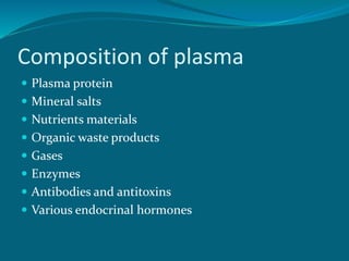
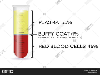
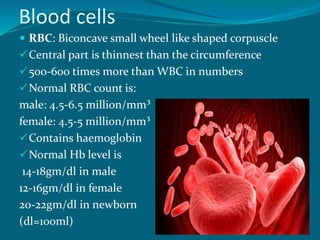
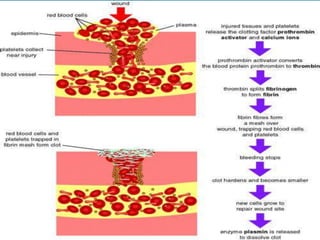
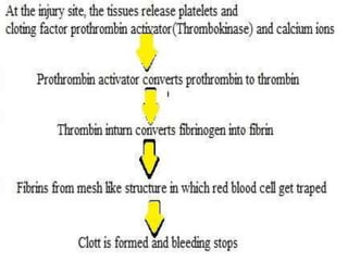
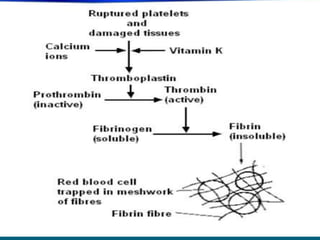
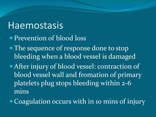
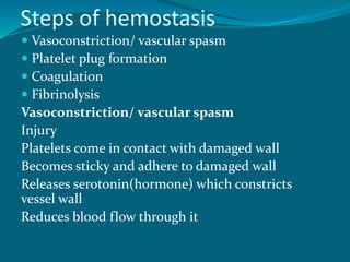
BLOOD
Introduction:
ļ‚— Liquid connective tissue found in the body
ļ‚— Consist of cells and fluid
ļ‚— Circulated in the rhythm with the
contraction of the heart through the heart,
arteries, capillaries and veins
ļ‚— Means of communication between cells and
external environment.
Characteristics of blood
ļ‚— Thick red fluid (bright in arties, dark red in veins)
ļ‚— Slightly alkaline in nature (pH 7.35-7.45)
ļ‚— Specific gravity 1.052 – 1.060
ļ‚— Volume 5-6 litres (7.8% of total body weight)
ļ‚— Viscocity 4.5 times more than water
ļ‚— Temperature 36 ̊C to 38 ̊C
ļ‚— Osmotic pressure 25mmof Hg
ļ‚— Taste salty
ļ‚— Normal output in one contraction of heart 70-80
ml.
Functions of blood
ļ‚— Carries oxygen from the lungs to tissue and carries
carbon dioxide from tissue to lungs
ļ‚— Transport nutrients from digestive tract to cells
ļ‚— Carries hormones, enzymes, vitamins and other
chemicals to particular organ
ļ‚— Regulates body temperature by distributing heat
ļ‚— Protects against infection (WBC)
ļ‚— Maintains water and electrolyte balance
ļ‚— Maintains acid base balance
ļ‚— Helps to prevents blood loss by clotting
mechanism
ļ‚— Helps in examination of health status
Composition of blood
Composed of blood cells (45%)and plasma
(55%)
ļ‚— Blood cells:
Red blood cells/erythrocytes (RBC)
White blood cells/ leucocytes (WBC)
Platelets / thrombocytes
ļ‚— Blood plasma:
Clear Yellow, alkaline fluid
Blood cells float in it
Composition of plasma
ļ‚— Plasma protein
ļ‚— Mineral salts
ļ‚— Nutrients materials
ļ‚— Organic waste products
ļ‚— Gases
ļ‚— Enzymes
ļ‚— Antibodies and antitoxins
ļ‚— Various endocrinal hormones
Blood cells
ļ‚— RBC: Biconcave small wheel like shaped corpuscle
Central part is thinnest than the circumference
500-600 times more than WBC in numbers
Normal RBC count is:
male: 4.5-6.5 million/mm³
female: 4.5-5 million/mm³
Contains haemoglobin
Normal Hb level is
14-18gm/dl in male
12-16gm/dl in female
20-22gm/dl in newborn
(dl=100ml)
Functions of RBC
ļ‚— Transport gases
ļ‚— Acid base balance
ļ‚— Iron balance
ļ‚— Maintenance of blood viscosity
ļ‚— Helps to detect blood grouping with its
antigen
Clinical problems
ļ‚— Anemia ( low hemoglobin /RBC due to infection or
low dietary intake)
ļ‚— Aplastic / hypo plastic anemia (due to alteration in
production of RBC from bone marrow)
ļ‚— Hemolytic anemia (infectious organism enter RBC
and reproduce until it bursts)
ļ‚— Megaloblastic anemia (deficiency of vitB12 and folate)
ļ‚— Pernicious anemia (deficiency /defects of vitB12
absorbing factors)
ļ‚— Jaundice (yellow discoloration of skin due to
destruction of RBC)
ļ‚— Polycythemia (large number of erythrocytes)
ļ‚— WBC:
colorless cell with nuclei
Slightly larger than RBC
Normal range is 400-11000/mm³
Formed in bone marrow
ļ‚— Functions of WBC
Leukocytosis ( To fight against
infection/microorganism causing increased
number of leucocytes during infection
Phagocytosis (To ingest and digest dead
tissue)
ļ‚— Types of WBC
Granular leukocytes (polymorpho-nuclear)
Neutrophil (rises in infections)
Basophil (rises in inflammations)
Eosinophil (allergic,infection)
Non granular leukocytes (mononuclear)
Lymphocyte
Monocyte
Clinical problems
ļ‚— Leukemia (uncontrolled production of leukocytes
eg. In malignancy)
ļ‚— Leukopenia (leukocyte count less than 4000mm³
eg. In Enteric fever)
ļ‚— Neutropenia (abnormal reduction of neutrophil
eg. In AML)
ļ‚— Leukocytosis (leukocyte count more than
11000mm³ eg. In CML, infections)
ļ‚— Eosinophilia (abnormal increased in number of
eosinophils eg. In allergic conditions)
AML= Acute myeloid leukemia (blood cancer)
ļ‚— Platelets
Tiny non- nucleated blood cells which help
in blood coagulation
They contains platelets/clotting factor III
and IV, serotonin (plasma protein)etc.
Normal count is 1,50,000-3,00,000 mm³
Thrombopoietin (glycoprotein hormone
produced by liver) helps to control
production of platelets
Life span is 8-11 days, unused platelets are
destroyed in spleen through macrophage
Process of coagulation
ļ‚— Coagulation, also known as clotting
ļ‚— The process by which blood changes from a
liquid to a gel, forming a blood clot. ...
ļ‚— Complicated process requiring several
substances:
Prothrombin (plasma protein)
Fibrinogen (plasma protein)
Calcium (mineral)
Thromboplastin (clotting factor)
Haemostasis
ļ‚— Prevention of blood loss
ļ‚— The sequence of response done to stop
bleeding when a blood vessel is damaged
ļ‚— After injury of blood vessel: contraction of
blood vessel wall and fromation of primary
platelets plug stops bleeding within 2-6
mins
ļ‚— Coagulation occurs with in 10 mins of injury
Steps of hemostasis
ļ‚— Vasoconstriction/ vascular spasm
ļ‚— Platelet plug formation
ļ‚— Coagulation
ļ‚— Fibrinolysis
Vasoconstriction/ vascular spasm
Injury
Platelets come in contact with damaged wall
Becomes sticky and adhere to damaged wall
Releases serotonin(hormone) which constricts
vessel wall
Reduces blood flow through it
ļ‚— Platelet plug formation:
Adherent platelets get more attached to each
other
Releases ADP(adenosine diphosphate) energy
compound which attracts more platelets to
damaged site
Forms a temporary seal known as platelets
plug within 60 secs
ļ‚— Coagulation:
clotting of blood
ļ‚— Fibrinolysis:
Process of removing the clot and heal the damaged
blood vessel tissue
Damaged endothelial cells releases plasmin activator
This activator changes inactive plasminogen present
in clot into enzyme plasmin (it breakdown plasma
proteins, fibrin clot)
Plasmin breakdown the fibrin clot to soluble
products as waste materials
Removed by phagocytosis (cell engulfing/absorbing)
Healing process is completed, vessel wall intensity is
restored
Natural anti-coagulants
ļ‚— Heparin
ļ‚— Anti thrombin III
ļ‚— Thrombin
Clinical problems
ļ‚— Hemophilia (prolonged coagulation time)
No blood clotting
Or very slow clotting
ļ‚— Thrombocytopenia (platelets counts less
than 30,000 mm³)
Blood grouping
ļ‚— A blood grouping is a classification
of blood, based on the presence and
absence of antibodies (immunoglobulin)
and inherited antigenic (foreign) substances
on the surface of red blood cells (RBCs).
ļ‚— There are 4 main blood groups: A,B,O,AB
ļ‚— Blood grouping is done by mixing an
individual’s red blood cells with anti-sera
(serum containing antibodies) and observe
the occurrence of agglutination (mixing).
ļ‚— Anti-sera: a serum containing antibodies
ļ‚— Agglutinogen: an antigenic substance
present in blood cells which stimulates the
formation of an agglutinin in blood serum.
ļ‚— Agglutinin: an antibody that causes
agglutination.
ļ‚— Agglutination: process of mixing antigen to
it’s corresponding antibodies
ABO blood group
ļ‚— Based on two glycolipid antigens i.e. A and B
ļ‚— RBCs display only antigen A have type A
blood
ļ‚— RBCs display only antigen B have type B
blood
ļ‚— RBCs display both antigen A and B have
type AB blood
ļ‚— RBCs display neither antigen have type O
blood
ļ‚—Blood plasma usually contains
antibodies called agglutinins that
reacts with antigen in RBC if mixed
ļ‚—These are anti-A and anti-B
ļ‚—If there is A antigen in RBC there will
be Anti-B in plasma and vise versa
ļ‚—If there is A and B antigen there will be
no antibodies
ļ‚—If there is None antigen there will
antibody ………………
The rhesus factor
ļ‚— The rhesus factor known as Rh factor inherited from
the Rhesus monkey .
ļ‚— Rh factor is Rh antigen/D antigen present in RBC
where as plasma do not contain Rh antibody
ļ‚— RBC having Rh antigen are Rh positive and vice versa
ļ‚— https://youtu.be/baGQJG1YGq8
Determining blood grouping
ļ‚— Three drops of blood is mixed with anti-A serum, anti-
B serum and Anti-D serum
ļ‚— If Anti-A agglutinates with antigen blood type is A
ļ‚— If Anti-B agglutinates with antigen blood type is B
ļ‚— If both Anti-A and Anti-B agglutinates with antigen
blood type is AB
ļ‚— If both Anti-A and Anti-B didn’t agglutinates with
antigen blood type is O
LANDSTEINER's LAW-
ļ‚— 1.If an agglutinogen is present on red blood
cell membrane ,the corresponding
agglutinin must be absent in the plasma.
ļ‚— 2.If an agglutinogen is absent on red blood
cell membrane, then corresponding
agglutinin must be present in the plasma.

Blood

  • 1.
    BLOOD Introduction: ļ‚— Liquid connectivetissue found in the body ļ‚— Consist of cells and fluid ļ‚— Circulated in the rhythm with the contraction of the heart through the heart, arteries, capillaries and veins ļ‚— Means of communication between cells and external environment.
  • 2.
    Characteristics of blood ļ‚—Thick red fluid (bright in arties, dark red in veins) ļ‚— Slightly alkaline in nature (pH 7.35-7.45) ļ‚— Specific gravity 1.052 – 1.060 ļ‚— Volume 5-6 litres (7.8% of total body weight) ļ‚— Viscocity 4.5 times more than water ļ‚— Temperature 36 ̊C to 38 ̊C ļ‚— Osmotic pressure 25mmof Hg ļ‚— Taste salty ļ‚— Normal output in one contraction of heart 70-80 ml.
  • 3.
    Functions of blood ļ‚—Carries oxygen from the lungs to tissue and carries carbon dioxide from tissue to lungs ļ‚— Transport nutrients from digestive tract to cells ļ‚— Carries hormones, enzymes, vitamins and other chemicals to particular organ ļ‚— Regulates body temperature by distributing heat ļ‚— Protects against infection (WBC) ļ‚— Maintains water and electrolyte balance ļ‚— Maintains acid base balance ļ‚— Helps to prevents blood loss by clotting mechanism ļ‚— Helps in examination of health status
  • 4.
    Composition of blood Composedof blood cells (45%)and plasma (55%) ļ‚— Blood cells: Red blood cells/erythrocytes (RBC) White blood cells/ leucocytes (WBC) Platelets / thrombocytes ļ‚— Blood plasma: Clear Yellow, alkaline fluid Blood cells float in it
  • 5.
    Composition of plasma ļ‚—Plasma protein ļ‚— Mineral salts ļ‚— Nutrients materials ļ‚— Organic waste products ļ‚— Gases ļ‚— Enzymes ļ‚— Antibodies and antitoxins ļ‚— Various endocrinal hormones
  • 7.
    Blood cells ļ‚— RBC:Biconcave small wheel like shaped corpuscle Central part is thinnest than the circumference 500-600 times more than WBC in numbers Normal RBC count is: male: 4.5-6.5 million/mm³ female: 4.5-5 million/mm³ Contains haemoglobin Normal Hb level is 14-18gm/dl in male 12-16gm/dl in female 20-22gm/dl in newborn (dl=100ml)
  • 8.
    Functions of RBC ļ‚—Transport gases ļ‚— Acid base balance ļ‚— Iron balance ļ‚— Maintenance of blood viscosity ļ‚— Helps to detect blood grouping with its antigen
  • 9.
    Clinical problems ļ‚— Anemia( low hemoglobin /RBC due to infection or low dietary intake) ļ‚— Aplastic / hypo plastic anemia (due to alteration in production of RBC from bone marrow) ļ‚— Hemolytic anemia (infectious organism enter RBC and reproduce until it bursts) ļ‚— Megaloblastic anemia (deficiency of vitB12 and folate) ļ‚— Pernicious anemia (deficiency /defects of vitB12 absorbing factors) ļ‚— Jaundice (yellow discoloration of skin due to destruction of RBC) ļ‚— Polycythemia (large number of erythrocytes)
  • 10.
    ļ‚— WBC: colorless cellwith nuclei Slightly larger than RBC Normal range is 400-11000/mm³ Formed in bone marrow
  • 11.
    ļ‚— Functions ofWBC Leukocytosis ( To fight against infection/microorganism causing increased number of leucocytes during infection Phagocytosis (To ingest and digest dead tissue)
  • 12.
    ļ‚— Types ofWBC Granular leukocytes (polymorpho-nuclear) Neutrophil (rises in infections) Basophil (rises in inflammations) Eosinophil (allergic,infection) Non granular leukocytes (mononuclear) Lymphocyte Monocyte
  • 13.
    Clinical problems ļ‚— Leukemia(uncontrolled production of leukocytes eg. In malignancy) ļ‚— Leukopenia (leukocyte count less than 4000mm³ eg. In Enteric fever) ļ‚— Neutropenia (abnormal reduction of neutrophil eg. In AML) ļ‚— Leukocytosis (leukocyte count more than 11000mm³ eg. In CML, infections) ļ‚— Eosinophilia (abnormal increased in number of eosinophils eg. In allergic conditions) AML= Acute myeloid leukemia (blood cancer)
  • 14.
    ļ‚— Platelets Tiny non-nucleated blood cells which help in blood coagulation They contains platelets/clotting factor III and IV, serotonin (plasma protein)etc. Normal count is 1,50,000-3,00,000 mm³ Thrombopoietin (glycoprotein hormone produced by liver) helps to control production of platelets Life span is 8-11 days, unused platelets are destroyed in spleen through macrophage
  • 16.
    Process of coagulation ļ‚—Coagulation, also known as clotting ļ‚— The process by which blood changes from a liquid to a gel, forming a blood clot. ... ļ‚— Complicated process requiring several substances: Prothrombin (plasma protein) Fibrinogen (plasma protein) Calcium (mineral) Thromboplastin (clotting factor)
  • 20.
    Haemostasis ļ‚— Prevention ofblood loss ļ‚— The sequence of response done to stop bleeding when a blood vessel is damaged ļ‚— After injury of blood vessel: contraction of blood vessel wall and fromation of primary platelets plug stops bleeding within 2-6 mins ļ‚— Coagulation occurs with in 10 mins of injury
  • 21.
    Steps of hemostasis ļ‚—Vasoconstriction/ vascular spasm ļ‚— Platelet plug formation ļ‚— Coagulation ļ‚— Fibrinolysis Vasoconstriction/ vascular spasm Injury Platelets come in contact with damaged wall Becomes sticky and adhere to damaged wall Releases serotonin(hormone) which constricts vessel wall Reduces blood flow through it
  • 22.
    ļ‚— Platelet plugformation: Adherent platelets get more attached to each other Releases ADP(adenosine diphosphate) energy compound which attracts more platelets to damaged site Forms a temporary seal known as platelets plug within 60 secs ļ‚— Coagulation: clotting of blood
  • 23.
    ļ‚— Fibrinolysis: Process ofremoving the clot and heal the damaged blood vessel tissue Damaged endothelial cells releases plasmin activator This activator changes inactive plasminogen present in clot into enzyme plasmin (it breakdown plasma proteins, fibrin clot) Plasmin breakdown the fibrin clot to soluble products as waste materials Removed by phagocytosis (cell engulfing/absorbing) Healing process is completed, vessel wall intensity is restored
  • 24.
    Natural anti-coagulants ļ‚— Heparin ļ‚—Anti thrombin III ļ‚— Thrombin
  • 25.
    Clinical problems ļ‚— Hemophilia(prolonged coagulation time) No blood clotting Or very slow clotting ļ‚— Thrombocytopenia (platelets counts less than 30,000 mm³)
  • 26.
    Blood grouping ļ‚— Ablood grouping is a classification of blood, based on the presence and absence of antibodies (immunoglobulin) and inherited antigenic (foreign) substances on the surface of red blood cells (RBCs). ļ‚— There are 4 main blood groups: A,B,O,AB ļ‚— Blood grouping is done by mixing an individual’s red blood cells with anti-sera (serum containing antibodies) and observe the occurrence of agglutination (mixing).
  • 27.
    ļ‚— Anti-sera: aserum containing antibodies ļ‚— Agglutinogen: an antigenic substance present in blood cells which stimulates the formation of an agglutinin in blood serum. ļ‚— Agglutinin: an antibody that causes agglutination. ļ‚— Agglutination: process of mixing antigen to it’s corresponding antibodies
  • 28.
    ABO blood group ļ‚—Based on two glycolipid antigens i.e. A and B ļ‚— RBCs display only antigen A have type A blood ļ‚— RBCs display only antigen B have type B blood ļ‚— RBCs display both antigen A and B have type AB blood ļ‚— RBCs display neither antigen have type O blood
  • 29.
    ļ‚—Blood plasma usuallycontains antibodies called agglutinins that reacts with antigen in RBC if mixed ļ‚—These are anti-A and anti-B ļ‚—If there is A antigen in RBC there will be Anti-B in plasma and vise versa ļ‚—If there is A and B antigen there will be no antibodies ļ‚—If there is None antigen there will antibody ………………
  • 30.
    The rhesus factor ļ‚—The rhesus factor known as Rh factor inherited from the Rhesus monkey . ļ‚— Rh factor is Rh antigen/D antigen present in RBC where as plasma do not contain Rh antibody ļ‚— RBC having Rh antigen are Rh positive and vice versa ļ‚— https://youtu.be/baGQJG1YGq8
  • 31.
    Determining blood grouping ļ‚—Three drops of blood is mixed with anti-A serum, anti- B serum and Anti-D serum ļ‚— If Anti-A agglutinates with antigen blood type is A ļ‚— If Anti-B agglutinates with antigen blood type is B ļ‚— If both Anti-A and Anti-B agglutinates with antigen blood type is AB ļ‚— If both Anti-A and Anti-B didn’t agglutinates with antigen blood type is O
  • 32.
    LANDSTEINER's LAW- ļ‚— 1.Ifan agglutinogen is present on red blood cell membrane ,the corresponding agglutinin must be absent in the plasma. ļ‚— 2.If an agglutinogen is absent on red blood cell membrane, then corresponding agglutinin must be present in the plasma.