Biosynthesis of selenium nanoparticles using lactic acid bacteria
1.
BIOSYNTHESIS, CHARACTERISATION, AND
BIOLOGICALACTIVITYOF SELENIUM
NANOPARTICLES SYNTHESISED USING
Lactobacillus sp.
NAME – Sanjana Yadav
GUIDE NAME – Dr. Snehal Gagre
CLASS – TY Bsc Biotechnology
ROLL.NO – 243231040
SEAT No. - 10101
2.
ABSTRACT
Selenium nanoparticles (SeNPs)are gaining attention for their potential in
medicine, agriculture, and environmental science. In this study, we used
Lactobacillus sp. to biosynthesize SeNPs, exploring how different sodium
selenite concentrations (2 mM, 4 mM, and 6 mM) affect their formation. A
striking color change from pale yellow to orange red signaled successful
synthesis, which was confirmed by UV-Vis spectroscopy. FTIR analysis
showed that biomolecules from the bacteria helped stabilize the
nanoparticles. The main objectives of the study were as follows :-
• To synthesize Selenium nanoparticles using Lactobacillus sp.
• Characterization of synthesized Se-NPs using bioanalytical techniques.
• To evaluate biological activity of biosynthesised selenium nanoparticles.
3.
INTRODUCTION
• Nanotechnology istransforming science by bringing together physics, chemistry, and
biology to create tiny materials with powerful properties. Among these, metal
nanomaterials like selenium have exciting applications in medicine, food safety, and
biosensors.
• However, traditional methods of making these nanoparticles often come with challenges
like toxicity and difficulty in large-scale production. That’s where biological or "green"
synthesis comes in—using natural processes to create safer, eco-friendly nanoparticles.
• Selenium is an essential nutrient that supports immunity and overall health, but too much
of it can be toxic. Interestingly, certain lactic acid bacteria (LAB), commonly found in
foods like curd, can naturally convert selenium salts into selenium nanoparticles (SeNPs).
• This study explores how Lactobacillus sp. from curd can be used to synthesize SeNPs
from sodium selenite. While these nanoparticles show great potential in medicine, their
safety and compatibility with the human body need further research to unlock their full
potential.
4.
MATERIALS AND METHODS
•MATERIALS –
1. Curd
2. De Man, Rogosa and Sharpe (MRS) Broth
3. Antibiotic (100mg/ml)
4. Escherichia coli culture
5. Staphylococcus aureus culture
6. Agar Powder (Bacteriology grade-HiMedia)
7. Sodium selenite (SRL )
5.
MATERIALS AND METHODS
•METHODS –
1. Isolation and Characterization of Lactobacillus
2. Synthesis of Selenium nanoparticles
3. Isolation and purification of Selenium nanoparticles
4. Antimicrobial activity of synthesized SeNPs
6.
RESULTS
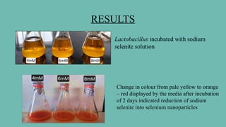
Lactobacillus incubated withsodium
selenite solution
Change in colour from pale yellow to orange
– red displayed by the media after incubation
of 2 days indicated reduction of sodium
selenite into selenium nanoparticles
7.
RESULTS
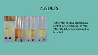
Tubes with positiveand negative
control for determining the MIC.
All of the tubes were observed to
be turbid.
8.
RESULTS
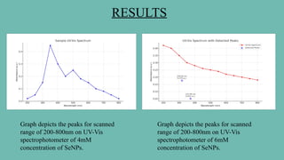
Graph depicts thepeaks for scanned
range of 200-800nm on UV-Vis
spectrophotometer of 4mM
concentration of SeNPs.
Graph depicts the peaks for scanned
range of 200-800nm on UV-Vis
spectrophotometer of 6mM
concentration of SeNPs.
9.
RESULTS
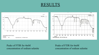
Peaks of FTIRfor 4mM
concentration of sodium selenite
Peaks of FTIR for 6mM
concentration of sodium selenite
10.
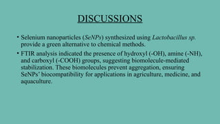
DISCUSSIONS
• Selenium nanoparticles(SeNPs) synthesized using Lactobacillus sp.
provide a green alternative to chemical methods.
• FTIR analysis indicated the presence of hydroxyl (-OH), amine (-NH),
and carboxyl (-COOH) groups, suggesting biomolecule-mediated
stabilization. These biomolecules prevent aggregation, ensuring
SeNPs’ biocompatibility for applications in agriculture, medicine, and
aquaculture.
11.
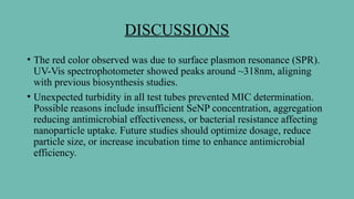
DISCUSSIONS
• The redcolor observed was due to surface plasmon resonance (SPR).
UV-Vis spectrophotometer showed peaks around ~318nm, aligning
with previous biosynthesis studies.
• Unexpected turbidity in all test tubes prevented MIC determination.
Possible reasons include insufficient SeNP concentration, aggregation
reducing antimicrobial effectiveness, or bacterial resistance affecting
nanoparticle uptake. Future studies should optimize dosage, reduce
particle size, or increase incubation time to enhance antimicrobial
efficiency.
12.
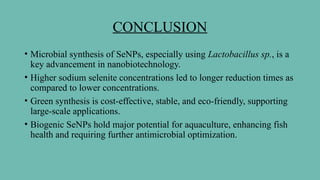
CONCLUSION
• Microbial synthesisof SeNPs, especially using Lactobacillus sp., is a
key advancement in nanobiotechnology.
• Higher sodium selenite concentrations led to longer reduction times as
compared to lower concentrations.
• Green synthesis is cost-effective, stable, and eco-friendly, supporting
large-scale applications.
• Biogenic SeNPs hold major potential for aquaculture, enhancing fish
health and requiring further antimicrobial optimization.
13.
REFRENCES
• Murugesan etal. (2019): Murugesan, G., Nagaraj, K., Sunmathi, D., & Subramani, K. (2019).
Methods involved in the synthesis of selenium nanoparticles and their different applications
—a review. European Journal of Biomedical and Pharmaceutical Sciences, 6(4), 189–194.
http://www.ejbps.com
• Visha et al. (2015):Visha, P., Nanjappan, K., Selvaraj, P., Jayachandran, S., Elango, A., &
Kumaresan, G. (2015). Biosynthesis and structural characteristics of selenium nanoparticles
using Lactobacillus acidophilus bacteria by wet sterilization process. International Journal of
Advanced Veterinary Science and Technology, 4(1), 178 183. http://scientific.cloud-
journals.com/index.php/IJAVST/article/view/Sci-312.
• Rao, V., & Poonia, A. (2024):Microbial biosynthesis of selenium nanoparticles using probiotic
strain and its characterization. Journal of Food Measurement and Characterization.
https://doi.org/10.1007/s11694-024-02581-z
• Martínez, F. G., Moreno-Martin, G., Pescuma, M., Madrid-Albarrán, Y., & Mozzi, F. :
Biotransformation of selenium by lactic acid bacteria: Formation of seleno-nanoparticles and
seleno-amino acids. VI International Symposium on Lactic Acid Bacteria. Retrieved from
https://bicyt.conicet.gov.ar/fichas/produccion/12227955
• Ingole, A. R., Thakare, S. R., Khati, N. T., Wankhade, A. V., & Burghate, D. K. (2010). Green
synthesis of selenium nanoparticles under ambient condition. Journal Name, 7(7), 485–489.