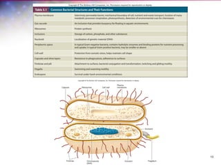
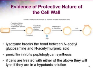
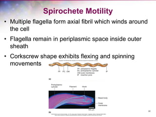
Bacterial Cell Structure
1
3
3.1 The “Prokaryote” Controversy
1. List the characteristics originally used to describe
prokaryotic cells
2. Form an opinion on the “prokaryote” controversy using
current evidence about bacterial cells
2
3.2 A Typical Bacterial Cell
1. Distinguish a typical bacterial cell from a typical plant
or animal cell in terms of cell shapes and
arrangements, size, and cell structures
2. Discuss the factors that determine the size and shape
of a bacterial cell.
3
4
Bacterial and Archaea Structure
and Function
• Prokaryotes differ from eukaryotes in size and
simplicity
– most lack internal membrane systems
– term prokaryotes is becoming blurred
– this text will use Bacteria and Archaea
– this chapter will cover Bacteria and their
structures
5
Size, Shape, and Arrangement
• Shape
– cocci and rods most common
– various others
• Arrangement
– determined by plane of division
– determined by separation or not
• Size - varies
6
Shape and Arrangement-1
• Cocci (s., coccus) – spheres
– diplococci (s., diplococcus) – pairs
– streptococci – chains
– staphylococci – grape-like clusters
– tetrads – 4 cocci in a square
– sarcinae – cubic configuration of 8 cocci
7
Shape and Arrangement-2
• bacilli (s., bacillus) – rods
– coccobacilli – very short rods
• vibrios – resemble rods, comma
shaped
• spirilla (s., spirillum) – rigid helices
• spirochetes – flexible helices
8
Shape and Arrangement-3
• mycelium – network of long, multinucleate filaments
• pleomorphic – organisms that are variable in shape
9
10
Size
• smallest – 0.3 μm
(Mycoplasma)
• average rod – 1.1
- 1.5 x 2 – 6 μm
(E. coli)
• very large – 600 x
80 μm
Epulopiscium
fishelsoni
11
Size – Shape Relationship
• important for nutrient uptake
• surface to volume ratio (S/V)
• small size may be protective mechanism from
predation
12
Bacterial Cell Organization
Common Features
– Cell envelope – 3 layers
– Cytoplasm
– External structures
13
3.3 Bacterial Plasma Membranes
1. Describe the fluid mosaic model of membrane
structure and identify the types of lipids typically found
in bacterial membranes.
2. Distinguish macroelements (macronutrients) from
micronutrients (trace elements) and provide examples
of each.
3. Provide examples of growth factors needed by some
microorganisms.
4. Compare and contrast passive diffusion, facilitated
diffusion, active transport, and group translocation, and
provide examples of each.
5. Discuss the difficulty of iron uptake and describe how
bacteria overcome this difficulty.
14
15
Bacterial Cell Envelope
• Plasma membrane
• Cell wall
• Layers outside the cell wall
16
Bacterial Plasma Membrane
• Absolute requirement for all living organisms
• Some bacteria also have internal membrane
systems
17
Plasma Membrane Functions
• Encompasses the cytoplasm
• Selectively permeable barrier
• Interacts with external environment
– receptors for detection of and response to
chemicals in surroundings
– transport systems
– metabolic processes
18
Fluid Mosaic Model of Membrane
Structure
• lipid bilayers with floating proteins
– amphipathic lipids
• polar ends (hydrophilic – interact with water)
• non-polar tails (hydrophobic – insoluble in water)
– membrane proteins
19
Membrane Proteins
• Peripheral
– loosely connected to membrane
– easily removed
• Integral
– amphipathic – embedded within membrane
– carry out important functions
– may exist as microdomains
20
Bacterial Lipids
• Saturation levels of
membrane lipids reflect
environmental
conditions such as
temperature
• Bacterial membranes
lack sterols but do
contain sterol-like
molecules, hopanoids
– stabilize membrane
– found in petroleum
21
Uptake of Nutrients – Getting Through
the Barrier
• Macroelements (macronutrients)
– C, O, H, N, S, P
• found in organic molecules such as proteins,
lipids, carbohydrates, and nucleic acids
– K, Ca, Mg, and Fe
• cations and serve in variety of roles including
enzymes, biosynthesis
– required in relatively large amounts
22
Uptake of Nutrients – Getting Through
the Barrier
• Micronutrients (trace elements)
– Mn, Zn, Co, Mo, Ni, and Cu
– required in trace amounts
– often supplied in water or in media
components
– ubiquitous in nature
– serve as enzymes and cofactors
• Some unique substances may be required
23
Uptake of Nutrients – Getting Through
the Barrier
• Growth factors
– organic compounds
– essential cell components (or their precursors)
that the cell cannot synthesize
– must be supplied by environment if cell is to
survive and reproduce
24
Classes of Growth Factors
• amino acids
– needed for protein synthesis
• purines and pyrimidines
– needed for nucleic acid synthesis
• vitamins
– function as enzyme cofactors
• heme
25
Uptake of Nutrients
• Microbes can only take in dissolved particles
across a selectively permeable membrane
• Some nutrients enter by passive diffusion
• Microorganisms use transport mechanisms
– facilitated diffusion – all microorganisms
– active transport – all microorganisms
– group translocation – Bacteria and Archaea
– endocytosis – Eukarya only
26
Passive Diffusion
• Molecules move from region of higher
concentration to one of lower concentration
between the cell’s interior and the exterior
• H2O, O2, and CO2 often move across
membranes this way
27
Facilitated Diffusion
• Similar to passive diffusion
– movement of molecules is not energy
dependent
– direction of movement is from high
concentration to low concentration
– size of concentration gradient impacts rate of
uptake
28
Facilitated
Diffusion…
• Differs from passive
diffusion
– uses membrane bound
carrier molecules
(permeases)
– smaller concentration
gradient is required for
significant uptake of
molecules
– effectively transports
glycerol, sugars, and amino
acids
• more prominent in
eukaryotic cells than in
bacteria or archaea
29
30
Active Transport
• energy-dependent process
– ATP or proton motive force used
• move molecules against the gradient
• concentrates molecules inside cell
• involves carrier proteins (permeases)
– carrier saturation effect is observed at high
solute concentrations
31
ABC Transporters
• Primary active transporters use ATP
• ATP-binding cassette (ABC) transporters
• Observed in Bacteria, Archaea, and eukaryotes
• Consist of
- 2 hydrophobic
membrane spanning
domains
- 2 cytoplasmic
associated ATP-binding
domains
- Substrate binding
domains
32
Secondary Active Transport
• Major facilitator superfamily (MFS)
• Use ion gradients to cotransport substances
– protons
– symport – two substances both move in the same
direction
– antiport – two substances move in opposite directions
33
34
Group Translocation
• Energy dependent transport that
chemically modifies molecule as it
is brought into cell
• Best known translocation system is
phosphoenolpyruvate: sugar
phosphotransferase system (PTS)
35
36
Iron Uptake
• Microorganisms
require iron
• Ferric iron is very
insoluble so uptake
is difficult
• Microorganisms
secrete siderophores
to aid uptake
• Siderophore
complexes with ferric
ion
• Complex is then
transported into cell
3.4 Bacterial Cell Walls
1. Describe peptidoglycan structure.
2. Compare and contrast the cell walls of typical Gram-
positive and Gram-negative bacteria.
3. Relate bacterial cell wall structure to the Gram-staining
reaction.
37
38
Bacterial Cell Wall
• Peptidoglycan (murein)
– rigid structure that lies just outside the cell
plasma membrane
– two types based on Gram stain
• Gram-positive: stain purple; thick peptidoglycan
• Gram-negative: stain pink or red; thin
peptidoglycan and outer membrane
39
Cell Wall Functions
• Maintains shape of the bacterium
– almost all bacteria have one
• Helps protect cell from osmotic lysis
• Helps protect from toxic materials
• May contribute to pathogenicity
40
Peptidoglycan
Structure
• Meshlike polymer of
identical subunits
forming long strands
– two alternating sugars
• N-acetylglucosamine
(NAG)
• N- acetylmuramic acid
– alternating D- and L-
amino acids
41
Strands Are Crosslinked
• Peptidoglycan strands
have a helical shape
• Peptidoglycan chains
are crosslinked by
peptides for strength
– interbridges may form
– peptidoglycan sacs –
interconnected
networks
– various structures
occur
42
43
Gram-Positive Cell
Walls
• Composed primarily of
peptidoglycan
• May also contain teichoic
acids (negatively charged)
– help maintain cell envelope
– protect from environmental
substances
– may bind to host cells
• some gram-positive bacteria
have layer of proteins on
surface of peptidoglycan
44
45
Periplasmic Space of Gram +
Bacteria
• Lies between plasma membrane and cell wall
and is smaller than that of Gram-negative
bacteria
• Periplasm has relatively few proteins
• Enzymes secreted by Gram-positive bacteria
are called exoenzymes
– aid in degradation of large nutrients
46
Gram-Negative Cell
Walls
• More complex than Gram-
positive
• Consist of a thin layer of
peptidoglycan surrounded by
an outer membrane
• Outer membrane composed
of lipids, lipoproteins, and
lipopolysaccharide (LPS)
• No teichoic acids
47
Gram-Negative Cell Walls
• Peptidoglycan is ~5-10% of cell wall weight
• Periplasmic space differs from that in Gram-
positive cells
– may constitute 20–40% of cell volume
– many enzymes present in periplasm
• hydrolytic enzymes, transport proteins and
other proteins
48
Gram-
Negative
Cell Walls
• outer membrane lies outside the thin peptidoglycan
layer
• Braun’s lipoproteins connect outer membrane to
peptidoglycan
• other adhesion sites reported
49
Lipopolysaccharide (LPS)
• Consists of three parts
– lipid A
– core polysaccharide
– O side chain (O
antigen)
• Lipid A embedded in
outer membrane
• Core polysaccharide, O
side chain extend out
from the cell
50
Importance of LPS
• contributes to negative charge on cell surface
• helps stabilize outer membrane structure
• may contribute to attachment to surfaces and
biofilm formation
• creates a permeability barrier
• protection from host defenses (O antigen)
• can act as an endotoxin (lipid A)
51
Gram-Negative Outer Membrane
Permeability
• More permeable than plasma membrane due to
presence of porin proteins and transporter proteins
– porin proteins form channels to let small molecules
(600–700 daltons) pass
52
Mechanism of Gram Stain Reaction
• Gram stain reaction due to nature of cell wall
• shrinkage of the pores of peptidoglycan layer
of Gram-positive cells
– constriction prevents loss of crystal violet
during decolorization step
• thinner peptidoglycan layer and larger pores
of Gram-negative bacteria does not prevent
loss of crystal violet
53
Osmotic Protection
• Hypotonic environments
– solute concentration outside the cell is less
than inside the cell
– water moves into cell and cell swells
– cell wall protects from lysis
• Hypertonic environments
– solute concentration outside the cell is greater
than inside
– water leaves the cell
– plasmolysis occurs
54
Evidence of Protective Nature of
the Cell Wall
• lysozyme breaks the bond between N-acetyl
glucosamine and N-acetylmuramic acid
• penicillin inhibits peptidoglycan synthesis
• if cells are treated with either of the above they will
lyse if they are in a hypotonic solution
55
Cells that Lose a Cell Wall May
Survive in Isotonic Environments
• Protoplasts
• Spheroplasts
• Mycoplasma
– does not produce a cell wall
– plasma membrane more resistant to osmotic
pressure
3.5 Cell Envelope Layers Outside the Cell
Wall
1. Compile a list of all the structures found in all the layers
of bacterial cell envelopes, noting the functions and the
major component molecules of each.
56
57
Components Outside of the Cell
Wall
• Outermost layer in the cell envelope
• Glycocalyx
– capsules and slime layers
– S layers
• Aid in attachment to solid surfaces
– e.g., biofilms in plants and animals
58
Capsules
• Usually composed of
polysaccharides
• Well organized and not
easily removed from cell
• Visible in light microscope
• Protective advantages
– resistant to phagocytosis
– protect from desiccation
– exclude viruses and
detergents
59
Slime Layers
• similar to capsules except diffuse,
unorganized and easily removed
• slime may aid in motility
60
S Layers
• Regularly structured
layers of protein or
glycoprotein that self-
assemble
– in Gram-negative
bacteria the S layer
adheres to outer
membrane
– in Gram-positive
bacteria it is associated
with the peptidoglycan
surface
61
S Layer Functions
• Protect from ion and pH fluctuations, osmotic
stress, enzymes, and predation
• Maintains shape and rigidity
• Promotes adhesion to surfaces
• Protects from host defenses
• Potential use in nanotechnology
– S layer spontaneously associates
3.6 Bacterial Cytoplasm
1. Create a table or concept map that identifies the
components of the bacterial cytoplasm and describes
their structure, molecular makeup, and functions.
62
63
Bacterial Cytoplasmic Structures
• Cytoskeleton
• Intracytoplasmic membranes
• Inclusions
• Ribosomes
• Nucleoid and plasmids
64
Protoplast and Cytoplasm
• Protoplast is plasma membrane and
everything within
• Cytoplasm - material bounded by the plasmid
membrane
65
The Cytoskeleton
• Homologs of all 3 eukaryotic cytoskeletal elements
have been identified in bacteria
• Functions are similar as in eukaryotes
66
Best Studied Examples
• FtsZ – many bacteria
– forms ring during septum
formation in cell division
• MreB – many rods
– maintains shape by
positioning peptidoglycan
synthesis machinery
• CreS – rare, maintains
curve shape
67
Intracytoplasmic Membranes
• Plasma membrane infoldings
– observed in many photosynthetic bacteria
– observed in many bacteria with high respiratory activity
• Anammoxosome in Planctomycetes
– organelle – site of anaerobic ammonia oxidation
68
Inclusions
• Granules of organic or inorganic material that
are stockpiled by the cell for future use
• Some are enclosed by a single-layered
membrane
– membranes vary in composition
– some made of proteins; others contain lipids
– may be referred to as microcompartments
69
Storage Inclusions
• Storage of nutrients, metabolic end products,
energy, building blocks
• Glycogen storage
• Carbon storage
– poly-β-hydroxybutyrate (PHB)
• Phosphate - Polyphosphate (Volutin)
• Amino acids - cyanophycin granules
70
Storage Inclusions
71
Microcompartments
• Not bound by
membranes but
compartmentalized for
a specific function
• Carboxysomes - CO2
fixing bacteria
– contain the enzyme
ribulose-1,5,-
bisphosphate
carboxylase (Rubisco),
enzyme used for CO2
fixation
72
Other Inclusions
• Gas vacuoles
– found in aquatic, photosynthetic bacteria and
archaea
– provide buoyancy in gas vesicles
73
Other Inclusions
• Magnetosomes
– found in aquatic bacteria
– magnetite particles for orientation
in Earth’s magnetic field
– cytoskeletal protein MamK
• helps form magnetosome chain
74
Ribosomes
• Complex protein/RNA structures
– sites of protein synthesis
– bacterial and archaea ribosome = 70S
– eukaryotic (80S) S = Svedburg unit
• Bacterial ribosomal RNA
– 16S small subunit
– 23S and 5S in large subunit
75
The Nucleoid
• Usually not membrane
bound (few exceptions)
• Location of chromosome
and associated proteins
• Usually 1 closed circular,
double-stranded DNA
molecule
• Supercoiling and nucleoid
proteins (different from
histones) aid in folding
76
Plasmids
• Extrachromosomal DNA
– found in bacteria, archaea, some fungi
– usually small, closed circular DNA molecules
• Exist and replicate independently of chromosome
– episomes – may integrate into chromosome
– inherited during cell division
• Contain few genes that are non-essential
– confer selective advantage to host (e.g., drug
resistance)
• Classification based on mode of existence, spread,
and function
77
3.7 External Structures
1. Distinguish pili (fimbriae) and flagella.
2. Illustrate the various patterns of flagella distribution.
78
79
External Structures
• Extend beyond the cell envelope in bacteria
• Function in protection, attachment to
surfaces, horizontal gene transfer, cell
movement
– pili and fimbriae
– flagella
80
Pili and Fimbriae
• Fimbriae (s., fimbria); pili (s.,
pilus)
– short, thin, hairlike,
proteinaceous appendages (up to
1,000/cell)
– can mediate attachment to
surfaces, motility, DNA uptake
• Sex pili (s., pilus)
– longer, thicker, and less
numerous (1-10/cell)
– genes for formation found on
plasmids
– required for conjugation
81
Flagella
• Threadlike, locomotor appendages extending
outward from plasma membrane and cell wall
• Functions
– motility and swarming behavior
– attachment to surfaces
– may be virulence factors
82
Bacterial Flagella
• Thin, rigid protein structures that cannot be
observed with bright-field microscope unless
specially stained
• Ultrastructure composed of three parts
• Pattern of flagellation varies
83
Patterns of Flagella Distribution
• Monotrichous – one flagellum
• Polar flagellum – flagellum at
end of cell
• Amphitrichous – one flagellum
at each end of cell
• Lophotrichous – cluster of
flagella at one or both ends
• Peritrichous – spread over
entire surface of cell
84
Three Parts of Flagella
• Filament
– extends from cell surface to the tip
– hollow, rigid cylinder of flagellin protein
• Hook
– links filament to basal body
• Basal body
– series of rings that drive flagellar motor
85
Flagellar Synthesis
• complex process involving many genes/gene
products
• new flagellin molecules transported through the
hollow filament using Type III-like secretion system
• filament subunits self-assemble with help of filament
cap at tip, not base
3.8 Bacterial Motility and Chemotaxis
1. Compare and contrast flagellar swimming motility,
spirochete flagellar motility, and twitching and gliding
motility.
2. State the source of energy that powers flagellar
motility.
3. Explain why bacterial chemotaxis is referred to as a
“biased random walk.”
86
87
Motility
• Flagellar movement
• Spirochete motility
• Twitching motility
• Gliding motility
88
Motility
• Bacteria and Archaea have directed
movement
• Chemotaxis
– move toward chemical attractants such as
nutrients, away from harmful substances
• Move in response to temperature, light,
oxygen, osmotic pressure, and gravity
89
Bacterial Flagellar
Movement
• Flagellum rotates like a
propeller
– very rapid rotation up to
1100 revolutions/sec
– in general, counterclockwise
(CCW) rotation causes forward
motion (run)
– in general, clockwise rotation
(CW) disrupts run causing cell
to stop and tumble
90
Mechanism of Flagellar Movement
• Flagellum is 2 part motor
producing torque
• Rotor
– C (FliG protein) ring and MS ring
turn and interact with stator
• Stator - Mot A and Mot B proteins
– form channel through plasma
membrane
– protons move through Mot A and
Mot B channels using energy of
proton motive force
– torque powers rotation of the
basal body and filament
91
Spirochete Motility
• Multiple flagella form axial fibril which winds around
the cell
• Flagella remain in periplasmic space inside outer
sheath
• Corkscrew shape exhibits flexing and spinning
movements
92
Twitching and Gliding Motility
• May involve Type IV pili and slime
• Twitching
– pili at ends of cell
– short, intermittent, jerky motions
– cells are in contact with each other and
surface
• Gliding
– smooth movements
93
Chemotaxis
• Movement toward a chemical attractant or
away from a chemical repellent
• Changing concentrations of chemical
attractants and chemical repellents bind
chemoreceptors of chemosensing system
94
95
Chemotaxis
• In presence of attractant (b) tumbling frequency is
intermittently reduced and runs in direction of
attractant are longer
• Behavior of bacterium is altered by temporal
concentration of chemical
• Chemotaxis away from repellent involves similar but
opposite responses
3.9 Bacterial Endospores
1. Describe the structure of the bacterial endospore.
2. Explain why the bacterial endospores are of particular
concern to the food industry and why endospore-
forming bacteria are important model organisms.
3. Describe in general terms the process of sporulation.
4. Describe those properties of endospores that are
thought to contribute to its resistance to environmental
stresses.
5. Describe the three stages that transform an endospore
into an active vegetative cell.
96
97
The Bacterial Endospore
• Complex, dormant structure formed by some
bacteria
• Various locations within the cell
• Resistant to numerous environmental conditions
– heat
– radiation
– chemicals
– desiccation
98
Endospore Structure
• Spore surrounded by thin covering called
exosporium
• Thick layers of protein form the spore coat
• Cortex, beneath the coat, thick peptidoglycan
• Core has nucleoid and ribosomes
99
What Makes an Endospore so
Resistant?
• Calcium (complexed with dipicolinic acid)
• Small, acid-soluble, DNA-binding proteins
(SASPs)
• Dehydrated core
• Spore coat and exosporium protect
100
Sporulation
• Process of endospore formation
• Occurs in a hours (up to 10 hours)
• Normally commences when growth ceases
because of lack of nutrients
• Complex multistage process
101
102
103
Formation of Vegetative Cell
• Activation
– prepares spores for germination
– often results from treatments like
heating
• Germination
– environmental nutrients are
detected
– spore swelling and rupture of
absorption of spore coat
– increased metabolic activity
• Outgrowth - emergence of
vegetative cell

Bacteria cell structure nd complete explanation of

  • 1.
  • 2.
    3.1 The “Prokaryote”Controversy 1. List the characteristics originally used to describe prokaryotic cells 2. Form an opinion on the “prokaryote” controversy using current evidence about bacterial cells 2
  • 3.
    3.2 A TypicalBacterial Cell 1. Distinguish a typical bacterial cell from a typical plant or animal cell in terms of cell shapes and arrangements, size, and cell structures 2. Discuss the factors that determine the size and shape of a bacterial cell. 3
  • 4.
    4 Bacterial and ArchaeaStructure and Function • Prokaryotes differ from eukaryotes in size and simplicity – most lack internal membrane systems – term prokaryotes is becoming blurred – this text will use Bacteria and Archaea – this chapter will cover Bacteria and their structures
  • 5.
    5 Size, Shape, andArrangement • Shape – cocci and rods most common – various others • Arrangement – determined by plane of division – determined by separation or not • Size - varies
  • 6.
    6 Shape and Arrangement-1 •Cocci (s., coccus) – spheres – diplococci (s., diplococcus) – pairs – streptococci – chains – staphylococci – grape-like clusters – tetrads – 4 cocci in a square – sarcinae – cubic configuration of 8 cocci
  • 7.
    7 Shape and Arrangement-2 •bacilli (s., bacillus) – rods – coccobacilli – very short rods • vibrios – resemble rods, comma shaped • spirilla (s., spirillum) – rigid helices • spirochetes – flexible helices
  • 8.
    8 Shape and Arrangement-3 •mycelium – network of long, multinucleate filaments • pleomorphic – organisms that are variable in shape
  • 9.
  • 10.
    10 Size • smallest –0.3 μm (Mycoplasma) • average rod – 1.1 - 1.5 x 2 – 6 μm (E. coli) • very large – 600 x 80 μm Epulopiscium fishelsoni
  • 11.
    11 Size – ShapeRelationship • important for nutrient uptake • surface to volume ratio (S/V) • small size may be protective mechanism from predation
  • 12.
    12 Bacterial Cell Organization CommonFeatures – Cell envelope – 3 layers – Cytoplasm – External structures
  • 13.
  • 14.
    3.3 Bacterial PlasmaMembranes 1. Describe the fluid mosaic model of membrane structure and identify the types of lipids typically found in bacterial membranes. 2. Distinguish macroelements (macronutrients) from micronutrients (trace elements) and provide examples of each. 3. Provide examples of growth factors needed by some microorganisms. 4. Compare and contrast passive diffusion, facilitated diffusion, active transport, and group translocation, and provide examples of each. 5. Discuss the difficulty of iron uptake and describe how bacteria overcome this difficulty. 14
  • 15.
    15 Bacterial Cell Envelope •Plasma membrane • Cell wall • Layers outside the cell wall
  • 16.
    16 Bacterial Plasma Membrane •Absolute requirement for all living organisms • Some bacteria also have internal membrane systems
  • 17.
    17 Plasma Membrane Functions •Encompasses the cytoplasm • Selectively permeable barrier • Interacts with external environment – receptors for detection of and response to chemicals in surroundings – transport systems – metabolic processes
  • 18.
    18 Fluid Mosaic Modelof Membrane Structure • lipid bilayers with floating proteins – amphipathic lipids • polar ends (hydrophilic – interact with water) • non-polar tails (hydrophobic – insoluble in water) – membrane proteins
  • 19.
    19 Membrane Proteins • Peripheral –loosely connected to membrane – easily removed • Integral – amphipathic – embedded within membrane – carry out important functions – may exist as microdomains
  • 20.
    20 Bacterial Lipids • Saturationlevels of membrane lipids reflect environmental conditions such as temperature • Bacterial membranes lack sterols but do contain sterol-like molecules, hopanoids – stabilize membrane – found in petroleum
  • 21.
    21 Uptake of Nutrients– Getting Through the Barrier • Macroelements (macronutrients) – C, O, H, N, S, P • found in organic molecules such as proteins, lipids, carbohydrates, and nucleic acids – K, Ca, Mg, and Fe • cations and serve in variety of roles including enzymes, biosynthesis – required in relatively large amounts
  • 22.
    22 Uptake of Nutrients– Getting Through the Barrier • Micronutrients (trace elements) – Mn, Zn, Co, Mo, Ni, and Cu – required in trace amounts – often supplied in water or in media components – ubiquitous in nature – serve as enzymes and cofactors • Some unique substances may be required
  • 23.
    23 Uptake of Nutrients– Getting Through the Barrier • Growth factors – organic compounds – essential cell components (or their precursors) that the cell cannot synthesize – must be supplied by environment if cell is to survive and reproduce
  • 24.
    24 Classes of GrowthFactors • amino acids – needed for protein synthesis • purines and pyrimidines – needed for nucleic acid synthesis • vitamins – function as enzyme cofactors • heme
  • 25.
    25 Uptake of Nutrients •Microbes can only take in dissolved particles across a selectively permeable membrane • Some nutrients enter by passive diffusion • Microorganisms use transport mechanisms – facilitated diffusion – all microorganisms – active transport – all microorganisms – group translocation – Bacteria and Archaea – endocytosis – Eukarya only
  • 26.
    26 Passive Diffusion • Moleculesmove from region of higher concentration to one of lower concentration between the cell’s interior and the exterior • H2O, O2, and CO2 often move across membranes this way
  • 27.
    27 Facilitated Diffusion • Similarto passive diffusion – movement of molecules is not energy dependent – direction of movement is from high concentration to low concentration – size of concentration gradient impacts rate of uptake
  • 28.
    28 Facilitated Diffusion… • Differs frompassive diffusion – uses membrane bound carrier molecules (permeases) – smaller concentration gradient is required for significant uptake of molecules – effectively transports glycerol, sugars, and amino acids • more prominent in eukaryotic cells than in bacteria or archaea
  • 29.
  • 30.
    30 Active Transport • energy-dependentprocess – ATP or proton motive force used • move molecules against the gradient • concentrates molecules inside cell • involves carrier proteins (permeases) – carrier saturation effect is observed at high solute concentrations
  • 31.
    31 ABC Transporters • Primaryactive transporters use ATP • ATP-binding cassette (ABC) transporters • Observed in Bacteria, Archaea, and eukaryotes • Consist of - 2 hydrophobic membrane spanning domains - 2 cytoplasmic associated ATP-binding domains - Substrate binding domains
  • 32.
    32 Secondary Active Transport •Major facilitator superfamily (MFS) • Use ion gradients to cotransport substances – protons – symport – two substances both move in the same direction – antiport – two substances move in opposite directions
  • 33.
  • 34.
    34 Group Translocation • Energydependent transport that chemically modifies molecule as it is brought into cell • Best known translocation system is phosphoenolpyruvate: sugar phosphotransferase system (PTS)
  • 35.
  • 36.
    36 Iron Uptake • Microorganisms requireiron • Ferric iron is very insoluble so uptake is difficult • Microorganisms secrete siderophores to aid uptake • Siderophore complexes with ferric ion • Complex is then transported into cell
  • 37.
    3.4 Bacterial CellWalls 1. Describe peptidoglycan structure. 2. Compare and contrast the cell walls of typical Gram- positive and Gram-negative bacteria. 3. Relate bacterial cell wall structure to the Gram-staining reaction. 37
  • 38.
    38 Bacterial Cell Wall •Peptidoglycan (murein) – rigid structure that lies just outside the cell plasma membrane – two types based on Gram stain • Gram-positive: stain purple; thick peptidoglycan • Gram-negative: stain pink or red; thin peptidoglycan and outer membrane
  • 39.
    39 Cell Wall Functions •Maintains shape of the bacterium – almost all bacteria have one • Helps protect cell from osmotic lysis • Helps protect from toxic materials • May contribute to pathogenicity
  • 40.
    40 Peptidoglycan Structure • Meshlike polymerof identical subunits forming long strands – two alternating sugars • N-acetylglucosamine (NAG) • N- acetylmuramic acid – alternating D- and L- amino acids
  • 41.
    41 Strands Are Crosslinked •Peptidoglycan strands have a helical shape • Peptidoglycan chains are crosslinked by peptides for strength – interbridges may form – peptidoglycan sacs – interconnected networks – various structures occur
  • 42.
  • 43.
    43 Gram-Positive Cell Walls • Composedprimarily of peptidoglycan • May also contain teichoic acids (negatively charged) – help maintain cell envelope – protect from environmental substances – may bind to host cells • some gram-positive bacteria have layer of proteins on surface of peptidoglycan
  • 44.
  • 45.
    45 Periplasmic Space ofGram + Bacteria • Lies between plasma membrane and cell wall and is smaller than that of Gram-negative bacteria • Periplasm has relatively few proteins • Enzymes secreted by Gram-positive bacteria are called exoenzymes – aid in degradation of large nutrients
  • 46.
    46 Gram-Negative Cell Walls • Morecomplex than Gram- positive • Consist of a thin layer of peptidoglycan surrounded by an outer membrane • Outer membrane composed of lipids, lipoproteins, and lipopolysaccharide (LPS) • No teichoic acids
  • 47.
    47 Gram-Negative Cell Walls •Peptidoglycan is ~5-10% of cell wall weight • Periplasmic space differs from that in Gram- positive cells – may constitute 20–40% of cell volume – many enzymes present in periplasm • hydrolytic enzymes, transport proteins and other proteins
  • 48.
    48 Gram- Negative Cell Walls • outermembrane lies outside the thin peptidoglycan layer • Braun’s lipoproteins connect outer membrane to peptidoglycan • other adhesion sites reported
  • 49.
    49 Lipopolysaccharide (LPS) • Consistsof three parts – lipid A – core polysaccharide – O side chain (O antigen) • Lipid A embedded in outer membrane • Core polysaccharide, O side chain extend out from the cell
  • 50.
    50 Importance of LPS •contributes to negative charge on cell surface • helps stabilize outer membrane structure • may contribute to attachment to surfaces and biofilm formation • creates a permeability barrier • protection from host defenses (O antigen) • can act as an endotoxin (lipid A)
  • 51.
    51 Gram-Negative Outer Membrane Permeability •More permeable than plasma membrane due to presence of porin proteins and transporter proteins – porin proteins form channels to let small molecules (600–700 daltons) pass
  • 52.
    52 Mechanism of GramStain Reaction • Gram stain reaction due to nature of cell wall • shrinkage of the pores of peptidoglycan layer of Gram-positive cells – constriction prevents loss of crystal violet during decolorization step • thinner peptidoglycan layer and larger pores of Gram-negative bacteria does not prevent loss of crystal violet
  • 53.
    53 Osmotic Protection • Hypotonicenvironments – solute concentration outside the cell is less than inside the cell – water moves into cell and cell swells – cell wall protects from lysis • Hypertonic environments – solute concentration outside the cell is greater than inside – water leaves the cell – plasmolysis occurs
  • 54.
    54 Evidence of ProtectiveNature of the Cell Wall • lysozyme breaks the bond between N-acetyl glucosamine and N-acetylmuramic acid • penicillin inhibits peptidoglycan synthesis • if cells are treated with either of the above they will lyse if they are in a hypotonic solution
  • 55.
    55 Cells that Losea Cell Wall May Survive in Isotonic Environments • Protoplasts • Spheroplasts • Mycoplasma – does not produce a cell wall – plasma membrane more resistant to osmotic pressure
  • 56.
    3.5 Cell EnvelopeLayers Outside the Cell Wall 1. Compile a list of all the structures found in all the layers of bacterial cell envelopes, noting the functions and the major component molecules of each. 56
  • 57.
    57 Components Outside ofthe Cell Wall • Outermost layer in the cell envelope • Glycocalyx – capsules and slime layers – S layers • Aid in attachment to solid surfaces – e.g., biofilms in plants and animals
  • 58.
    58 Capsules • Usually composedof polysaccharides • Well organized and not easily removed from cell • Visible in light microscope • Protective advantages – resistant to phagocytosis – protect from desiccation – exclude viruses and detergents
  • 59.
    59 Slime Layers • similarto capsules except diffuse, unorganized and easily removed • slime may aid in motility
  • 60.
    60 S Layers • Regularlystructured layers of protein or glycoprotein that self- assemble – in Gram-negative bacteria the S layer adheres to outer membrane – in Gram-positive bacteria it is associated with the peptidoglycan surface
  • 61.
    61 S Layer Functions •Protect from ion and pH fluctuations, osmotic stress, enzymes, and predation • Maintains shape and rigidity • Promotes adhesion to surfaces • Protects from host defenses • Potential use in nanotechnology – S layer spontaneously associates
  • 62.
    3.6 Bacterial Cytoplasm 1.Create a table or concept map that identifies the components of the bacterial cytoplasm and describes their structure, molecular makeup, and functions. 62
  • 63.
    63 Bacterial Cytoplasmic Structures •Cytoskeleton • Intracytoplasmic membranes • Inclusions • Ribosomes • Nucleoid and plasmids
  • 64.
    64 Protoplast and Cytoplasm •Protoplast is plasma membrane and everything within • Cytoplasm - material bounded by the plasmid membrane
  • 65.
    65 The Cytoskeleton • Homologsof all 3 eukaryotic cytoskeletal elements have been identified in bacteria • Functions are similar as in eukaryotes
  • 66.
    66 Best Studied Examples •FtsZ – many bacteria – forms ring during septum formation in cell division • MreB – many rods – maintains shape by positioning peptidoglycan synthesis machinery • CreS – rare, maintains curve shape
  • 67.
    67 Intracytoplasmic Membranes • Plasmamembrane infoldings – observed in many photosynthetic bacteria – observed in many bacteria with high respiratory activity • Anammoxosome in Planctomycetes – organelle – site of anaerobic ammonia oxidation
  • 68.
    68 Inclusions • Granules oforganic or inorganic material that are stockpiled by the cell for future use • Some are enclosed by a single-layered membrane – membranes vary in composition – some made of proteins; others contain lipids – may be referred to as microcompartments
  • 69.
    69 Storage Inclusions • Storageof nutrients, metabolic end products, energy, building blocks • Glycogen storage • Carbon storage – poly-β-hydroxybutyrate (PHB) • Phosphate - Polyphosphate (Volutin) • Amino acids - cyanophycin granules
  • 70.
  • 71.
    71 Microcompartments • Not boundby membranes but compartmentalized for a specific function • Carboxysomes - CO2 fixing bacteria – contain the enzyme ribulose-1,5,- bisphosphate carboxylase (Rubisco), enzyme used for CO2 fixation
  • 72.
    72 Other Inclusions • Gasvacuoles – found in aquatic, photosynthetic bacteria and archaea – provide buoyancy in gas vesicles
  • 73.
    73 Other Inclusions • Magnetosomes –found in aquatic bacteria – magnetite particles for orientation in Earth’s magnetic field – cytoskeletal protein MamK • helps form magnetosome chain
  • 74.
    74 Ribosomes • Complex protein/RNAstructures – sites of protein synthesis – bacterial and archaea ribosome = 70S – eukaryotic (80S) S = Svedburg unit • Bacterial ribosomal RNA – 16S small subunit – 23S and 5S in large subunit
  • 75.
    75 The Nucleoid • Usuallynot membrane bound (few exceptions) • Location of chromosome and associated proteins • Usually 1 closed circular, double-stranded DNA molecule • Supercoiling and nucleoid proteins (different from histones) aid in folding
  • 76.
    76 Plasmids • Extrachromosomal DNA –found in bacteria, archaea, some fungi – usually small, closed circular DNA molecules • Exist and replicate independently of chromosome – episomes – may integrate into chromosome – inherited during cell division • Contain few genes that are non-essential – confer selective advantage to host (e.g., drug resistance) • Classification based on mode of existence, spread, and function
  • 77.
  • 78.
    3.7 External Structures 1.Distinguish pili (fimbriae) and flagella. 2. Illustrate the various patterns of flagella distribution. 78
  • 79.
    79 External Structures • Extendbeyond the cell envelope in bacteria • Function in protection, attachment to surfaces, horizontal gene transfer, cell movement – pili and fimbriae – flagella
  • 80.
    80 Pili and Fimbriae •Fimbriae (s., fimbria); pili (s., pilus) – short, thin, hairlike, proteinaceous appendages (up to 1,000/cell) – can mediate attachment to surfaces, motility, DNA uptake • Sex pili (s., pilus) – longer, thicker, and less numerous (1-10/cell) – genes for formation found on plasmids – required for conjugation
  • 81.
    81 Flagella • Threadlike, locomotorappendages extending outward from plasma membrane and cell wall • Functions – motility and swarming behavior – attachment to surfaces – may be virulence factors
  • 82.
    82 Bacterial Flagella • Thin,rigid protein structures that cannot be observed with bright-field microscope unless specially stained • Ultrastructure composed of three parts • Pattern of flagellation varies
  • 83.
    83 Patterns of FlagellaDistribution • Monotrichous – one flagellum • Polar flagellum – flagellum at end of cell • Amphitrichous – one flagellum at each end of cell • Lophotrichous – cluster of flagella at one or both ends • Peritrichous – spread over entire surface of cell
  • 84.
    84 Three Parts ofFlagella • Filament – extends from cell surface to the tip – hollow, rigid cylinder of flagellin protein • Hook – links filament to basal body • Basal body – series of rings that drive flagellar motor
  • 85.
    85 Flagellar Synthesis • complexprocess involving many genes/gene products • new flagellin molecules transported through the hollow filament using Type III-like secretion system • filament subunits self-assemble with help of filament cap at tip, not base
  • 86.
    3.8 Bacterial Motilityand Chemotaxis 1. Compare and contrast flagellar swimming motility, spirochete flagellar motility, and twitching and gliding motility. 2. State the source of energy that powers flagellar motility. 3. Explain why bacterial chemotaxis is referred to as a “biased random walk.” 86
  • 87.
    87 Motility • Flagellar movement •Spirochete motility • Twitching motility • Gliding motility
  • 88.
    88 Motility • Bacteria andArchaea have directed movement • Chemotaxis – move toward chemical attractants such as nutrients, away from harmful substances • Move in response to temperature, light, oxygen, osmotic pressure, and gravity
  • 89.
    89 Bacterial Flagellar Movement • Flagellumrotates like a propeller – very rapid rotation up to 1100 revolutions/sec – in general, counterclockwise (CCW) rotation causes forward motion (run) – in general, clockwise rotation (CW) disrupts run causing cell to stop and tumble
  • 90.
    90 Mechanism of FlagellarMovement • Flagellum is 2 part motor producing torque • Rotor – C (FliG protein) ring and MS ring turn and interact with stator • Stator - Mot A and Mot B proteins – form channel through plasma membrane – protons move through Mot A and Mot B channels using energy of proton motive force – torque powers rotation of the basal body and filament
  • 91.
    91 Spirochete Motility • Multipleflagella form axial fibril which winds around the cell • Flagella remain in periplasmic space inside outer sheath • Corkscrew shape exhibits flexing and spinning movements
  • 92.
    92 Twitching and GlidingMotility • May involve Type IV pili and slime • Twitching – pili at ends of cell – short, intermittent, jerky motions – cells are in contact with each other and surface • Gliding – smooth movements
  • 93.
    93 Chemotaxis • Movement towarda chemical attractant or away from a chemical repellent • Changing concentrations of chemical attractants and chemical repellents bind chemoreceptors of chemosensing system
  • 94.
  • 95.
    95 Chemotaxis • In presenceof attractant (b) tumbling frequency is intermittently reduced and runs in direction of attractant are longer • Behavior of bacterium is altered by temporal concentration of chemical • Chemotaxis away from repellent involves similar but opposite responses
  • 96.
    3.9 Bacterial Endospores 1.Describe the structure of the bacterial endospore. 2. Explain why the bacterial endospores are of particular concern to the food industry and why endospore- forming bacteria are important model organisms. 3. Describe in general terms the process of sporulation. 4. Describe those properties of endospores that are thought to contribute to its resistance to environmental stresses. 5. Describe the three stages that transform an endospore into an active vegetative cell. 96
  • 97.
    97 The Bacterial Endospore •Complex, dormant structure formed by some bacteria • Various locations within the cell • Resistant to numerous environmental conditions – heat – radiation – chemicals – desiccation
  • 98.
    98 Endospore Structure • Sporesurrounded by thin covering called exosporium • Thick layers of protein form the spore coat • Cortex, beneath the coat, thick peptidoglycan • Core has nucleoid and ribosomes
  • 99.
    99 What Makes anEndospore so Resistant? • Calcium (complexed with dipicolinic acid) • Small, acid-soluble, DNA-binding proteins (SASPs) • Dehydrated core • Spore coat and exosporium protect
  • 100.
    100 Sporulation • Process ofendospore formation • Occurs in a hours (up to 10 hours) • Normally commences when growth ceases because of lack of nutrients • Complex multistage process
  • 101.
  • 102.
  • 103.
    103 Formation of VegetativeCell • Activation – prepares spores for germination – often results from treatments like heating • Germination – environmental nutrients are detected – spore swelling and rupture of absorption of spore coat – increased metabolic activity • Outgrowth - emergence of vegetative cell