This document discusses how ATP acts as a biological hydrotrope at millimolar concentrations in cells. It shows that ATP can prevent the formation of and dissolve protein aggregates at physiological concentrations between 5-10 mM. This ability is similar to classical hydrotropes that increase the solubility of hydrophobic molecules in water. The document provides evidence that ATP has hydrotrope-like properties through experiments demonstrating it can solubilize hydrophobic molecules and alter a dye's fluorescence in a manner similar to known hydrotropes. It suggests ATP may act as a biological hydrotrope to help keep proteins soluble in cells, which could partially explain its high cellular concentrations.
![temperatures, egg proteins lose their native con-
formation, thus exposing hidden hydrophobic
patches within the proteins, which drive the
aggregation of egg-white proteins. ATP inhibited
the aggregation of boiled egg white in a dose-
dependent manner (Fig. 4, A to C; fig. S4, A and
B; and movie S1). Thus, ATP appears to prevent
aggregation of egg white by stabilizing the native
globular state (fig. S4C).
Taken together, our experiments suggest that
ATP has two different chemical properties. At
micromolar concentrations, it acts as an energy
source to drive chemical reactions, whereas at mil-
limolar concentrations it acts to solubilize proteins.
The action of ATP at millimolar concentra-
tions to solubilize proteins is reminiscent of the
known activity of a hydrotrope. Since their descrip-
tion by Neuberg (16), hydrotropes have been
widely employed in industry to achieve highly
concentrated aqueous formulations of hydro-
phobic compounds. They modify viscosity and
cloud point, minimize phase separation at am-
bient temperatures, and reduce foaming. They
reduce long-range ordering of amphiphilic or
hydrophobic molecules by forming a stacked,
highly dynamic layer around the solute molecules
to produce a continuous isotropic solution (15).
The data showing that the hydrophobic adenosine
ring of ATP strongly enhances the effect of the
charged tripolyphosphate moiety on protein sta-
bility are consistent with the hypothesis that ATP
also acts as a hydrotrope, where the aromatic
group could form dynamic clusters around hy-
drophobic solute molecules (17, 19, 21). However,
the precise molecular mechanism underlying the
action of ATP to stabilize biological molecules
remains to be elucidated (23, 27–30). Recently,
rigorous statistical thermodynamic theories in-
dicate that it is the preferential nonstoichiomet-
ric binding/clustering of hydrotrope molecules
around a solute that could explain molecular
mechanisms behind hydrotrope action (19–21).
ATP might be a suitable biological hydrotrope
because of interactions of both its charged and
hydrophobic moieties with the amino acids of
proteins.
The high concentration of ATP in cells has long
been a puzzle, because ATP-dependent enzymes
require micromolar concentrations of ATP. Why,
then, would a cell invest so much energy into
maintaining its cytoplasmic ATP concentration
at ~5 mM? One explanation is that the free-energy
difference between ATP and ADP is required to
drive ATP-dependent reactions and that the
Patel et al., Science 356, 753–756 (2017) 19 May 2017 2 of 4
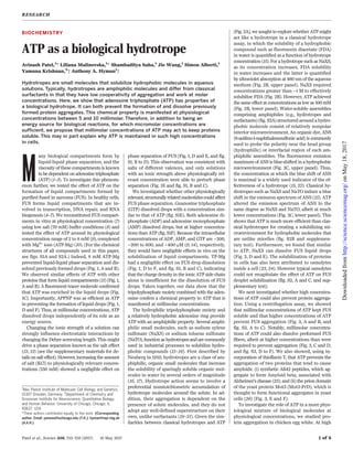
Fig. 1. ATP inhibits the phase separation of unstructured proteins.
(A) Fluorescent images of phase-separated protein drops treated with
ATP-Mg at different concentrations. FUS-GFP was used at 5 mM,
TAF15-Snap-Alexa Fluor 546 was used at 1 mM, hnRNPA3-GFP was used
at 25 mM, and PGL-3-GFP was used at 2.5 mM. Phase-separated drops
formed in low-salt buffer (70 mM). Increasing concentration of ATP-Mg
inhibited phase separation. Scale bar, 10 mm. (B) Quantification of
phase separation. FUS-GFP, TAF15-Snap-Alexa Fluor 546, hnRNPA3-
GFP, and PGL-3-GFP were used at 7 mM, 1 mM, 25 mM, and 2.5 mM,
respectively. Phase separation is determined as the ratio of protein
inside drops (Iin) to protein outside drops (Iout). Total fluorescence
intensity of protein is taken as a measure for protein amount.
Decreasing Iin/Iout ratios reflect inhibition of phase separation. Lines
represent fitted dose-response curves [log(concentration) versus Iin/Iout
ratio]. MC[ATP-Mg] is the minimal concentration of ATP-Mg needed to
inhibit phase separation. Error bars, mean ± SD (N = 36). The
physiological concentration range of ATP-Mg is highlighted in gray.
(C) ATP enrichment inside the phase-separated FUS droplet phase.
2,4,6-trinitrophenol-ATP (ATP-TNP) (1 mM) was used as fluorescent
tracer for ATP in a solution of phase-separated FUS protein. Line scan
of fluorescent intensity of ATP-TNP (plotted across the magenta
line) indicates fourfold enrichment inside the droplet phase compared
with the surrounding solution. (D) Fluorescent images of phase-
separated FUS-GFP protein drops treated with ATP-Mg, APPNP-Mg,
salt (KCl), and magnesium tripolyphosphate (Mg-TP). FUS-GFP was
used at 5 mM. The range of concentrations of all added reagents
was adjusted to match the range of ionic strength of ATP-Mg (see
the supplementary materials for details). Phase separation is
inhibited by 8 mM of ATP-Mg and APPNP-Mg. Scale, bar 10 mm.
(E) Quantification of phase separation [conditions as in (D)] with
FUS-GFP used at 7 mM. MC is the minimal concentration needed to
inhibit phase separation. Error bars, mean ± SD (N = 36).
(F) Quantification of minimum ionic strength needed to disrupt phase
separation of FUS-GFP in the presence of different reagents. FUS-GFP
was used at 7 mM. Ionic strength was calculated using the shown
equation, where c is concentration, z is the charge of the ion, and
n is the number of ions in a solution (see the supplementary materials
for details).
RESEARCH | REPORT
onMay18,2017http://science.sciencemag.org/Downloadedfrom](https://image.slidesharecdn.com/atpasabiologicalhydrotrope-180315214104/85/Atp-as-a-biological-hydrotrope-2-320.jpg)
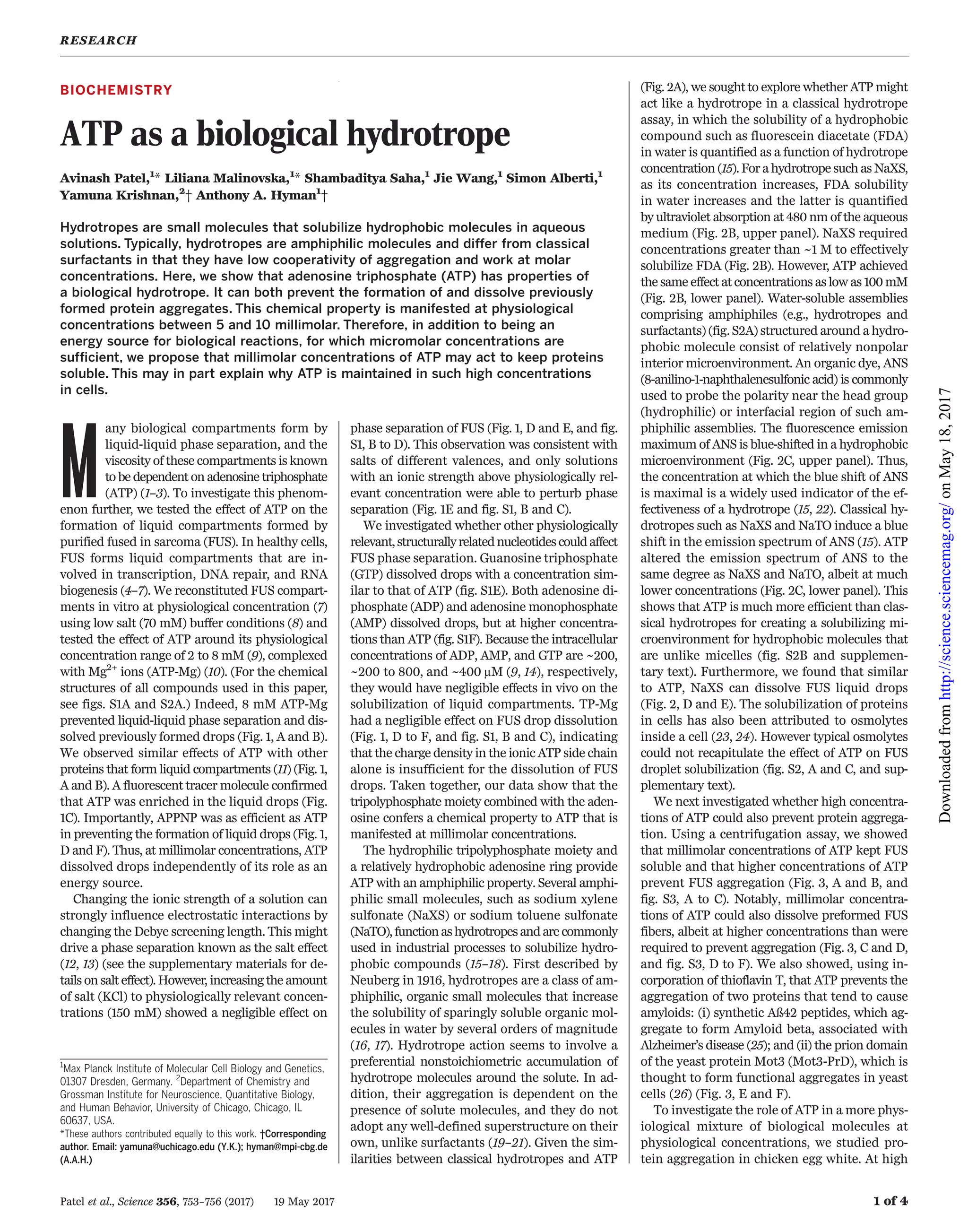
![Patel et al., Science 356, 753–756 (2017) 19 May 2017 3 of 4
Fig. 2. Hydrotrope-
like properties of
ATP. (A) Comparison
of chemical structure
of ATP, NaXS, and
NaTO.The molecules
have a hydrophobic
aromatic ring (purple)
and a short hydro-
philic, charged residue
(blue). (B) Effect of
ATP-Mg, NaXS, NaTO,
and salt on the solubil-
ity of FDA in aqueous
solutions at room tem-
perature as measured
by the absorbance at
480 nm. If in solution,
FDA absorbs light at
480 nm, while
aggregated FDA
scatters light (sche-
matics). Lines repre-
sent fitted dose-response curves [log(concentration) versus absorption of FDA
at 480 nm]. Error bars, mean ± SD (N = 3). (C) ATP-Mg solutions offer a less polar
environment than water.The variation of the fluorescence spectral band
maximum of the probe ANS is shown as a function of the concentration of ATP-Mg
and NaXS. ATP-Mg solutions exhibit the most prominent blue shift. Lines represent
fitted dose-response curves [log(concentration) versus emission maxima
of ANS]. Error bars, mean ± SD (N = 3). (D) Fluorescent images of phase-
separated FUS-GFP protein drops treated with ATP-Mg and NaXS. FUS-GFP
was used at 5 mM. Scale bar, 10 mm. (E) Quantification of phase
separation [conditions as in (D)] with FUS-GFP used at 7 mM. The range of
concentrations of NaXS was adjusted to match the range of ionic
strength of ATP-Mg (see the supplementary materials for details). MC is
the minimal concentration needed to inhibit phase separation. Error bars,
mean ± SD (N = 36).
Fig. 3. Inhibition of
protein aggregation
by hydrotropes.
(A) Hydrotropes
enhance the solubility
of FUS-GFP. FUS-GFP
(input) at high concen-
tration (40 mM) had a
limited solubility in low-
salt buffer conditions
(150 mM) after cleav-
age of the tagged solu-
bilizing domain
[maltose binding pro-
tein (MBP)]. Soluble
FUS-GFP in the super-
natant after centrifuga-
tion of aggregated
FUS-GFP was visual-
ized on a SDS–
polyacrylamide gel
electrophoresis
(SDS-PAGE) gel
stained with Coomassie
blue. ATP-Mg and its nonhydrolysable analog, APPNP, enhanced the solubility
of FUS-GFP to a similar extent as NaXS.Cleaved MBP tag served as the loading
control. (B) Dose-response curve of FUS-GFP solubility in the presence of
ATP-Mg, APPNP, and NaXS. Lines represent fitted dose-response curves
[log(concentration) versus soluble fraction with respect to total input of
FUS-GFP]. Error bars, mean ± SD (N = 3). (C) ATP dissolves preformed FUS
aggregates. Mutant FUSG156E-GFP (5 mM) was aged to induce aggregation.
Hydrotropes were added after aggregate formation and incubated for
12 hours. Soluble fractions were visualized on a SDS-PAGE stained with
Coomassie blue. ATP and NaXS dissolve the FUS aggregates at
high concentrations (32 mM and 128 mM, respectively). Lysozyme was
spiked into the assay as a loading control. (D) Quantification of the solubilization
of FUS aggregates [conditions as in (C)]. Lines represent fitted dose-response
curves [log(concentration) versus soluble fraction with respect to total input
of FUS-GFP]. Error bars, mean ± SD (N = 3). (E and F) ATP-Mg prevents fiber
formation.Thioflavin T (ThT) was incorporated in amyloid fibers and
displays an enhanced fluorescence (Ex 440nm/Em 480nm). ATP-Mg or
nonhydrolysable APPCP-Mg prevented formation of amyloid fibers of Aß42
peptide (E) and the prion domain of the yeast Mot3 protein (Mot3-PrD)
(F) in a concentration-dependent manner. Lines represent fitted dose-response
curves [log(concentration) versus aggregated protein assessed by ThT
emission]. Error bars, mean ± SD (N = 3).The range of concentrations
of all added reagents was adjusted to match the range of ionic strength of
ATP-Mg (see the supplementary materials for details).
RESEARCH | REPORT
onMay18,2017http://science.sciencemag.org/Downloadedfrom](https://image.slidesharecdn.com/atpasabiologicalhydrotrope-180315214104/85/Atp-as-a-biological-hydrotrope-3-320.jpg)
![50-fold higher ATP/ADP ratio is necessary to
fuel the myriad metabolic reactions taking place
simultaneously in a cell. However, cytoplasm can
have protein concentrations over 100 mg/mL
(31–33), and it is extremely difficult to maintain
such high protein concentrations in a test tube
without spontaneous aggregation. The hydro-
trope activity of ATP may help keep proteins sol-
uble in the cytoplasm (34) and provide another,
but not mutually exclusive, explanation for high
ATP concentrations in cells. Possibly also, as the
levels of ATP decline with age or mitochondrial
impairment, this could lead to increased aggre-
gation and consequently neurodegenerative de-
cline during aging. Our work in this paper has
focused on the role of ATP in keeping unstruc-
tured proteins soluble, because these are the
types of proteins that have a propensity to form
pathological aggregates (35). It will be interesting
to examine the role of high ATP concentrations in
stability and function of multimolecular protein
machines.
More generally, during evolution, the produc-
tion of complex macromolecules would have im-
mediately presented the problem of aggregation.
As one of the basic building blocks in RNA and
DNA, ATP may have been coopted early in evo-
lution to prevent such aggregation. It is ideal for
this purpose, due to the high activation energy re-
quired to hydrolyze the polyphosphate bonds in
an ATP-Mg-water complex. ATP could later have
been adopted to provide the basic energy source
for metabolism, which is the hydrolysis of ATP
to ADP.
REFERENCES AND NOTES
1. C. P. Brangwynne, T. J. Mitchison, A. A. Hyman, Proc. Natl.
Acad. Sci. U.S.A. 108, 4334–4339 (2011).
2. S. Jain et al., Cell 164, 487–498 (2016).
3. A. A. Hyman, C. A. Weber, F. Jülicher, Annu. Rev. Cell Dev. Biol.
30, 39–58 (2014).
4. M. Polymenidou et al., Brain Res. 1462, 3–15 (2012).
5. W. Y. Wang et al., Nat. Neurosci. 16, 1383–1391 (2013).
6. X. Wang et al., Nature 454, 126–130 (2008).
7. A. Patel et al., Cell 162, 1066–1077 (2015).
8. K. A. Burke, A. M. Janke, C. L. Rhine, N. L. Fawzi, Mol. Cell 60,
231–241 (2015).
9. T. W. Traut, Mol. Cell. Biochem. 140, 1–22 (1994).
10. K. Clarke et al., J. Biol. Chem. 271, 21142–21150
(1996).
11. S. Saha et al., Cell 166, 1572–1584 (2016).
12. T. J. Nott et al., Mol. Cell 57, 936–947 (2015).
13. P. Debye, E. Hückel, Phys. Z. 24, 185–206
(1923).
14. E. Gout, F. Rébeillé, R. Douce, R. Bligny, Proc. Natl. Acad.
Sci. U.S.A. 111, E4560–E4567 (2014).
15. D. Balasubramanian, V. Srinivas, V. G. Gaikar, M. M. Sharma,
J. Phys. Chem. 93, 3865–3870 (1989).
16. C. Neuberg, Biochem. Z. 76, 107–176 (1916).
17. J. Eastoe, M. H. Hatzopoulos, P. J. Dowding, Soft Matter 7,
5917–5925 (2011).
18. C. V. Subbarao, I. P. K. Chakravarthy, A. V. S. L. Sai Bharadwaj,
K. M. M. K. Prasad, Chem. Eng. Technol. 35, 225–237
(2012).
19. J. J. Booth, S. Abbott, S. Shimizu, J. Phys. Chem. B 116,
14915–14921 (2012).
20. J. J. Booth, M. Omar, S. Abbott, S. Shimizu, Phys. Chem. Chem.
Phys. 17, 8028–8037 (2015).
21. S. Shimizu, N. Matubayasi, J. Phys. Chem. B 118, 10515–10524
(2014).
22. V. Srinivas, D. Balasubramanian, Langmuir 14, 6658–6661
(1998).
23. D. W. Bolen, I. V. Baskakov, J. Mol. Biol. 310, 955–963
(2001).
24. P. H. Yancey, M. E. Clark, S. C. Hand, R. D. Bowlus,
G. N. Somero, Science 217, 1214–1222 (1982).
25. G. G. Glenner, C. W. Wong, Biochem. Biophys. Res. Commun.
120, 885–890 (1984).
26. R. Halfmann et al., Nature 482, 363–368
(2012).
27. S. N. Timasheff, Biochemistry 41, 13473–13482
(2002).
28. S. Shimizu, N. Matubayasi, J. Phys. Chem. B 118, 3922–3930
(2014).
29. S. Shimizu, D. J. Smith, J. Chem. Phys. 121, 1148–1154
(2004).
30. S. Shimizu, Proc. Natl. Acad. Sci. U.S.A. 101, 1195–1199
(2004).
31. R. Milo, BioEssays 35, 1050–1055 (2013).
32. B. J. Zeskind et al., Nat. Methods 4, 567–569
(2007).
33. S. B. Zimmerman, S. O. Trach, J. Mol. Biol. 222, 599–620
(1991).
34. B. R. Parry et al., Cell 156, 183–194 (2014).
35. S. Alberti, A. A. Hyman, BioEssays 38, 959–968
(2016).
ACKNOWLEDGMENTS
We particularly thank W. Kunz and colleagues for their help,
support, advice, and discussions during the preparation of
the manuscript. We thank D. Drechsel, A. Nadler, K. Sandhoff,
D. Tang, and members of the Hyman, Krishnan, and Alberti
laboratories for helpful discussions; B. Bogdanovo and
R. Lemaitre for help with protein expression and purification;
B. Lombardot and R. Hasse from the Scientific Computing
facility for image analysis; and B. Nitzsche and B. Schroth-Diez
for help with light microscopy. We gratefully acknowledge
funding from the Alexander von Humboldt Foundation (GRO/
1156614 STP-2 to A.P. and USA/1153678 STP to S.S.), EMBO
ALTF (608-2013 to S.S.), and German Federal Ministry of
Research and Education (BMBF 031A359A Max Syn Bio). Y.K.
acknowledges the Scientific Innovation Award from the Brain
Research Foundation (BRF SIA-2016-01) and start-up funds
from the University of Chicago. A.A.H. and A.P. are inventors on
patent application 1305-5403-MSG-ZE, submitted by the Max
Planck Society, which covers nucleotides as hydrotropes.
SUPPLEMENTARY MATERIALS
www.sciencemag.org/content/356/6339/753/suppl/DC1
Materials and Methods
Supplementary Text
Figs. S1 to S4
Table S1
Movie S1
References (36–41)
14 March 2016; accepted 24 March 2017
10.1126/science.aaf6846
Patel et al., Science 356, 753–756 (2017) 19 May 2017 4 of 4
Fig. 4. ATP enhances protein stability. (A) Heat denaturation of crude egg white can be
inhibited by addition of ATP-Mg. Crude egg white was heated at 60°C in a water bath in the
presence of equimolar (10 mM) amounts of ATP-Mg, nonhydrolysable APPCP-Mg, and NaXS.
NaCl (40 mM) was used to match the ionic strength of ATP-Mg. Over 30 min, the aggregation
of egg white is abolished in the presence of ATP-Mg and APPCP-Mg. (B) The stabilization of
heat-denatured egg white is concentration dependent. The kinetic traces of egg-white
aggregation [conditions as in (A)] in the presence of increasing concentrations of ATP-Mg
over 60 min. The amount of egg-white aggregation is assessed by changes of the pixel value
(integrated density). Four mM of ATP-Mg blocked aggregation by 50%, whereas 12 mM of
ATP-Mg completely abolished aggregation. The shaded area represents the range of the
standard error (n = 3). (C) The dose response for stabilization of heat-denatured egg white. The
amount of egg-white aggregation was assessed by turbidity measurement in a 96-well plate.
The aggregation of egg white decreases with increasing concentrations. Lines represent fitted
dose-response curves [log(concentration versus aggregated material assessed by turbidity
measurement]. Error bars, mean ± SD (N = 3). The range of concentrations of all added reagents
was adjusted to match the range of ionic strength of ATP-Mg (see the supplementary materials
for details).
RESEARCH | REPORT
onMay18,2017http://science.sciencemag.org/Downloadedfrom](https://image.slidesharecdn.com/atpasabiologicalhydrotrope-180315214104/85/Atp-as-a-biological-hydrotrope-4-320.jpg)
![(6339), 753-756. [doi: 10.1126/science.aaf6846]356Science
2017)
18,Simon Alberti, Yamuna Krishnan and Anthony A. Hyman (May
Avinash Patel, Liliana Malinovska, Shambaditya Saha, Jie Wang,
ATP as a biological hydrotrope
Editor's Summary
, this issue p. 753; see also p. 701Science
aggregation that occur in disease or liquid-liquid phase separations that occur within cells.
results raise the possibility that ATP concentrations could influence processes such as protein
concentrations found in cells could act as a hydrotrope to help solubilize hydrophobic proteins. The
Rosen). Protein concentrations in cells can exceed 100 mg/ml. The authors found that ATP at
explain why such high concentrations of ATP are maintained in cells (see the Perspective by Rice and
find that ATP may also enhance protein solubility, which could helpet al.reactions within cells. Patel
Adenosine triphosphate (ATP) has well-characterized roles in providing energy for biochemical
ATP boosts protein solubility
This copy is for your personal, non-commercial use only.
Article Tools
http://science.sciencemag.org/content/356/6339/753
article tools:
Visit the online version of this article to access the personalization and
Permissions
http://www.sciencemag.org/about/permissions.dtl
Obtain information about reproducing this article:
is a registered trademark of AAAS.ScienceAdvancement of Science; all rights reserved. The title
Avenue NW, Washington, DC 20005. Copyright 2016 by the American Association for the
in December, by the American Association for the Advancement of Science, 1200 New York
(print ISSN 0036-8075; online ISSN 1095-9203) is published weekly, except the last weekScience
onMay18,2017http://science.sciencemag.org/Downloadedfrom](https://image.slidesharecdn.com/atpasabiologicalhydrotrope-180315214104/85/Atp-as-a-biological-hydrotrope-5-320.jpg)


![Polymer [ बहुलक ] Chemistry Notes PDF - Irfanullah Mehar - JJ Sir Chemistry.pdf](https://cdn.slidesharecdn.com/ss_thumbnails/polymerchemistrynotespdf-irfanullahmehar-jjsirchemistry-260210172118-3f9b37f7-thumbnail.jpg?width=640&height=640&fit=bounds)


