SlideShare a Scribd company logo
1 of 1
Download to read offline
The regulation of microRNA activity by ATM in the DNA damage response
David W. Salzman1, Frank Slack2, Joanne B. Weidhaas1
1Department of Therapeutic Radiology, Yale University School of Medicine
2Department of Molecular, Cellular and Developmental Biology, Yale University
MicroRNAs are a class of non-coding, 18-24nucleotide, trans-acting RNAs
that negatively regulate gene expression at the posttranscriptional level by
annealing to cis-regulatory element(s) in a target mRNA and inhibiting
protein translation. The temporal and spatial expression of microRNAs
proceeds through several regulatory checkpoints mainly, transcription and
processing of the pri-microRNA and pre-microRNA precursors by the
RNaseIII enzymes Drosha and Dicer.
Previous work indicates that miR-34 is regulated at the transcriptional level
following DNA damage in a P53-dependent manor and protects cells
against DNA damage-induced apoptosis by targeting a number of critical
genes required for cell cycle progression. However, data from several
laboratories (including our own) suggests that there is a pool of
ubiquitously expressed miR-34 in cells, the expression of which is
independent of both P53 and DNA damage.
The role of P53/DNA damage-independent miR-34 is not understood. To
study this, we established a luciferase reporter assay to measure miR-34
gene silencing activity. Our data indicated that the pre-existing pool of
miR-34 is inactive (not competent to silence gene expression). However,
the pool of miR-34 was rapidly activated by ionizing radiation (IR) which
occurred several hours before P53-mediated transcription.
Introduction
0
2
4
6
8
10
12
14
16
let-7 miR-34
microRNAactivity
(folddifferenceMT/WT)
MCF-7
0
2
4
6
8
10
12
14
16
18
let-7 miR-34
microRNAactivity
(folddifferenceMT/WT)
H460
0
2
4
6
8
10
12
14
let-7 miR-34
microRNAactivity
(folddifferenceMT/WT)
A549
0
1
2
3
4
5
6
7
8
9
10
let-7 miR-34
microRNAactivity
(folddifferenceMT/WT)
HeLa
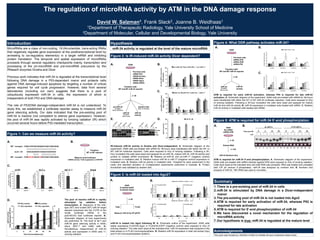
Figure 1: Can we measure miR-34 activity?
Hypothesis
WT target CGACCGUCACAGAAUCGACCAACAGA
||||||||||||||||||||||
miR-34 UGGCAGUGUCUUAGCUGGUUGU
| |||| |||||||||||
MT target CGAGGCACACACUAUCGACCAACAGA
A. B.
Transfect:
WT or MT reporter
Measure dual-luciferase
miRNA activity = fold suppression of MT/WT
C.
D. The pool of inactive miR-34 is rapidly
stimulated by radiation before
transcription. A. Sequence of the wild-
type (WT) and mutant (MT) miR-34 target
element subcloned into the 3’UTR of the
renilla luciferase mRNA in the
psiCHECK2 dual luciferase reporter. B.
Schematic diagram of how experiments
are preformed. C. The level of let-7 and
miR-34 gene silencing activity in A549,
HeLa, MCF-7 and H460 cells, D.
Simultaneous measurement of miR-34
activity and expression in A549 cells +/-
ionizing radiation.
miR-34 activity is regulated at the level of the mature microRNA
Figure 2: Is IR-induced miR-34 activity Dicer dependent?
0
2
4
6
8
10
No siRNA siP53 siDROSHA siDICER siAGO2 siGAPD
pri-microRNAexpression
(foldchange)
pri-miR-34a pri-miR-34b/c pri-miR-17
0
2
4
6
8
10
No siRNA siP53 siDROSHA siDICER siAGO2 siGAPD
microRNAexpression
(foldchange)
miR-34a miR-34b miR-34c miR-17
0
5
10
15
20
No siRNA siP53 siDROSHA siDICER siAGO2 siGAPD
miR-34activity
-IR +IR
Pre-treat cells:
siRNA-P53
siRNA-Drosha
siRNA-Dicer
siRNA-Ago2
siRNA-GAPD
Transfect with:
WT and MT miR-34 reporter
A. A549
-/+ IR
pri-miR-34
RT-qPCR
Norm. to B-Actin
miR-34
RT-qPCR
Norm. to U6
miR-34
activity
western blot
Norm. to Vimentin
B.
E.
VIM
Drosha
P53
_ +
_
+
_
+
_
+
Vim
Dicer
Ago2
IR
GAPD
_ +
_
+
_
+ IR
IR-induced miR-34 activity is Drosha and Dicer-independent. A. Schematic diagram of the
experiment. A549 cells pre-treated with siRNA for 36-hours were transfected with either the WT or
MT miR-34 luciferase reporters. Cells were exposed to 4Gy of ionizing radiation. Following a 30-
hour incubation the cells were lysed and assayed for pri-miR-34, mature miR-34, miR-34 activity and
protein to validate siRNA knockdown. B. Relative pri-miR-34 and pri-miR-17 (negative control)
expression in irradiated cells. C. Relative mature miR-34 or miR-17 (negative control) expression in
irradiated cells. D. Relative miR-34 activity in irradiated cells. *Graphed for each experiment is the
mean and standard deviation of 2-independent experiments preformed in triplicate. E. Protein
knockdown was validated by western blot.
C.
D.
Summary
1. There is a pre-existing pool of miR-34 in cells
2. miR-34 is stimulated by DNA damage in a Dicer-independent
manor
3. The pre-existing pool of miR-34 is not loaded into Ago2
4. ATM is required for early activation of miR-34, whereas P53 is
required for late activation
5. ATM is required for 5’-end phosphorylation of miR-34
6. We have discovered a novel mechanism for the regulation of
microRNA activity
7. Our hypothesis is true: miR-34 is regulated at the mature level
Figure 4: What DDR pathway activates miR-34?
Pre-treat cells:
siRNA-ATR
siRNA-ATM
siRNA-P53
Transfect with:
WT and MT miR-34 reporter
A549
-/+ IR
miR-34
RT-qPCR
Norm. to U6
miR-34
activity 0
1
2
3
4
5
6
7
no siRNA ATR siRNA ATM siRNA P53 siRNA
RelativemiR-34activity
0hr 4hr 12hr 36hr
0
1
2
3
4
5
no siRNA ATR siRNA ATM siRNA P53 siRNA
RelativemiR-34aexpression
0hr 4hr 12hr 36hr
B.A.
ATM is required for early miR-34 activation, whereas P53 is required for late miR-34
activation. A. Schematic diagram of the experiment. A549 cells pre-treated with siRNA for 36-hours
were transfected with either the WT or MT miR-34 luciferase reporters. Cells were exposed to 4Gy
of ionizing radiation. Following a 30-hour incubation the cells were lysed and assayed for mature
miR-34 and miR-34 activity. B. miR-34 expression in irradiated cells treated with siRNA. C. Relative
miR-34 activity in irradiated cells treated with siRNA.
C.
Figure 3: Is miR-34 loaded into Ago2?
Measure miR-34 by RT-qPCR
A549
FLAG/HA-EGFP
FLAG/HA-AGO2
-/+ IR
FLAGTotal
A.
miR-34 is loaded into Ago2 following IR. A. Schematic outline of the experiment. A549 cells
stably expressing FLAG/HA-Ago2 or FLAG/HA-EGFP (negative control) were exposed to 4Gy of
ionizing radiation. The cells were lysed at the indicated time. miR-34 expression was analyzed in the
total extract or in FLAG immunoprecipitates. B. Relative miR-34 expression in total cell extract (top)
and FLAG immunoprecipitates (bottom).
B.
Acknoledgements
This work was funded by: NIH/NCI 5 K08 CA124484-05 and a Kalimeris Seed Grant.
Figure 5: ATM is required for miR-34 5’-end phosphorylation
miR-34 Northern blot
A549
No siRNA
siRNA-ATM
-/+ IR
Extract RNA
-/+ CIP
A. B.
ATM is required for miR-34 5’-end phosphorylation. A. Schematic diagram of the experiment.
A549 cells pre-treated with siRNA directed against ATM were exposed to 2Gy of ionizing radiation.
Cells were lysed at the indicated time and total RNA was extracted. RNA was treated and untreated
with CIP (to remove 5’-phosphates). miR-34 was analyzed by northern blot. B. Northern blot
analysis of miR-34. 18S rRNA was used to normalize.

More Related Content

What's hot

Signaling events triggered by inactivation of the TSC1-2 Complex: Implication...
Signaling events triggered by inactivation of the TSC1-2 Complex: Implication...Signaling events triggered by inactivation of the TSC1-2 Complex: Implication...
Signaling events triggered by inactivation of the TSC1-2 Complex: Implication...dgotto
 
study of EGFR protein expression and mutation
study of EGFR protein expression and mutation study of EGFR protein expression and mutation
study of EGFR protein expression and mutation premvarma064
 
Strong reversal of the lung fibrosis disease signature by autotaxin inhibitor...
Strong reversal of the lung fibrosis disease signature by autotaxin inhibitor...Strong reversal of the lung fibrosis disease signature by autotaxin inhibitor...
Strong reversal of the lung fibrosis disease signature by autotaxin inhibitor...Maté Ongenaert
 
REU Final PowerPoint
REU Final PowerPointREU Final PowerPoint
REU Final PowerPointAlyssa Castle
 
Inhibition of the mtorc pathway in the antiphospholipid
Inhibition of the mtorc pathway in the antiphospholipidInhibition of the mtorc pathway in the antiphospholipid
Inhibition of the mtorc pathway in the antiphospholipidSaris Arango
 
Cancer signal-transduction
Cancer signal-transductionCancer signal-transduction
Cancer signal-transductionDeepika Tripathi
 
A high copy number screen of the trs23Δ99C mutant
A high copy number screen of the trs23Δ99C mutantA high copy number screen of the trs23Δ99C mutant
A high copy number screen of the trs23Δ99C mutantGabriel Bendavit
 
Stat family:stat 3 in skin disorders by yousry
Stat family:stat 3 in skin disorders  by yousryStat family:stat 3 in skin disorders  by yousry
Stat family:stat 3 in skin disorders by yousryM.YOUSRY Abdel-Mawla
 
RNA splicing mutations and human disease: Pompe disease.
RNA splicing mutations and human disease: Pompe disease.RNA splicing mutations and human disease: Pompe disease.
RNA splicing mutations and human disease: Pompe disease.CentroMalattieRareFVG
 
Glucocorticoids modulate micro rna expression and processing during lymphocyt...
Glucocorticoids modulate micro rna expression and processing during lymphocyt...Glucocorticoids modulate micro rna expression and processing during lymphocyt...
Glucocorticoids modulate micro rna expression and processing during lymphocyt...wangdong2336
 
The mammalian target of rapamycin a cell key modulator of ageing
The mammalian target of rapamycin a cell key modulator of ageingThe mammalian target of rapamycin a cell key modulator of ageing
The mammalian target of rapamycin a cell key modulator of ageingXavier Viñals Gonzalez
 

What's hot (19)

Signaling events triggered by inactivation of the TSC1-2 Complex: Implication...
Signaling events triggered by inactivation of the TSC1-2 Complex: Implication...Signaling events triggered by inactivation of the TSC1-2 Complex: Implication...
Signaling events triggered by inactivation of the TSC1-2 Complex: Implication...
 
nihms557954
nihms557954nihms557954
nihms557954
 
study of EGFR protein expression and mutation
study of EGFR protein expression and mutation study of EGFR protein expression and mutation
study of EGFR protein expression and mutation
 
In press
In pressIn press
In press
 
Strong reversal of the lung fibrosis disease signature by autotaxin inhibitor...
Strong reversal of the lung fibrosis disease signature by autotaxin inhibitor...Strong reversal of the lung fibrosis disease signature by autotaxin inhibitor...
Strong reversal of the lung fibrosis disease signature by autotaxin inhibitor...
 
Final Paper
Final PaperFinal Paper
Final Paper
 
REU Final PowerPoint
REU Final PowerPointREU Final PowerPoint
REU Final PowerPoint
 
Inhibition of the mtorc pathway in the antiphospholipid
Inhibition of the mtorc pathway in the antiphospholipidInhibition of the mtorc pathway in the antiphospholipid
Inhibition of the mtorc pathway in the antiphospholipid
 
Jewellrgb
JewellrgbJewellrgb
Jewellrgb
 
Cancer signal-transduction
Cancer signal-transductionCancer signal-transduction
Cancer signal-transduction
 
Role of Mirn 155 in thr regulation of lymhocyte
Role of Mirn 155 in thr regulation of lymhocyteRole of Mirn 155 in thr regulation of lymhocyte
Role of Mirn 155 in thr regulation of lymhocyte
 
PNS 0502002625
PNS 0502002625PNS 0502002625
PNS 0502002625
 
A high copy number screen of the trs23Δ99C mutant
A high copy number screen of the trs23Δ99C mutantA high copy number screen of the trs23Δ99C mutant
A high copy number screen of the trs23Δ99C mutant
 
Stat family:stat 3 in skin disorders by yousry
Stat family:stat 3 in skin disorders  by yousryStat family:stat 3 in skin disorders  by yousry
Stat family:stat 3 in skin disorders by yousry
 
RNA splicing mutations and human disease: Pompe disease.
RNA splicing mutations and human disease: Pompe disease.RNA splicing mutations and human disease: Pompe disease.
RNA splicing mutations and human disease: Pompe disease.
 
EASD 2009
EASD 2009EASD 2009
EASD 2009
 
Glucocorticoids modulate micro rna expression and processing during lymphocyt...
Glucocorticoids modulate micro rna expression and processing during lymphocyt...Glucocorticoids modulate micro rna expression and processing during lymphocyt...
Glucocorticoids modulate micro rna expression and processing during lymphocyt...
 
The mammalian target of rapamycin a cell key modulator of ageing
The mammalian target of rapamycin a cell key modulator of ageingThe mammalian target of rapamycin a cell key modulator of ageing
The mammalian target of rapamycin a cell key modulator of ageing
 
Micro RNA's
Micro RNA'sMicro RNA's
Micro RNA's
 

Viewers also liked

Presentación PROMO R&H 2012
Presentación PROMO R&H 2012 Presentación PROMO R&H 2012
Presentación PROMO R&H 2012 Alberto Rojas
 
Barr Memorial Library Catalog
Barr Memorial Library CatalogBarr Memorial Library Catalog
Barr Memorial Library Cataloglneal3
 
William Barr, PhD
William Barr, PhDWilliam Barr, PhD
William Barr, PhDNYU FACES
 
R tools for micro-RNA pathway analysis
R tools for micro-RNA pathway analysisR tools for micro-RNA pathway analysis
R tools for micro-RNA pathway analysisAnwesha Bohler
 
Certificate C Vem8 Khvhhn
Certificate C Vem8 KhvhhnCertificate C Vem8 Khvhhn
Certificate C Vem8 Khvhhnkevinskow
 
6th Grade lesson 1
6th Grade lesson 1 6th Grade lesson 1
6th Grade lesson 1 ivolenec
 
Producing Amazing Audio Content In-House
Producing Amazing Audio Content In-HouseProducing Amazing Audio Content In-House
Producing Amazing Audio Content In-HouseHeather Marie Wells
 
Regression of dalton’s lymphoma in vivo via decline
Regression of dalton’s lymphoma in vivo via declineRegression of dalton’s lymphoma in vivo via decline
Regression of dalton’s lymphoma in vivo via declinerkkoiri
 
2burwick.neila.il ltutorial
2burwick.neila.il ltutorial2burwick.neila.il ltutorial
2burwick.neila.il ltutorialNeila Burwick
 
Finkelstein encore catalog-revised
Finkelstein encore catalog-revisedFinkelstein encore catalog-revised
Finkelstein encore catalog-revisedZeke Kilbride
 
E books & eaudiobooks 1.26
E books & eaudiobooks 1.26E books & eaudiobooks 1.26
E books & eaudiobooks 1.26skoebel
 
Regression of dalton’s lymphoma in vivo via decline
Regression of dalton’s lymphoma in vivo via declineRegression of dalton’s lymphoma in vivo via decline
Regression of dalton’s lymphoma in vivo via declinerkkoiri
 
Mass-use models for OER
Mass-use models for OERMass-use models for OER
Mass-use models for OERtbirdcymru
 
Sequential Folding Model of RNA secondary structure
Sequential Folding Model of RNA secondary structureSequential Folding Model of RNA secondary structure
Sequential Folding Model of RNA secondary structureLi Tai Fang
 

Viewers also liked (18)

Presentación PROMO R&H 2012
Presentación PROMO R&H 2012 Presentación PROMO R&H 2012
Presentación PROMO R&H 2012
 
Find Books @ the Library
Find Books @ the LibraryFind Books @ the Library
Find Books @ the Library
 
Barr Memorial Library Catalog
Barr Memorial Library CatalogBarr Memorial Library Catalog
Barr Memorial Library Catalog
 
William Barr, PhD
William Barr, PhDWilliam Barr, PhD
William Barr, PhD
 
FISV2008
FISV2008FISV2008
FISV2008
 
R tools for micro-RNA pathway analysis
R tools for micro-RNA pathway analysisR tools for micro-RNA pathway analysis
R tools for micro-RNA pathway analysis
 
Certificate C Vem8 Khvhhn
Certificate C Vem8 KhvhhnCertificate C Vem8 Khvhhn
Certificate C Vem8 Khvhhn
 
6th Grade lesson 1
6th Grade lesson 1 6th Grade lesson 1
6th Grade lesson 1
 
Producing Amazing Audio Content In-House
Producing Amazing Audio Content In-HouseProducing Amazing Audio Content In-House
Producing Amazing Audio Content In-House
 
Regression of dalton’s lymphoma in vivo via decline
Regression of dalton’s lymphoma in vivo via declineRegression of dalton’s lymphoma in vivo via decline
Regression of dalton’s lymphoma in vivo via decline
 
Podcasting101
Podcasting101Podcasting101
Podcasting101
 
Ensayo 2 b 7 de junio
Ensayo 2 b 7 de junioEnsayo 2 b 7 de junio
Ensayo 2 b 7 de junio
 
2burwick.neila.il ltutorial
2burwick.neila.il ltutorial2burwick.neila.il ltutorial
2burwick.neila.il ltutorial
 
Finkelstein encore catalog-revised
Finkelstein encore catalog-revisedFinkelstein encore catalog-revised
Finkelstein encore catalog-revised
 
E books & eaudiobooks 1.26
E books & eaudiobooks 1.26E books & eaudiobooks 1.26
E books & eaudiobooks 1.26
 
Regression of dalton’s lymphoma in vivo via decline
Regression of dalton’s lymphoma in vivo via declineRegression of dalton’s lymphoma in vivo via decline
Regression of dalton’s lymphoma in vivo via decline
 
Mass-use models for OER
Mass-use models for OERMass-use models for OER
Mass-use models for OER
 
Sequential Folding Model of RNA secondary structure
Sequential Folding Model of RNA secondary structureSequential Folding Model of RNA secondary structure
Sequential Folding Model of RNA secondary structure
 

Similar to ATM

Mi rna functional analysis 2013
Mi rna functional analysis 2013Mi rna functional analysis 2013
Mi rna functional analysis 2013Elsa von Licy
 
CSUN SYMPOSIUM Final
CSUN SYMPOSIUM Final CSUN SYMPOSIUM Final
CSUN SYMPOSIUM Final Alina Adamian
 
Thesis for resume presentation
Thesis for resume presentationThesis for resume presentation
Thesis for resume presentationaimlue
 
2016-08-20-srep31431
2016-08-20-srep314312016-08-20-srep31431
2016-08-20-srep31431Arpad Palfi
 
The Role Of Mir 125A And Mir 223
The Role Of Mir 125A And Mir 223The Role Of Mir 125A And Mir 223
The Role Of Mir 125A And Mir 223Jenny Richardson
 
Ttp Lab Tech Talk 051810
Ttp Lab Tech Talk 051810Ttp Lab Tech Talk 051810
Ttp Lab Tech Talk 051810Neil Kubica
 
Poster Presentation
Poster PresentationPoster Presentation
Poster PresentationChunghee Kim
 
Role of MicroRNA in Phosphorus Defficiency
Role of MicroRNA in Phosphorus DefficiencyRole of MicroRNA in Phosphorus Defficiency
Role of MicroRNA in Phosphorus DefficiencyHarsh Kishore Mishra
 
The 5' terminal uracil of let-7a is critical for the recruitment of mRNA to A...
The 5' terminal uracil of let-7a is critical for the recruitment of mRNA to A...The 5' terminal uracil of let-7a is critical for the recruitment of mRNA to A...
The 5' terminal uracil of let-7a is critical for the recruitment of mRNA to A...David W. Salzman
 
Generation of MRP2 Efflux Transporter Knock-Out in HepaRG Cell Line
Generation of MRP2 Efflux Transporter Knock-Out in HepaRG Cell LineGeneration of MRP2 Efflux Transporter Knock-Out in HepaRG Cell Line
Generation of MRP2 Efflux Transporter Knock-Out in HepaRG Cell Linemdmitc
 
Non coding RNA as targets in drug discovery.pptx
Non coding RNA as targets in drug discovery.pptxNon coding RNA as targets in drug discovery.pptx
Non coding RNA as targets in drug discovery.pptxLijoMani
 
IL23R splice mutations
IL23R splice mutationsIL23R splice mutations
IL23R splice mutationsShih-hsin Kan
 

Similar to ATM (20)

Mi rna functional analysis 2013
Mi rna functional analysis 2013Mi rna functional analysis 2013
Mi rna functional analysis 2013
 
Mirna and its applications
Mirna and its applicationsMirna and its applications
Mirna and its applications
 
CSUN SYMPOSIUM Final
CSUN SYMPOSIUM Final CSUN SYMPOSIUM Final
CSUN SYMPOSIUM Final
 
Seminario Biologia Molecular
Seminario Biologia Molecular Seminario Biologia Molecular
Seminario Biologia Molecular
 
Thesis for resume presentation
Thesis for resume presentationThesis for resume presentation
Thesis for resume presentation
 
2016-08-20-srep31431
2016-08-20-srep314312016-08-20-srep31431
2016-08-20-srep31431
 
Pathways07 mi rna
Pathways07 mi rnaPathways07 mi rna
Pathways07 mi rna
 
Croce
CroceCroce
Croce
 
The Role Of Mir 125A And Mir 223
The Role Of Mir 125A And Mir 223The Role Of Mir 125A And Mir 223
The Role Of Mir 125A And Mir 223
 
Ttp Lab Tech Talk 051810
Ttp Lab Tech Talk 051810Ttp Lab Tech Talk 051810
Ttp Lab Tech Talk 051810
 
Scheibner
ScheibnerScheibner
Scheibner
 
Poster Presentation
Poster PresentationPoster Presentation
Poster Presentation
 
Role of MicroRNA in Phosphorus Defficiency
Role of MicroRNA in Phosphorus DefficiencyRole of MicroRNA in Phosphorus Defficiency
Role of MicroRNA in Phosphorus Defficiency
 
The 5' terminal uracil of let-7a is critical for the recruitment of mRNA to A...
The 5' terminal uracil of let-7a is critical for the recruitment of mRNA to A...The 5' terminal uracil of let-7a is critical for the recruitment of mRNA to A...
The 5' terminal uracil of let-7a is critical for the recruitment of mRNA to A...
 
Dr. Croce on Causes and Consequences of microRNA Dysregulation in Cancer
Dr. Croce on Causes and Consequences of microRNA Dysregulation in CancerDr. Croce on Causes and Consequences of microRNA Dysregulation in Cancer
Dr. Croce on Causes and Consequences of microRNA Dysregulation in Cancer
 
Generation of MRP2 Efflux Transporter Knock-Out in HepaRG Cell Line
Generation of MRP2 Efflux Transporter Knock-Out in HepaRG Cell LineGeneration of MRP2 Efflux Transporter Knock-Out in HepaRG Cell Line
Generation of MRP2 Efflux Transporter Knock-Out in HepaRG Cell Line
 
Aacr2009 rn ai
Aacr2009 rn aiAacr2009 rn ai
Aacr2009 rn ai
 
Non coding RNA as targets in drug discovery.pptx
Non coding RNA as targets in drug discovery.pptxNon coding RNA as targets in drug discovery.pptx
Non coding RNA as targets in drug discovery.pptx
 
Seminar 1104
Seminar 1104Seminar 1104
Seminar 1104
 
IL23R splice mutations
IL23R splice mutationsIL23R splice mutations
IL23R splice mutations
 

ATM

  • 1. The regulation of microRNA activity by ATM in the DNA damage response David W. Salzman1, Frank Slack2, Joanne B. Weidhaas1 1Department of Therapeutic Radiology, Yale University School of Medicine 2Department of Molecular, Cellular and Developmental Biology, Yale University MicroRNAs are a class of non-coding, 18-24nucleotide, trans-acting RNAs that negatively regulate gene expression at the posttranscriptional level by annealing to cis-regulatory element(s) in a target mRNA and inhibiting protein translation. The temporal and spatial expression of microRNAs proceeds through several regulatory checkpoints mainly, transcription and processing of the pri-microRNA and pre-microRNA precursors by the RNaseIII enzymes Drosha and Dicer. Previous work indicates that miR-34 is regulated at the transcriptional level following DNA damage in a P53-dependent manor and protects cells against DNA damage-induced apoptosis by targeting a number of critical genes required for cell cycle progression. However, data from several laboratories (including our own) suggests that there is a pool of ubiquitously expressed miR-34 in cells, the expression of which is independent of both P53 and DNA damage. The role of P53/DNA damage-independent miR-34 is not understood. To study this, we established a luciferase reporter assay to measure miR-34 gene silencing activity. Our data indicated that the pre-existing pool of miR-34 is inactive (not competent to silence gene expression). However, the pool of miR-34 was rapidly activated by ionizing radiation (IR) which occurred several hours before P53-mediated transcription. Introduction 0 2 4 6 8 10 12 14 16 let-7 miR-34 microRNAactivity (folddifferenceMT/WT) MCF-7 0 2 4 6 8 10 12 14 16 18 let-7 miR-34 microRNAactivity (folddifferenceMT/WT) H460 0 2 4 6 8 10 12 14 let-7 miR-34 microRNAactivity (folddifferenceMT/WT) A549 0 1 2 3 4 5 6 7 8 9 10 let-7 miR-34 microRNAactivity (folddifferenceMT/WT) HeLa Figure 1: Can we measure miR-34 activity? Hypothesis WT target CGACCGUCACAGAAUCGACCAACAGA |||||||||||||||||||||| miR-34 UGGCAGUGUCUUAGCUGGUUGU | |||| ||||||||||| MT target CGAGGCACACACUAUCGACCAACAGA A. B. Transfect: WT or MT reporter Measure dual-luciferase miRNA activity = fold suppression of MT/WT C. D. The pool of inactive miR-34 is rapidly stimulated by radiation before transcription. A. Sequence of the wild- type (WT) and mutant (MT) miR-34 target element subcloned into the 3’UTR of the renilla luciferase mRNA in the psiCHECK2 dual luciferase reporter. B. Schematic diagram of how experiments are preformed. C. The level of let-7 and miR-34 gene silencing activity in A549, HeLa, MCF-7 and H460 cells, D. Simultaneous measurement of miR-34 activity and expression in A549 cells +/- ionizing radiation. miR-34 activity is regulated at the level of the mature microRNA Figure 2: Is IR-induced miR-34 activity Dicer dependent? 0 2 4 6 8 10 No siRNA siP53 siDROSHA siDICER siAGO2 siGAPD pri-microRNAexpression (foldchange) pri-miR-34a pri-miR-34b/c pri-miR-17 0 2 4 6 8 10 No siRNA siP53 siDROSHA siDICER siAGO2 siGAPD microRNAexpression (foldchange) miR-34a miR-34b miR-34c miR-17 0 5 10 15 20 No siRNA siP53 siDROSHA siDICER siAGO2 siGAPD miR-34activity -IR +IR Pre-treat cells: siRNA-P53 siRNA-Drosha siRNA-Dicer siRNA-Ago2 siRNA-GAPD Transfect with: WT and MT miR-34 reporter A. A549 -/+ IR pri-miR-34 RT-qPCR Norm. to B-Actin miR-34 RT-qPCR Norm. to U6 miR-34 activity western blot Norm. to Vimentin B. E. VIM Drosha P53 _ + _ + _ + _ + Vim Dicer Ago2 IR GAPD _ + _ + _ + IR IR-induced miR-34 activity is Drosha and Dicer-independent. A. Schematic diagram of the experiment. A549 cells pre-treated with siRNA for 36-hours were transfected with either the WT or MT miR-34 luciferase reporters. Cells were exposed to 4Gy of ionizing radiation. Following a 30- hour incubation the cells were lysed and assayed for pri-miR-34, mature miR-34, miR-34 activity and protein to validate siRNA knockdown. B. Relative pri-miR-34 and pri-miR-17 (negative control) expression in irradiated cells. C. Relative mature miR-34 or miR-17 (negative control) expression in irradiated cells. D. Relative miR-34 activity in irradiated cells. *Graphed for each experiment is the mean and standard deviation of 2-independent experiments preformed in triplicate. E. Protein knockdown was validated by western blot. C. D. Summary 1. There is a pre-existing pool of miR-34 in cells 2. miR-34 is stimulated by DNA damage in a Dicer-independent manor 3. The pre-existing pool of miR-34 is not loaded into Ago2 4. ATM is required for early activation of miR-34, whereas P53 is required for late activation 5. ATM is required for 5’-end phosphorylation of miR-34 6. We have discovered a novel mechanism for the regulation of microRNA activity 7. Our hypothesis is true: miR-34 is regulated at the mature level Figure 4: What DDR pathway activates miR-34? Pre-treat cells: siRNA-ATR siRNA-ATM siRNA-P53 Transfect with: WT and MT miR-34 reporter A549 -/+ IR miR-34 RT-qPCR Norm. to U6 miR-34 activity 0 1 2 3 4 5 6 7 no siRNA ATR siRNA ATM siRNA P53 siRNA RelativemiR-34activity 0hr 4hr 12hr 36hr 0 1 2 3 4 5 no siRNA ATR siRNA ATM siRNA P53 siRNA RelativemiR-34aexpression 0hr 4hr 12hr 36hr B.A. ATM is required for early miR-34 activation, whereas P53 is required for late miR-34 activation. A. Schematic diagram of the experiment. A549 cells pre-treated with siRNA for 36-hours were transfected with either the WT or MT miR-34 luciferase reporters. Cells were exposed to 4Gy of ionizing radiation. Following a 30-hour incubation the cells were lysed and assayed for mature miR-34 and miR-34 activity. B. miR-34 expression in irradiated cells treated with siRNA. C. Relative miR-34 activity in irradiated cells treated with siRNA. C. Figure 3: Is miR-34 loaded into Ago2? Measure miR-34 by RT-qPCR A549 FLAG/HA-EGFP FLAG/HA-AGO2 -/+ IR FLAGTotal A. miR-34 is loaded into Ago2 following IR. A. Schematic outline of the experiment. A549 cells stably expressing FLAG/HA-Ago2 or FLAG/HA-EGFP (negative control) were exposed to 4Gy of ionizing radiation. The cells were lysed at the indicated time. miR-34 expression was analyzed in the total extract or in FLAG immunoprecipitates. B. Relative miR-34 expression in total cell extract (top) and FLAG immunoprecipitates (bottom). B. Acknoledgements This work was funded by: NIH/NCI 5 K08 CA124484-05 and a Kalimeris Seed Grant. Figure 5: ATM is required for miR-34 5’-end phosphorylation miR-34 Northern blot A549 No siRNA siRNA-ATM -/+ IR Extract RNA -/+ CIP A. B. ATM is required for miR-34 5’-end phosphorylation. A. Schematic diagram of the experiment. A549 cells pre-treated with siRNA directed against ATM were exposed to 2Gy of ionizing radiation. Cells were lysed at the indicated time and total RNA was extracted. RNA was treated and untreated with CIP (to remove 5’-phosphates). miR-34 was analyzed by northern blot. B. Northern blot analysis of miR-34. 18S rRNA was used to normalize.