1. Determining Effect of DNA Demethylation of Glutamate Receptor, Ionotropic, N-Methyl-D-Asparate
2D (GRIN2D) on Protein Expression and Cellular Phenotype
DNA methylation is identified as an essential epigenetic modification regulating gene
expression in normal development and has critical roles in genomic imprinting and X-
chromosome inactivation. Genome wide loss of DNA methylation is characteristic of
many invasive cancers; aberrant demethylation of CpG islands in DNA promoters in
human colon cancers can be a mechanism to upregulate oncogenic gene expression.
We employed reduced representation bisulfite sequencing (RRBS) to identify areas of
differential methylation between normal and neoplastic colon tissue. Through this
comparison, we identified glutamate receptor, ionotropic, N-methyl D-aspartate 2D
(GRIN2D) as differentially demethylated in a subset (approximately 60%) of studied
colon cancer cell lines. In order to determine the significance of GRIN2D demethylation
in this significant fraction of cancer cell lines, we assessed whether or not the
epigenetic alteration resulted in upregulated gene expression by quantifying GRIN2D
protein expression. We performed western blotting with antibody synthesized with
isolated GRIN2D immunogen and found differential expression of GRIN2D peptides in
demethylated versus methylated lines. To investigate the role of differential GRIN2D
protein expression in the cancer phenotype, we constructed small hairpin RNA
(shRNA) knockdown and clustered regularly interspaced short palindromic
repeats/CRISPR associated protein (CRISPR/Cas) knockout vectors. These vectors
were introduced to GRIN2D expressing RKO human colon cancer cells via viral
transfection. We found in one knockdown RKO cell line, sh_467, GRIN2D peptide
expression was effectively reduced which corresponded to reduced rates of cell
growth. This data with future replication suggests that demethylation events in tumor
cell oncogenesis of GRIN2D are implicated in the tumor cell phenotype.
Presenter: David Dornblaser
Collaborators: Ryan Fecteau, Clifford Ivester, Helen Moinova, Sanford Markowitz
Case Comprehensive Cancer Center, Case Western Reserve University, Cleveland, OH
Abstract Figure 1. Distribution of GRIN2D
methylation status in human colon
cancer cell lines. A 90-100% range of
CpG methylation defined methylation
status of ten cell lines (V1001, V429,
V1059, V947, V9M, V670, V871, V241,
V9P, V852) as “methylated” and a 0-
9.99% range defined fifty-seven cell lines
as “demethylated”. Demethylated cell
lines accounted for approximately 60% of
the human colon cancer cell lines
included in the reduced representation
bisulfite sequencing (RRBS) for GRIN2D
(57 out of 91 cell lines).
Cell culture. Human colon cancer cells (RKO) were obtained from the American Type Culture
Collection. The RKO cell line was grown in monolayer culture. It was maintained in Dulbecco’s
modified Eagle’s medium (DMEM). All media were supplemented with fetal bovine serum (FBS), L-
glutamine, and gentamicin. The cultures were kept at 37˚C in a humidified atmosphere of 95% air and
5% CO2.
Transfection and establishment of stable transfectants. Viral transfection media was created using
the Lipofectamine 2000 protocol. Retroviral supernatant containing small-hairpin RNA vectors targeting
GRIN2D expression were produced and harvested from 293T cell line. Parental RKO human colon
cancer cell population were plated at a density of 106 cell per 10 cm dish. Cells were incubated in
12.5% viral transfection media for 24 hours. To establish stable transfection, viral vectors carried a
puromycin resistance gene; at 48 hours, puromycin was added to select for stably transfected cells.
Preparation of protein extracts. The cultured cells were harvested, washed with cold PBS and lysed
in RIPA buffer (0.1 % SDS) and protease inhibitor cocktail (Roche). After 45 min incubation period at
4˚C, cell lysates were centrifuged at 15,000 × g for 15 min. Total protein content was measured in the
supernatant fractions by Bicinchoninic Acid Assay Protein Quantification Kit (Thermo Scientific; Pierce).
Western blotting. Western blotting analysis was used to detect shRNA-mediated inhibition of
GRIN2D. Equal amounts of cellular proteins (25 or 50 μg) were solubilized in 2 × Laemmli sample
buffer (26.3% glycerol, 2.1% SDS, 65.8 mM Tris-HCl, pH 6.8, 5% β-mercaptoethanol, 0.01%
Bromphenol blue) and boiled for 5 min at 95˚C. Protein samples were electrophoretically separated on
a 4-12% SDS-polyacrylamide gel under reducing conditions and transferred by semi-dry gel transfer
apparatus onto a PVDF membrane. Membranes were blocked for one hour at room temperature with
5% milk, Tris Buffered Saline plus Tween 20 (TTBS) and then incubated with primary antibody in
blocking solution for 1-2 hours at room temperature. The conditions of the primary antibody were the
following: GRIN2D (1:1000; Santa Cruz Biotechnologies Inc.), GRIN2D (1:1000; Sigma-Aldrich Corp.)
in blocking buffer. Horseradish peroxidase (HRP)-conjugated immunoglobulins were used as
secondary antibody (1:2000) in blocking buffer, and proteins were detected using ECL system and
autoradiography film (Pierce ECL Western Blotting Substrate).
Cell viability assay. The transfected and selected cell populations were plated on 96-well microplates
at a density of 2.5×10⁴ cells per well in culture medium for 72 hours. Cell viability was determined by
CellTiter-Glo Luminescent Cell Viability Assay (Promega Corp.) which utilizes an “add-mix measure”
format to lyse cells and generate a luminescent signal proportional to the amount of ATP present which
is directionally proportional to the number of cells present in culture. Luminescent signal with a 5-hour
half-life relies on the proprietary thermostable luciferase.
Materials and Methods
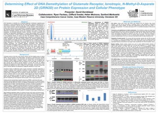
Figure 4. Protein expression and cell viability in shRNA knockdown RKO cells. (a) Western blotting with Sigma-Aldrich primary
anti-GRIN2D antibody (1:1000 dilution in blocking solution). Lysates from RKO cell lines virally transfected with vectors containing small-
hairpin RNA interfering with GRIN2D protein expression were isolated: sh_465, sh_467, sh_575. One RKO cell line was transfected with
a vector containing shRNA targeting green fluorescent protein transcript (sh_GFP) serving as a negative control. Experimental lysates
were blotted with parental RKO human colon cancer cell line lysate (red). (b) CellTiter-Glo Luminescent Cell Viability Assay of transfected
line measuring growth relative to sh_GFP negative control.
Colorectal cancer is the third most common cancer and third leading cause of cancer
death in men and women in the United States. The decline in colorectal cancer mortality
can be partially attributed to the introduction and dissemination of screening tests to
diagnose at earlier stages of disease. However, currently only 40% of patients with
colorectal cancer are diagnosed when the disease is at a local stage, for which the 5-
year survival rate is 90.3%. Survival declines to 70.4% and 12.5% for patients
diagnosed with regional and distant-stage disease, respectively1. Improvements in early
screening and detection are vital to continue to lower mortality. One area of focus has
been to improve detection by assessing for epigenetic alterations in tumor cells.
Inheritance based on differential levels of gene expression constitutes epigenetics in
contrast to genetics which describes inheritance of gene sequences. An important
epigenetic modification is methylation of cytosines at CpG dinucleotides. The distribution
of these sites of methylation can be localized to specific tissue types in the body in a
series of patches known as CpG islands. These patches can function normally to
prevent the expression of genes not required of a differentiated cell type. However,
global genomic hypomethylation of cancer cell genomes may occur that promotes
genomic instability and carcinogenesis without proper control of gene expression2-3.
Utilizing reduced representation bisulfite sequencing (RRBS), a technique that uses
restriction enzyme digest and bisulfite conversion of genomic DNA on a reduced fraction
of the genome with high CpG content, we validated a demethylation event occurring in a
potential proto-oncogene, Glutamate Receptor, Ionotropic, N-methyl-D-aspartate or
GRIN2D (Figure 1).. This gene normally expresses one subunit of a heteromer N-
methyl-D-aspartate (NMDA) receptor that functions in long-term potentiation increasing
the efficiency of synaptic transmission thought to be responsible for learning and
memory. However, emerging experimental evidence implicates a role for glutamate
receptor subunit expression in tumor growth in a variety of cancer types including colon4.
After identifying methylation status of a subset of human colon cancer cell lines through
RRBS, we performed real-time polymerase chain reaction (RT-PCR) assays to correlate
methylation status of GRIN2D with levels of gene expression. Based on the RRBS and
RT-PCR data, we selected human colon cancer cell line RKO as a demethylated high
expressor of GRIN2D. Functional studies indicate inhibition of NMDA subunit expression
can result in negative regulation of cell viability and proliferation5. We developed viral
transfection vectors that utilize knockdown and knockout biochemistry to introduce to
GRIN2D expressing human colon cancer cells. The knockdown model incorporated a
small hairpin RNA (shRNA), an interfering RNA that targeted exon 6 of the GRIN2D
transcript to prevent protein expression at the translational level. We also developed a
vector to utilize CRISPR-RNA (crRNA)-guided interference. This knockout vector
expressed 4 different guide RNAs to localize Cas9 mediated excision of the GRIN2D
gene at exons 2 and 12. In this study we explored the functional role of GRIN2D DNA
demethylation and protein expression in human colon cancer cells.
Background
References
Results and Future Direction
1. Siegel, R., DeSantis, C., & Jemal, A. (2014). Colorectal cancer statistics, 2014. CA: a cancer
journal for clinicians, 64(2), 104-117.
2. Esteller, M., & Herman, J. G. (2002). Cancer as an epigenetic disease: DNA methylation and
chromatin alterations in human tumours. The Journal of pathology, 196(1), 1-7.
3. Baylin S. B., Herman J. G., Graff J. R., Vertino, P. M., & Issa, J. P. (1997). Alterations in DNA
methylation: a fundamental aspect of neoplasia. Advances in cancer research, 72, 141–196.
4. Yoo BC., Jeon E., Hong SH., Shin YK., Chang HJ., & Park JG. (2004) Metabotropic glutamate
receptor 4-mediated 5-fluorouracil resistance in a human colon cancer cell line. Clin cancer res
10: 4176-4184.
5. Luksch, H., Uckermann, O., Stepulak, A., Hendruschk, S., Marzahn, J., Bastian, S., Staufner, C.,
Temme, A., & Ikonomidou, C. (2011). Silencing of selected glutamate receptor subunits
modulates cancer growth. Anticancer research. 31: 3181-3192.
6. R.D. Finn, A. Bateman, J. Clements, P. Coggill, R.Y. Eberhardt, S.R. Eddy, A. Heger, K.
Hetherington, L. Holm, J. Mistry, E.L.L. Sonnhammer, J. Tate, M. Punta. (2014). The Pfam
protein families database. Nucleic acids research. 42:D222-D230.
Figure 2. Protein subunits of GRIN2D highlighting
immunogens for western blotting primary antibodies.
The Pfam protein families database6 identified three
subunits from the GRIN2D amino acid sequence: (1) N-
terminal signal peptide (2) ANF Receptor - an extracellular
ligand binding domain (3) Ligand channel binding site -
luminal domain just upstream of the first transmembrane
region, M1; binds L-glutamate and and glycine (4) Ligand
channel: ionotropic glutamate membrane channel with 3
transmembrane domains. Western blotting primary
antibodies included: (1) Sigma Aldrich anti-GRIN2D
antibody produced in rabbit binding a ~50 amino acid
immunogen within one of the extracellular domains of the
ligand channel (2) Santa Cruz anti-GRIN2D antibody
produced in goat binding at the cytoplasmic C-terminus.
Figure 3. Western blotting for GRIN2D expression in differentially methylated human colon cancer cell lines. (a) Western
blotting with Santa Cruz primary anti-GRIN2D antibody (1:1000 dilution in blocking solution). (b) Western blotting with Sigma-Aldrich
primary anti-GRIN2D antibody (1:1000 dilution in blocking solution). Demethylated high GRIN2D expressing human colon cancer cell
lines included RKO, V703, V400, and V451 (red). Methylated low GRIN2D expressing human colon cancer cell lines included V429 and
V670 (green). V9M (yellow) was identified as demethylated by RRBS without corresponding upregulated GRIN2D gene expression
based on RT-PCR. IMR32 (blue) was an ordered protein lysate (Santa Cruz) from a neuroblastoma cell line.
0
0.2
0.4
0.6
0.8
1
1.2
465 467 575 GFP
CellViabilityRelativetoGFP
shRNA Line
Relative Growth of shGRIN2D Knockdown Lines
(a) (b)
(a) (b)
Here we show one instance of RKO transfected cell line sh_467 when knockdown expression of
a potential proto-oncogene, GRIN2D demethylated in human colon cancer cell lines, with a small
hairpin interfering RNA functional model correlated in decreased protein expression confirmed by
western blot. Additionally, correlated reduced cell viability was confirmed with cell viability assay.
This result suggests there may be a biochemical model that describes how a demethylation event
can result in upregulated protein expression and uncontrolled cell growth in a human colon
cancer phenotype. However, many additional undertakings are required to investigate this
possibility. First, we need to identify a positive control to verify that the primary antibodies
included in this study are in fact binding to GRIN2D moieties. The IMR-32 neuroblastoma cell line
was ordered as a potential positive control, but we have not demonstrated that our anti-GRIN2D
antibodies bind to an immunogen in its protein lysate. Second, western blotting revealed size
discrepancies for the GRIN2D protein. Electrophoretic separation should indicate GRIN2D as a
143 kDA; however, the Santa Cruz antibody identified a ~50kDa peptide while the Sigma-Aldrich
antibody identified a protein closer to expected size, ~170kDa. We hypothesize that because
GRIN2D has three transmembrane domains, preparation of protein extract requires a higher
fraction of SDS detergent to isolate the entire protein from the cellular membrane. Finally, cell
viability and colony forming assays on knockdown lines must be performed in repetition to
confirm the functional model we have developed.