Download as PDF, PPTX


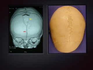
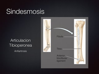
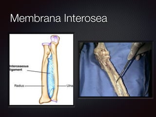


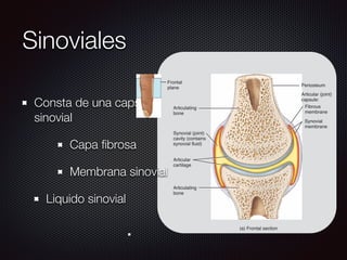
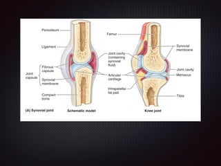
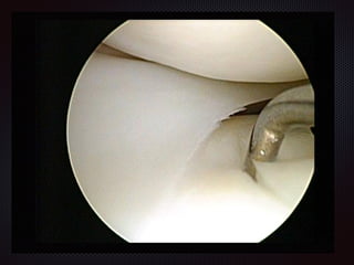

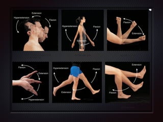
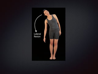
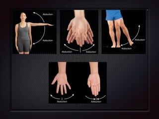
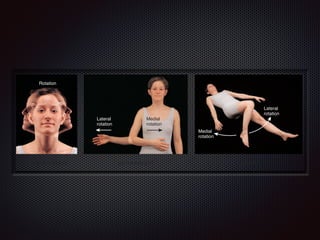
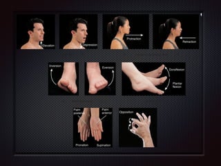


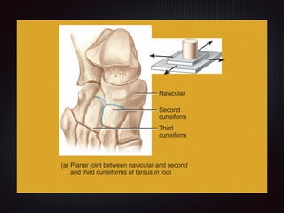
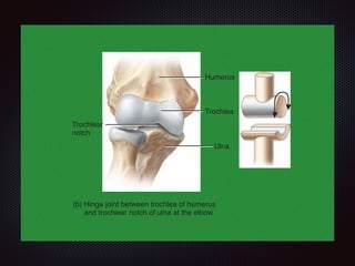

There are three main types of joints based on structure and movement: synarthroses which do not allow movement, amphiarthroses which allow slight movement, and diarthroses which are freely movable. Diarthroses are further classified into fibrous, cartilaginous, and synovial joints. Synovial joints are the most complex and include the shoulder, elbow, wrist, hip, knee, ankle, and small joints of the hands and feet. They contain a joint cavity lined with synovial membrane which produces synovial fluid for lubrication.


