Download to read offline


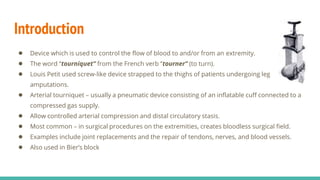


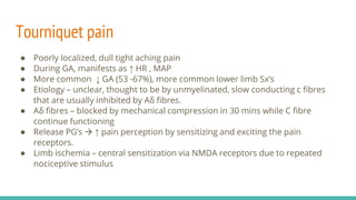
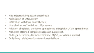

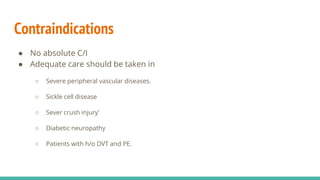


The document discusses anesthetic concerns regarding tourniquet application. It covers the components and use of pneumatic tourniquets, including cuff selection and inflation pressure. Physiological effects of tourniquets are also reviewed, such as local and systemic impacts. Potential complications are outlined. Tourniquet pain mechanisms and management are described. Contraindications for tourniquet use focus on vascular conditions.


![ONFH[AVN HIP] -TRIPLE REGIME -A NOVAL SURGICAL CONCEPT .pptx](https://cdn.slidesharecdn.com/ss_thumbnails/onfhavnhip2026koaconcalicutdrgokuldevdrmashraf-260210064517-213ec005-thumbnail.jpg?width=640&height=640&fit=bounds)


