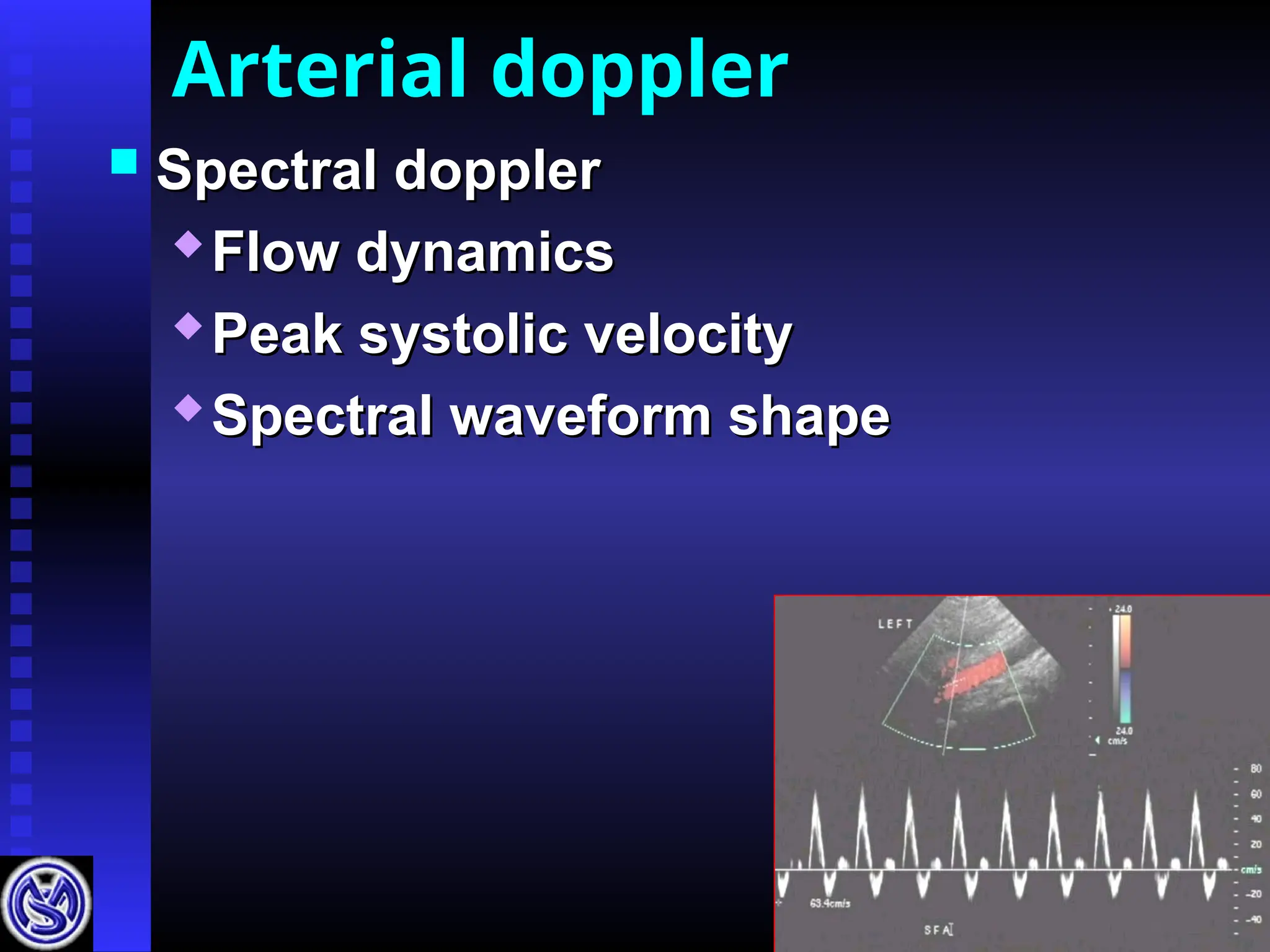
Arterial hemodynamics
Propagationspeed increases
Propagation speed increases
with stiffness of wall
with stiffness of wall
Arteries stiffer from aorta to
Arteries stiffer from aorta to
periphery
periphery
Pulse pressure incr. by
Pulse pressure incr. by
vasoconstriction & decr. by
vasoconstriction & decr. by
vasodilatation
vasodilatation
Pressure waves
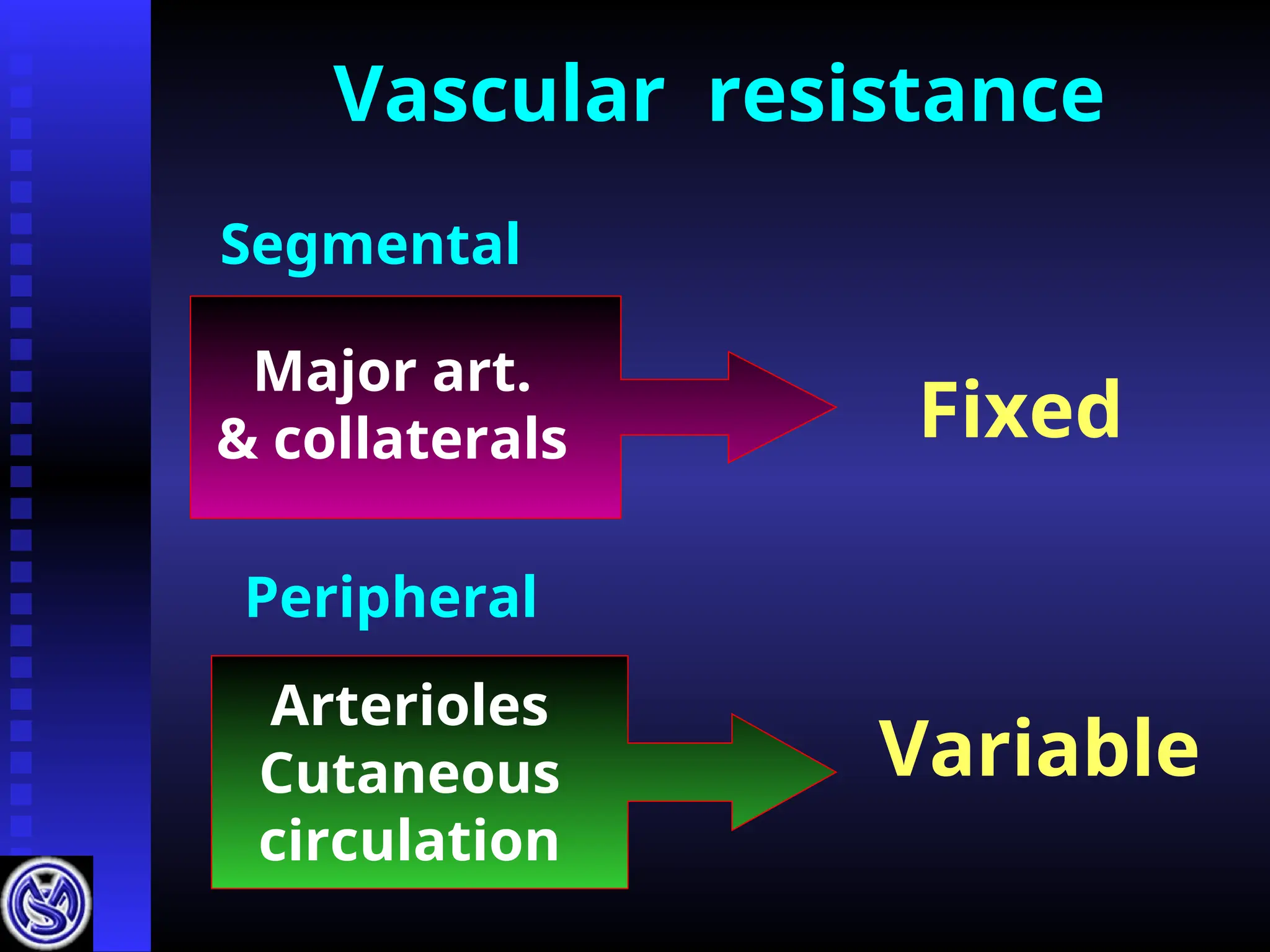
Hemodynamic resistance
Resting state
Restingstate
Segmental resistance
Segmental resistance Low
Low
Peripheral resistance
Peripheral resistance High
High
Pressure drop across
Pressure drop across
major segmental artery
major segmental artery Low
Low
7.
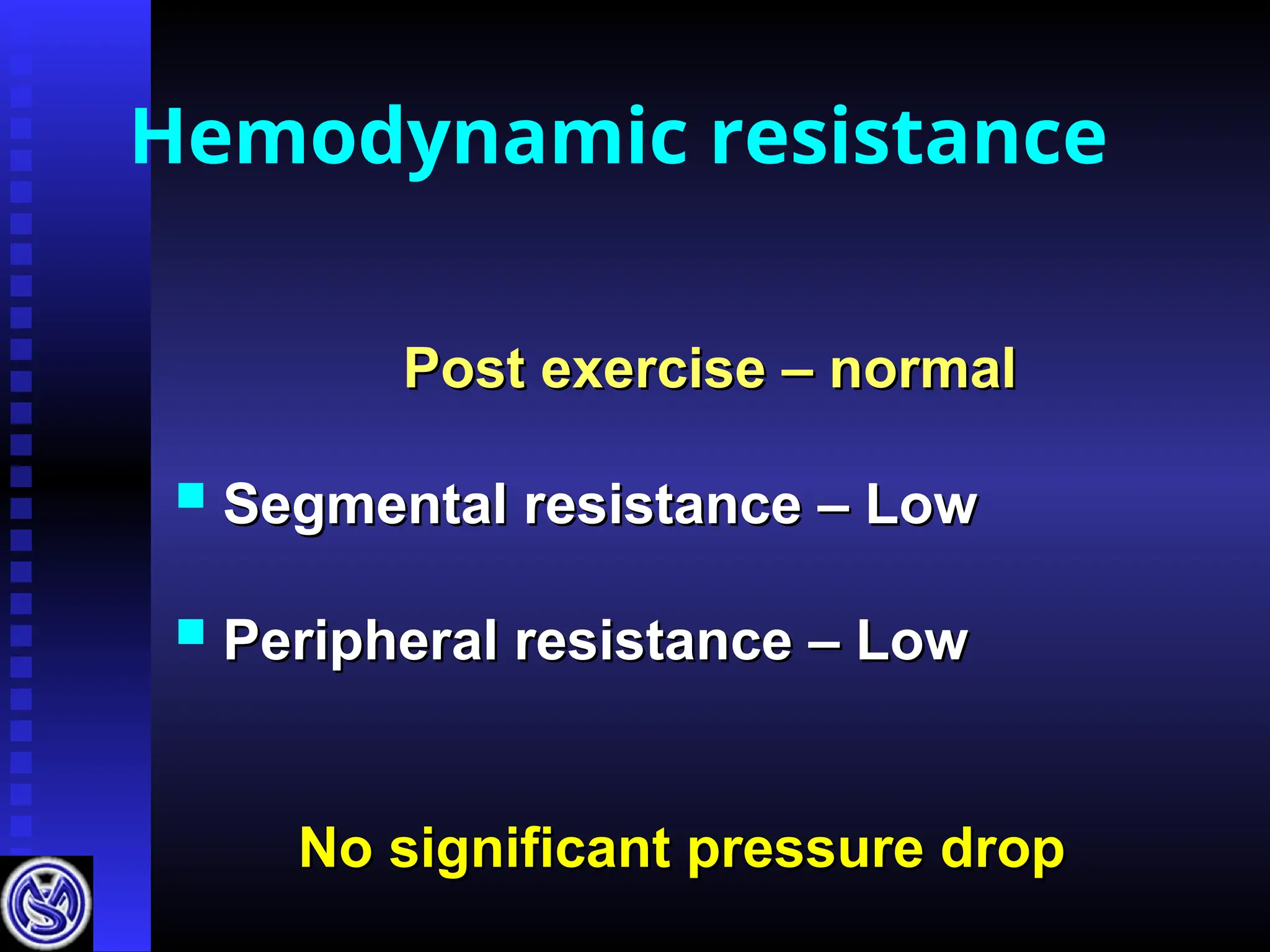
Hemodynamic resistance
Post exercise– normal
Post exercise – normal
Segmental resistance – Low
Segmental resistance – Low
Peripheral resistance – Low
Peripheral resistance – Low
No significant pressure drop
No significant pressure drop
8.
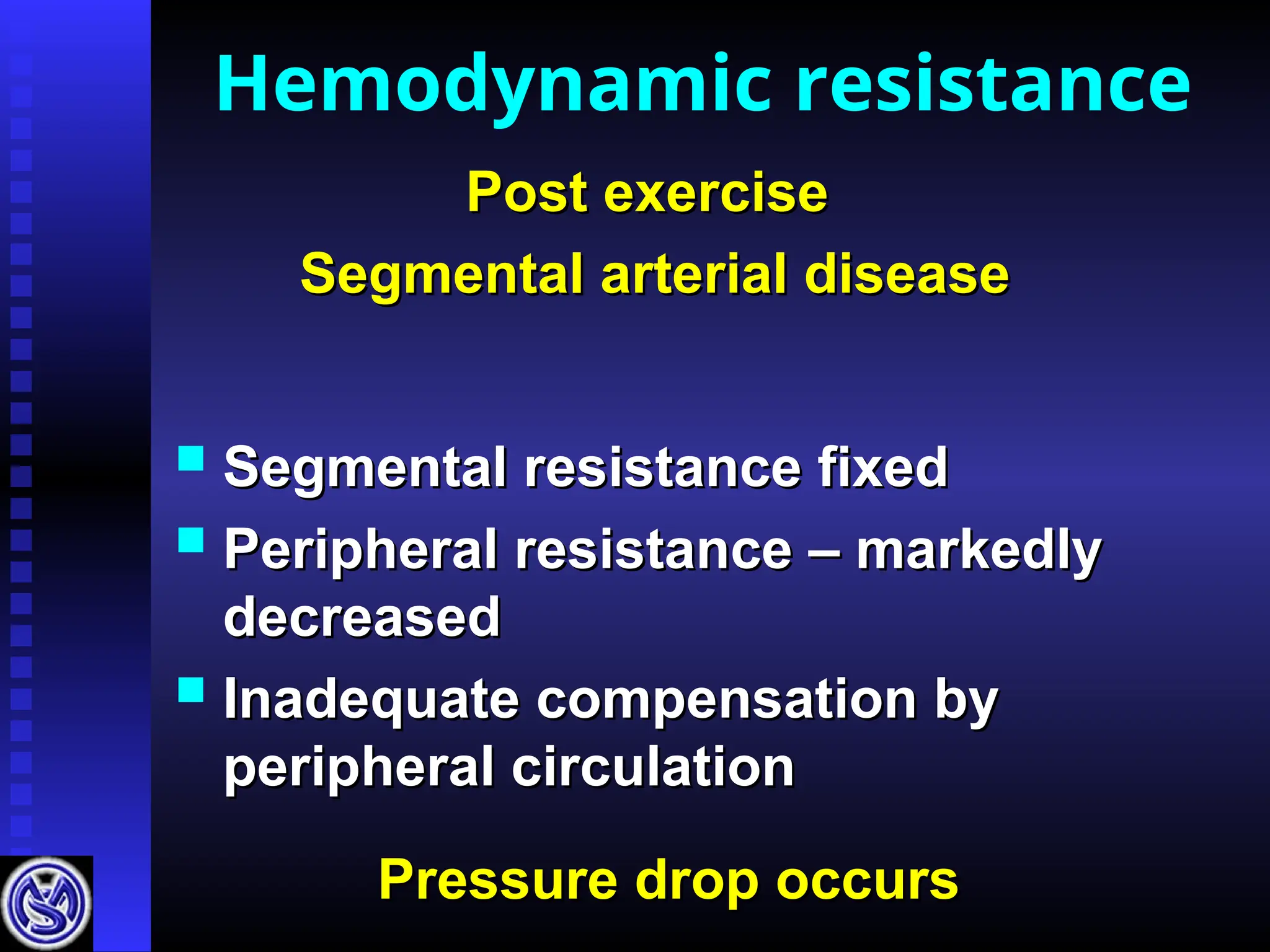
Segmental resistancefixed
Segmental resistance fixed
Peripheral resistance – markedly
Peripheral resistance – markedly
decreased
decreased
Inadequate compensation by
Inadequate compensation by
peripheral circulation
peripheral circulation
Hemodynamic resistance
Post exercise
Post exercise
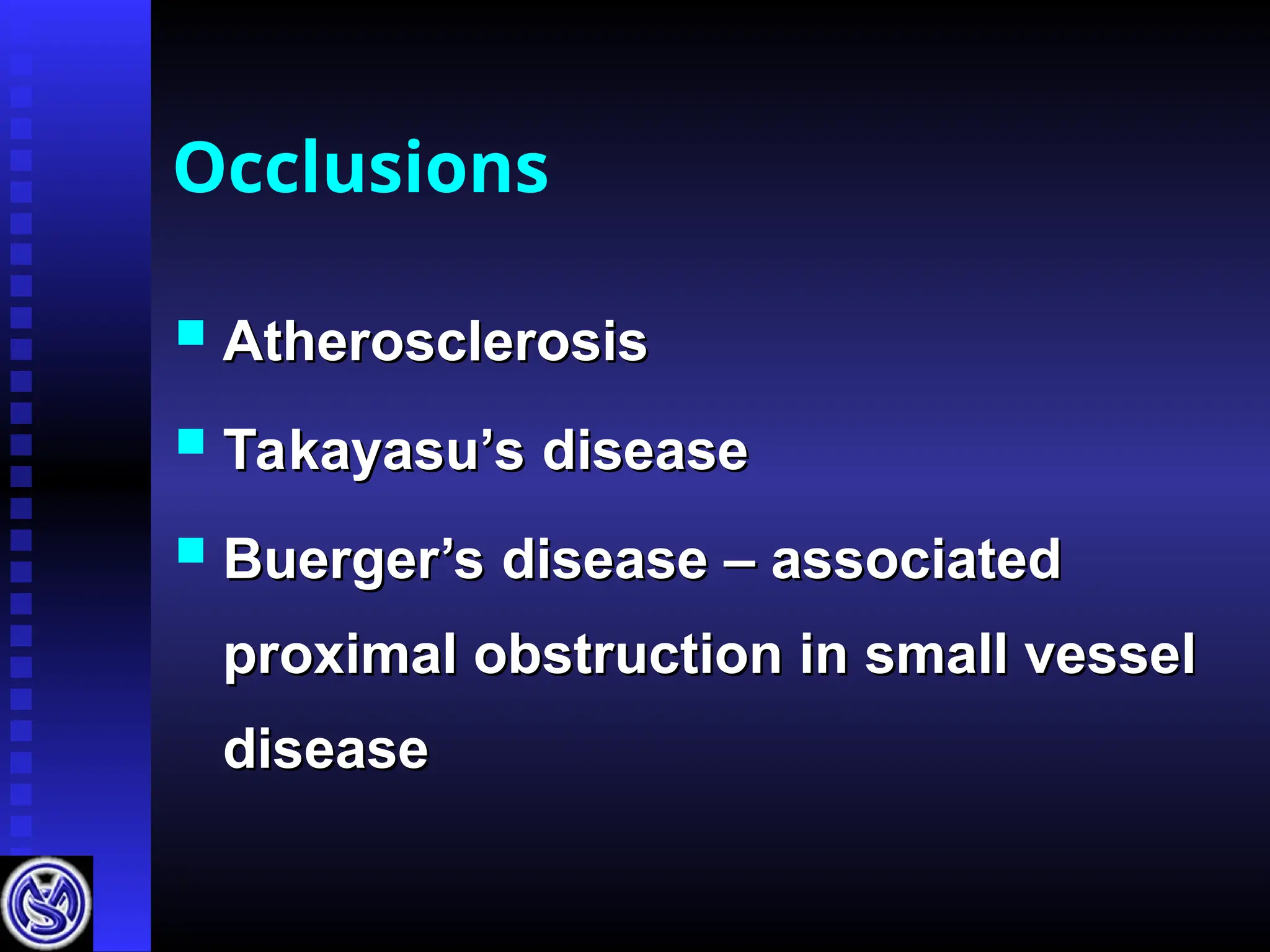
Segmental arterial disease
Segmental arterial disease
Pressure drop occurs
Pressure drop occurs
EXERCISE TESTING
Treadmillexercise test
Treadmill exercise test
12% grade at 2 km/ hr.
12% grade at 2 km/ hr.
Exercise time 5 minutes
Exercise time 5 minutes
Stop exercise if pain develops
Stop exercise if pain develops
Contraindications
Contraindications
Cardiac impairment
Cardiac impairment
Physical disability
Physical disability
13.
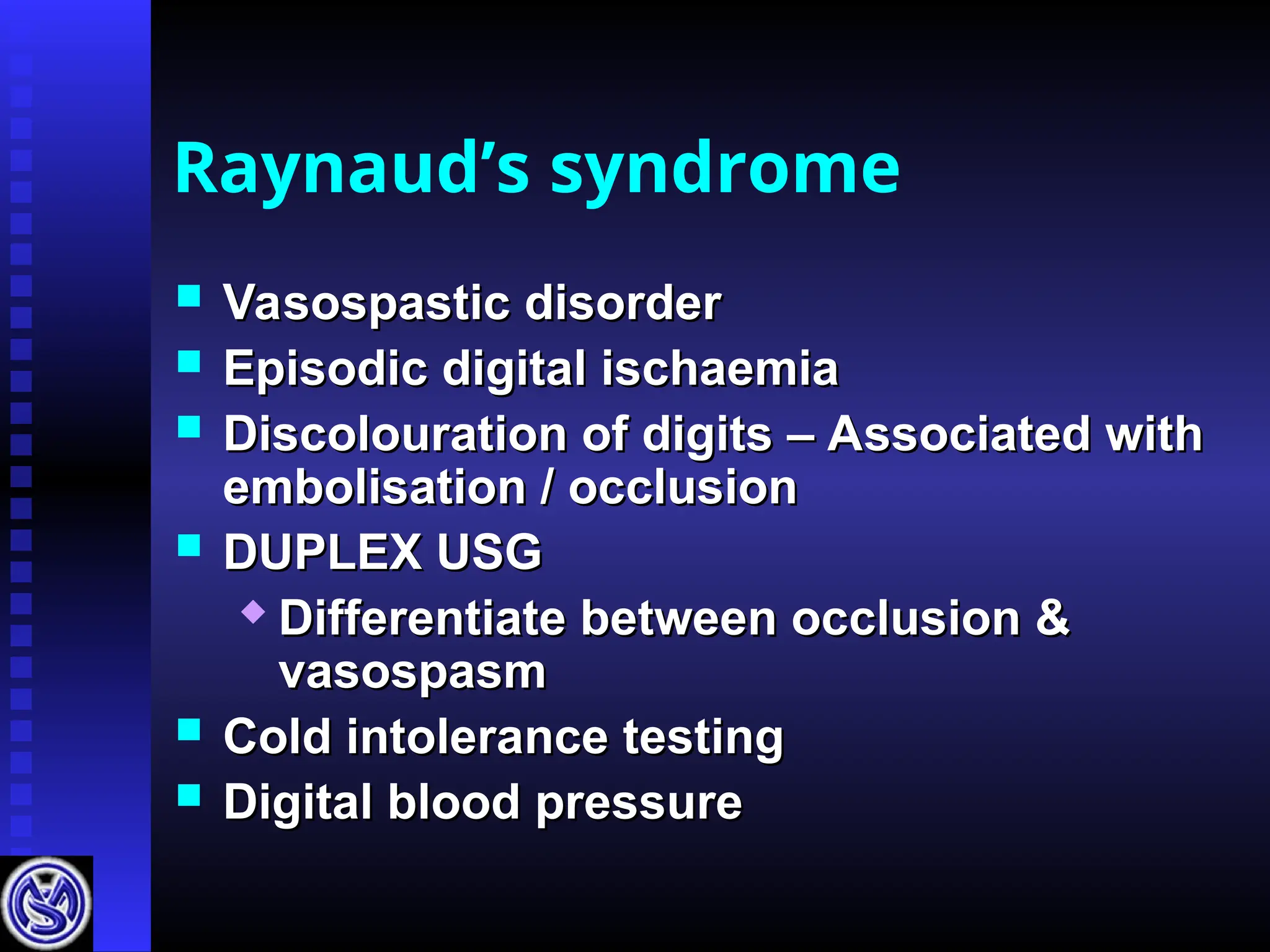
Reactive hyperemia
Patients withcardiac problems and
Patients with cardiac problems and
physical disability
physical disability
Increase systolic pressure
Increase systolic pressure
Maintain for 3-5 min. – Vasodilatation
Maintain for 3-5 min. – Vasodilatation
Release and take pressure
Release and take pressure
Occlusion – Single level 50% drop
Occlusion – Single level 50% drop
- Multi level > 50%
- Multi level > 50%
17-34% overlap of normal
and abnormal values
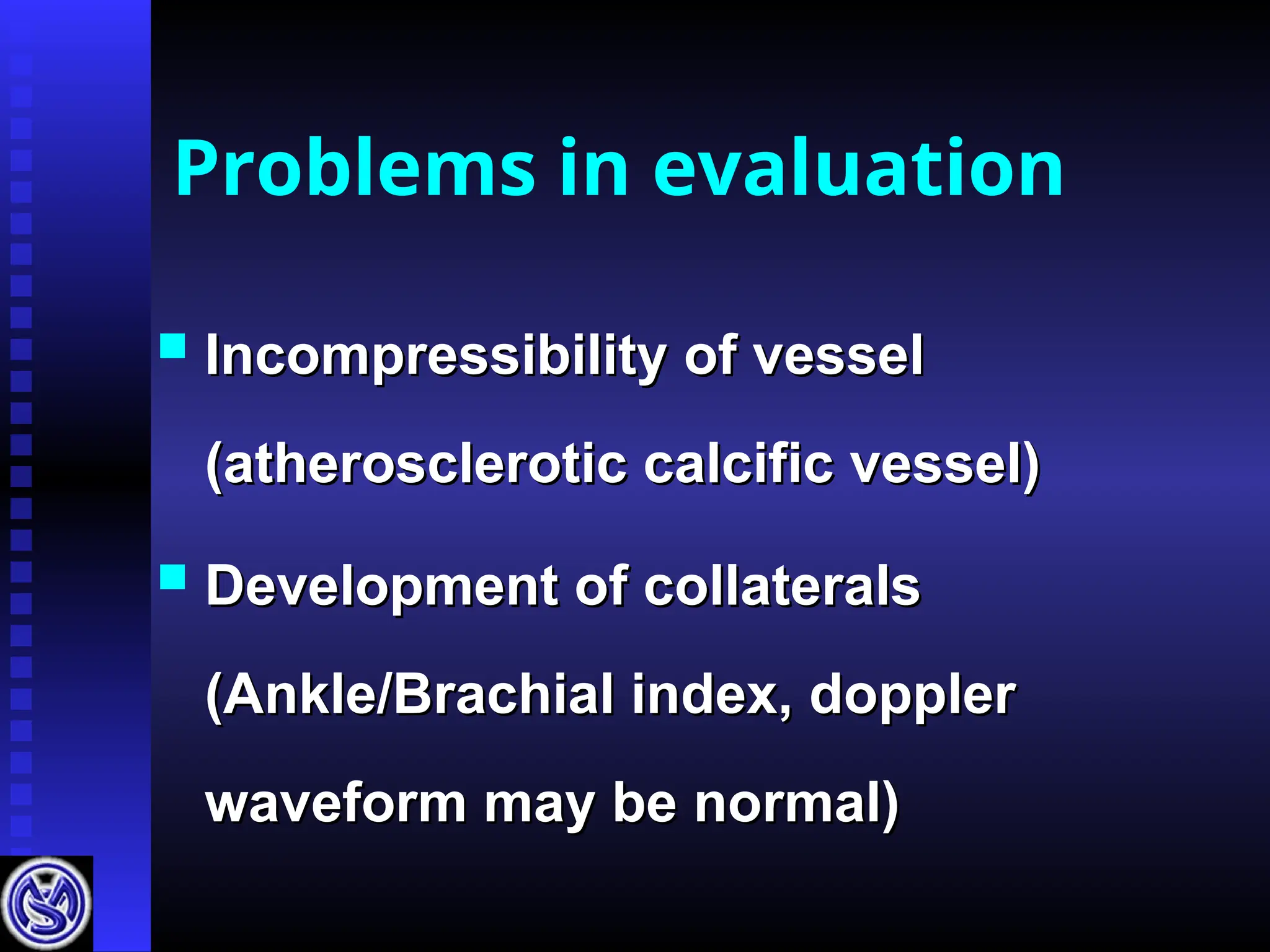
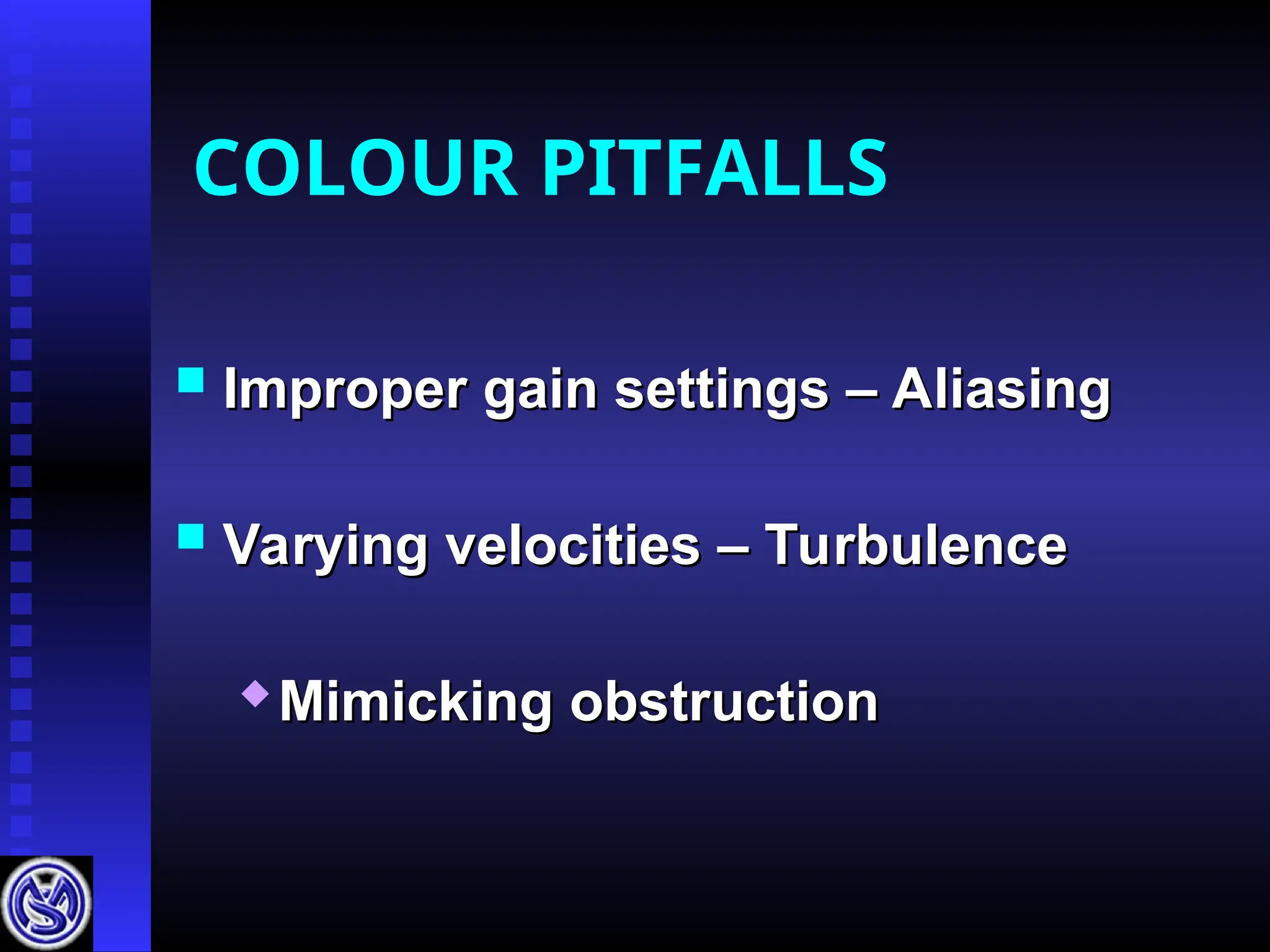
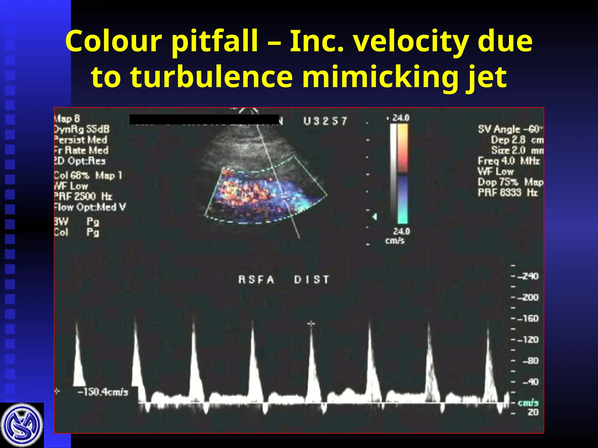
Problems in evaluation
Incompressibility of vessel
Incompressibility of vessel
(atherosclerotic calcific vessel)
(atherosclerotic calcific vessel)
Development of collaterals
Development of collaterals
(Ankle/Brachial index, doppler
(Ankle/Brachial index, doppler
waveform may be normal)
waveform may be normal)
Pitfalls
Resting period
Restingperiod
Cuff width
Cuff width
Quick deflation
Quick deflation
Distance between the measurement
Distance between the measurement
area & probe position
area & probe position
Subclavian obstruction
Subclavian obstruction
Atherosclerosis
Atherosclerosis
High segmental pressures
Right
Brachial- 150
Left
Brachial - 160
222
208
184
170
164 162
160
172
182
200
AA Index 1.1 1.01
0
20
40
60
80
100
120
140
160
180
200
2 4 6 8 10
TIME (Minutes)
PRESSU
RE
(m
m
H
g
)
Right
Left
Atherosclerotic
disease
20.
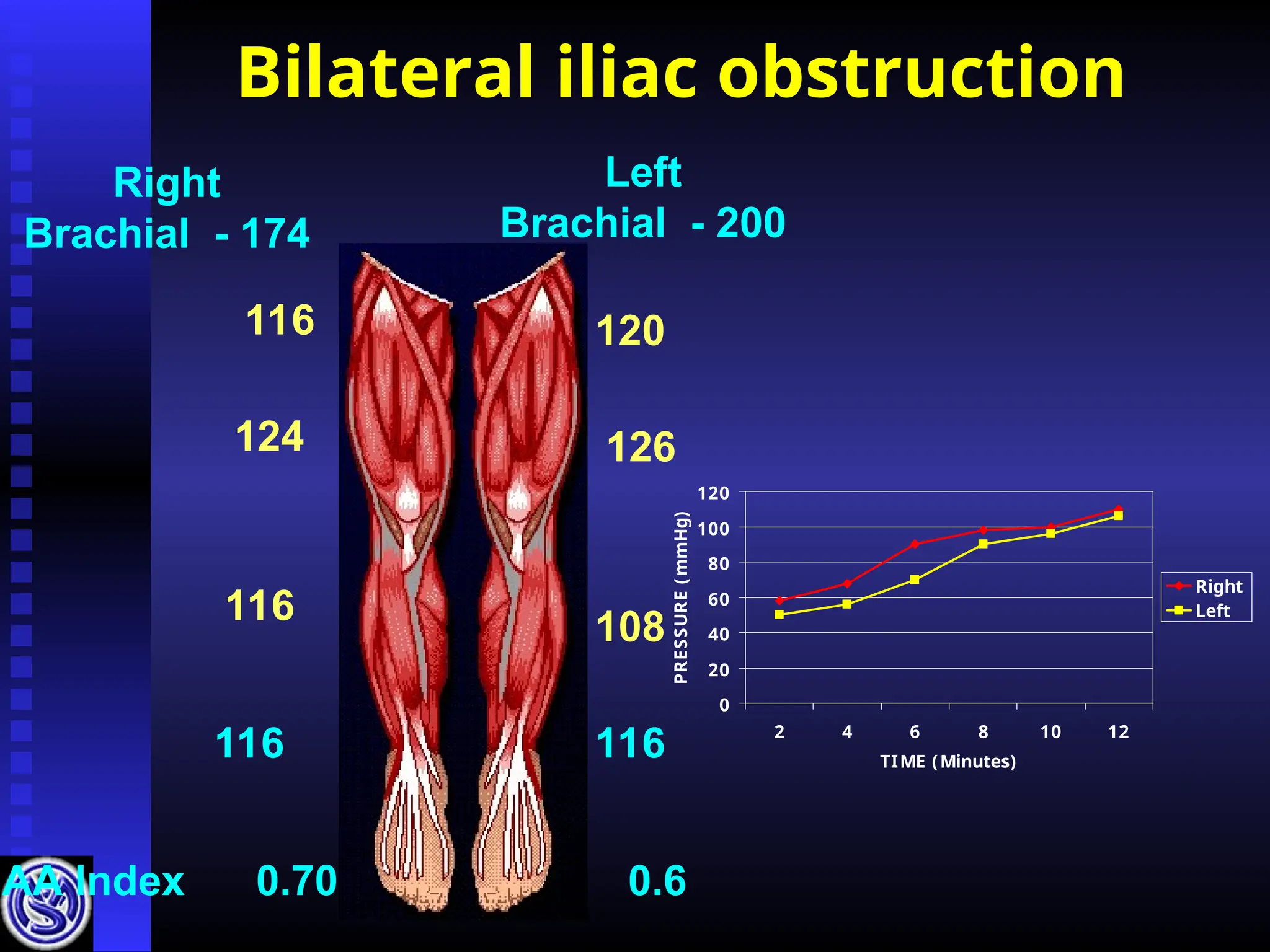
Bilateral iliac obstruction
Right
Brachial- 174
Left
Brachial - 200
116
124
116
116 116
108
126
120
AA Index 0.70 0.6
0
20
40
60
80
100
120
2 4 6 8 10 12
TIME (Minutes)
PRESSURE
(
mmHg)
Right
Left
21.
Left iliac obstruction
Right
Brachial- 120
Left
Brachial - 120
150
124
130
120
116 66
72
70
83
96
AA Index 1.0 0.60
0
20
40
60
80
100
120
140
160
2 4 6 8 10 12
TIME (Minutes)
PRESSURE
(m
m
Hg)
Right
Left
22.
Multisegmental obstruction -Right
Right
Brachial - 115
Left
Brachial - 116
90
62
50
48 122
130
134
146
AA Index
0.42 1.06
0
20
40
60
80
100
120
140
160
2 4 6 8 10 12
TIME (Minutes)
PRESSURE
(m
m
H
g)
Left
Right
23.
Multisegmental obstruction
Right
Brachial -165
Left
Brachial - 159
130
110
94
72
76 100
102
110
152
164
Index 0.44 0.64
30
40
50
60
70
80
90
100
4 6 8 10 12
TIME (Minutes)
PRESSU
RE
(m
m
H
g)
Right
Left
24.
Rt. Popliteal art.embolism
Right
Brachial - 124
Left
Brachial - 120
160
140
130
70
70 119
124
130
134
140
AA Index 0.58 1.03
25.
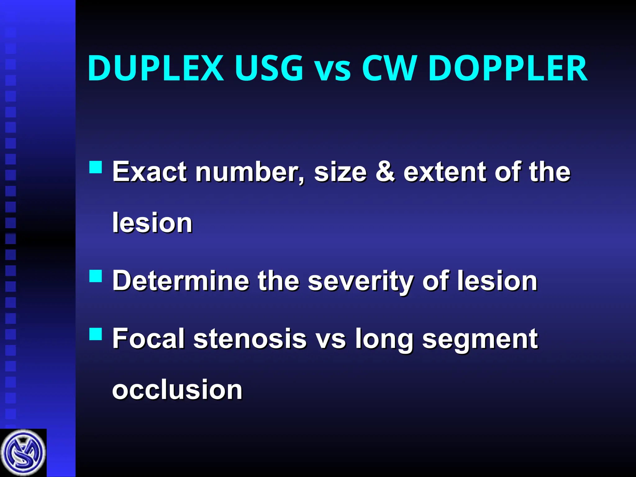
DUPLEX USG vsCW DOPPLER
Exact number, size & extent of the
Exact number, size & extent of the
lesion
lesion
Determine the severity of lesion
Determine the severity of lesion
Focal stenosis vs long segment
Focal stenosis vs long segment
occlusion
occlusion
26.
PERIPHERAL ARTERIAL
DOPPLER -INDICATIONS
Occlusive arterial disease
Occlusive arterial disease
Follow up after bypass graft,
Follow up after bypass graft,
angioplasty
angioplasty
Diagnosis & follow up of aneurysms
Diagnosis & follow up of aneurysms
Diagnosis & treatment of
Diagnosis & treatment of
pseudoaneurysm
pseudoaneurysm
Evaluation of pulsatile swelling
Evaluation of pulsatile swelling
Assessment of dialysis shunts
Assessment of dialysis shunts
Classify peripheralarterial disease
Classify peripheral arterial disease
Locate stenotic sites
Locate stenotic sites
Grading of severity of stenosis
Grading of severity of stenosis
Negative study excludes
Negative study excludes
hemodynamically significant disease
hemodynamically significant disease
APPLICATIONS OF
ARTERIAL DUPLEX
30.
ARTERIAL DOPPLER
Protocol
Imagefrom abdominal aorta to distal vessels
Image from abdominal aorta to distal vessels
Aortic bifurcation
Aortic bifurcation
Common iliacs
Common iliacs
External iliacs
External iliacs
(internal il.)
(internal il.)
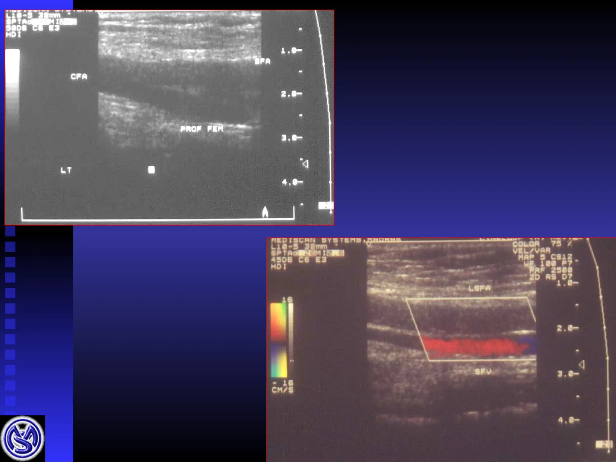
Common femoral
Common femoral
Profunda
Profunda
Superficial femoral A
Superficial femoral A
Popliteal A
Popliteal A
Ant tib. / post tib / peroneal A
Ant tib. / post tib / peroneal A
ARTERIAL DOPPLER
Surveyof the arteries
Survey of the arteries
Diameter, wall pathology
Diameter, wall pathology
Study plaque morphology
Study plaque morphology
Selection of patients for
Selection of patients for
angiography
angiography
ARTERIOGRAM IS GOLD STANDARD
DOPPLER CRITERIA FORFLOW
OBSTRUCTIVE LESIONS
Tri – or biphasic
Tri – or biphasic
Systolic window +
Systolic window +
no spectral broadening
no spectral broadening
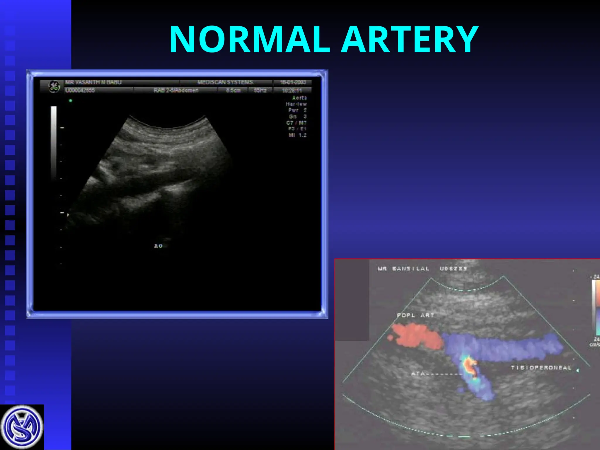
NORMAL
NORMAL
41.
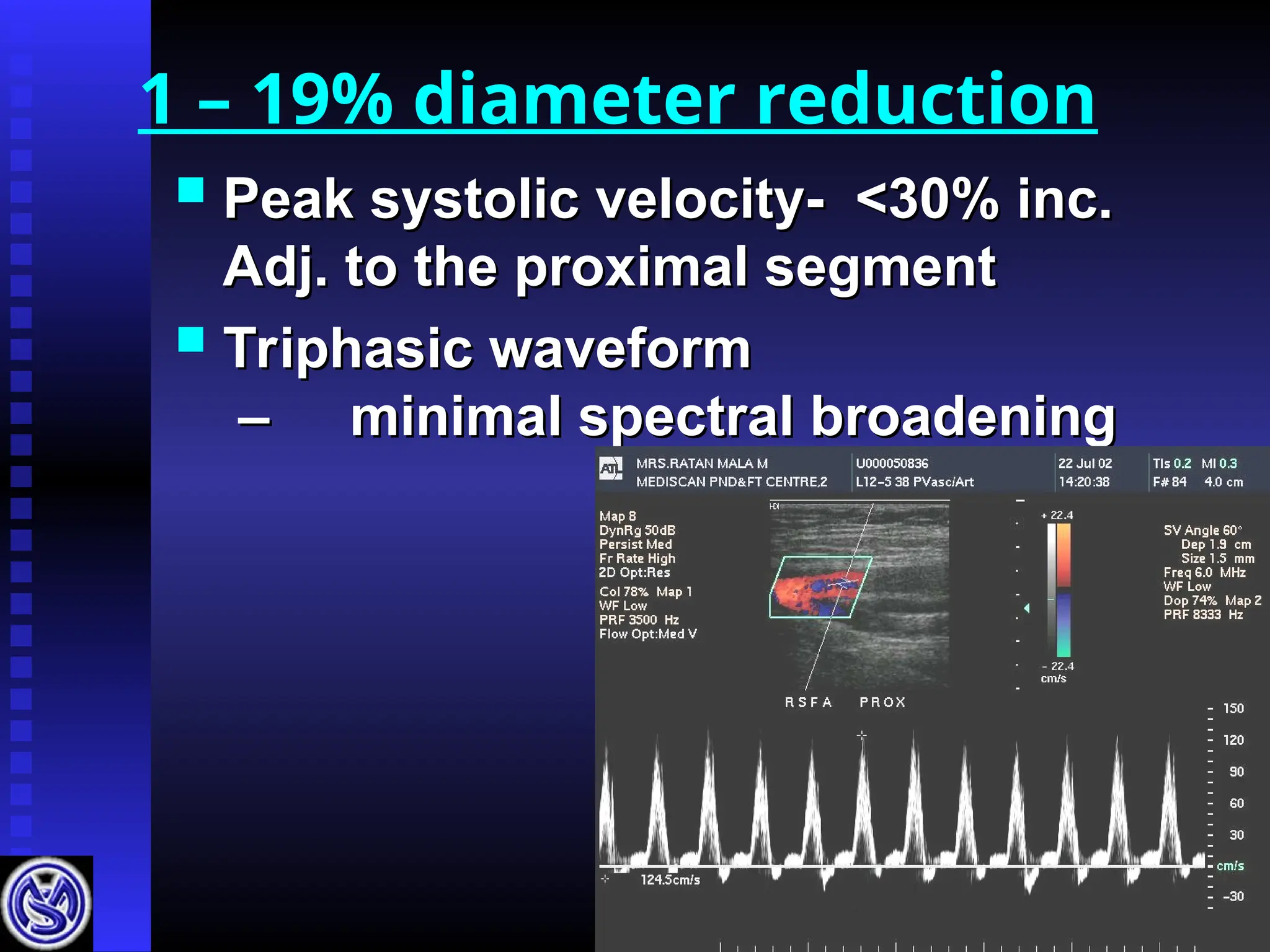
1 – 19%diameter reduction
Peak systolic velocity- <30% inc.
Peak systolic velocity- <30% inc.
Adj. to the proximal segment
Adj. to the proximal segment
Triphasic waveform
Triphasic waveform
–
– minimal spectral broadening
minimal spectral broadening
Stenosis calculation
Ratio=
Ratio = PSV at stenosis
PSV at stenosis
Normal PSV prox. to stenosis
Normal PSV prox. to stenosis
3.7 – 75% stenosis
3.7 – 75% stenosis
7.0 – 90% stenosis
7.0 – 90% stenosis
50.
Multiple tandem lesions
Cumulative effect seen distally
Cumulative effect seen distally
“
“resistances in series”
resistances in series”
51.
COLOUR DOPPLER
Identifiescourse of vessels
Identifies course of vessels
calcified vessels – better seen
calcified vessels – better seen
Detects :
Detects :
Isoechoic lesions
Isoechoic lesions
Areas of stenosis and occlusion
Areas of stenosis and occlusion
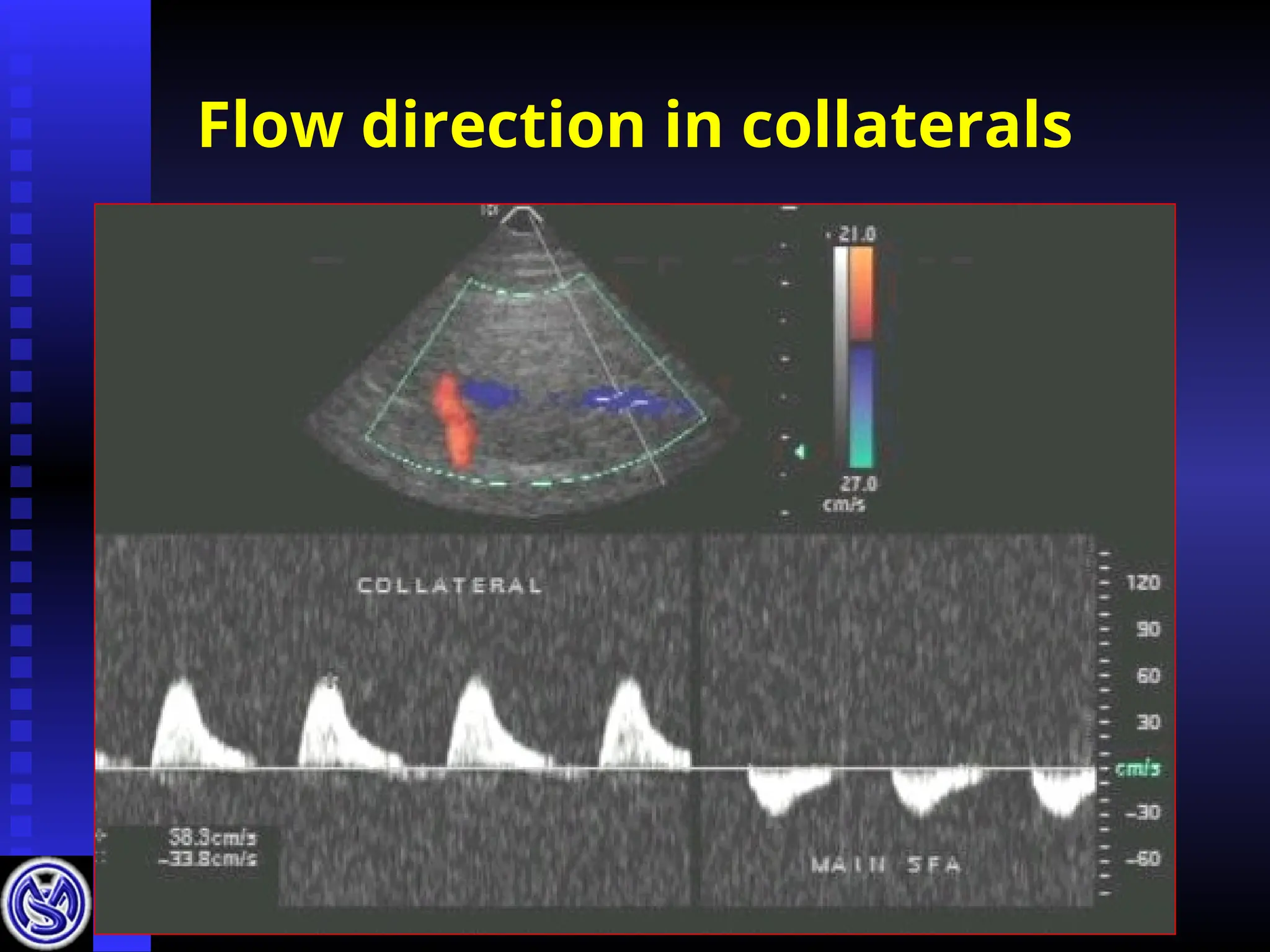
Identifying collaterals
Identifying collaterals
Reduces examination time
Colour doppler of
Aneurysms
Superior to arteriography
Superior to arteriography
Identifying intramural thrombus
Identifying intramural thrombus
Serial USG in aneurysm < 2 cms
Serial USG in aneurysm < 2 cms
in diameter
in diameter
Increase in the size (to decide
Increase in the size (to decide
intervention)
intervention)
Embolism in distal arteries
Embolism in distal arteries
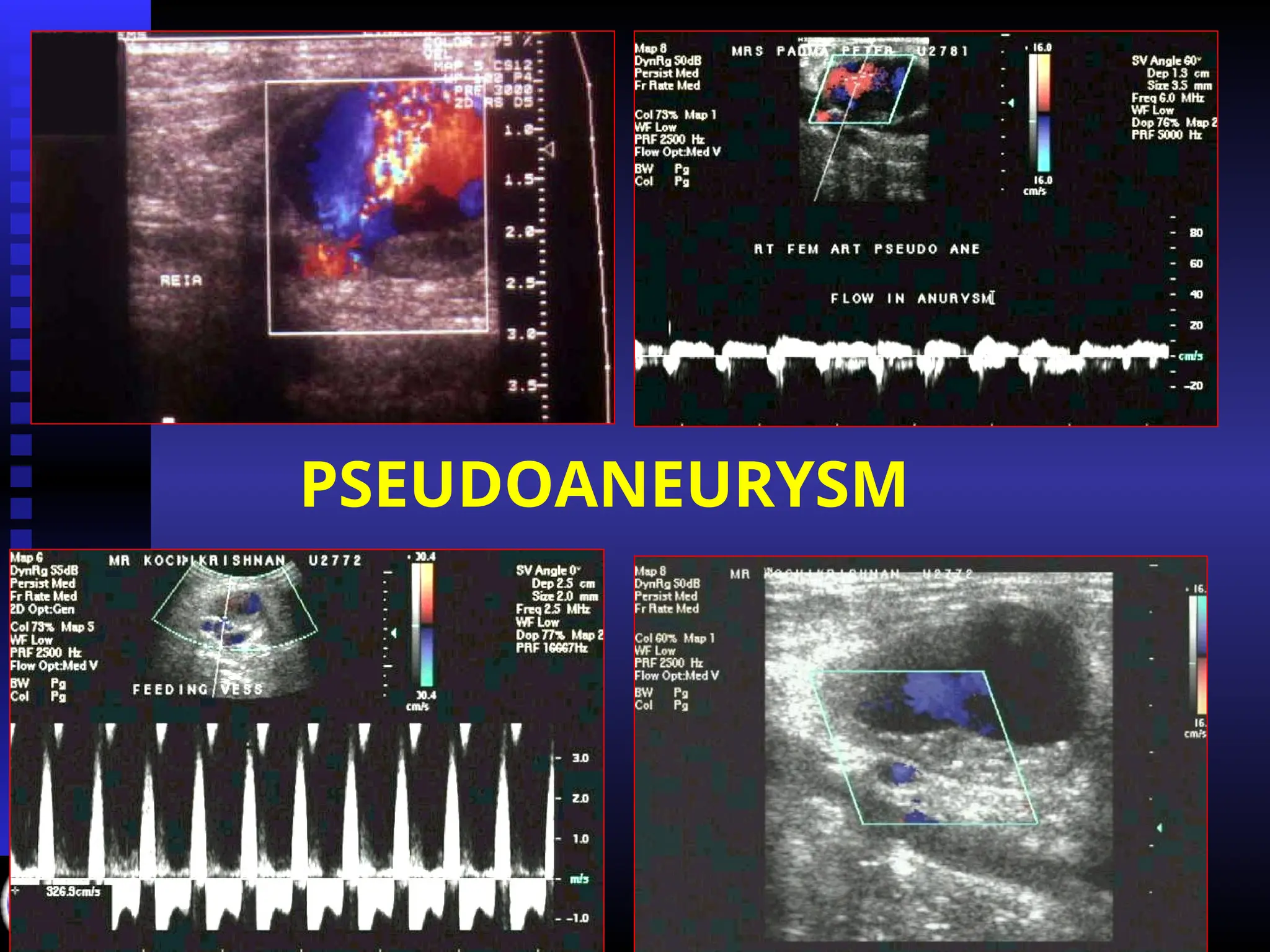
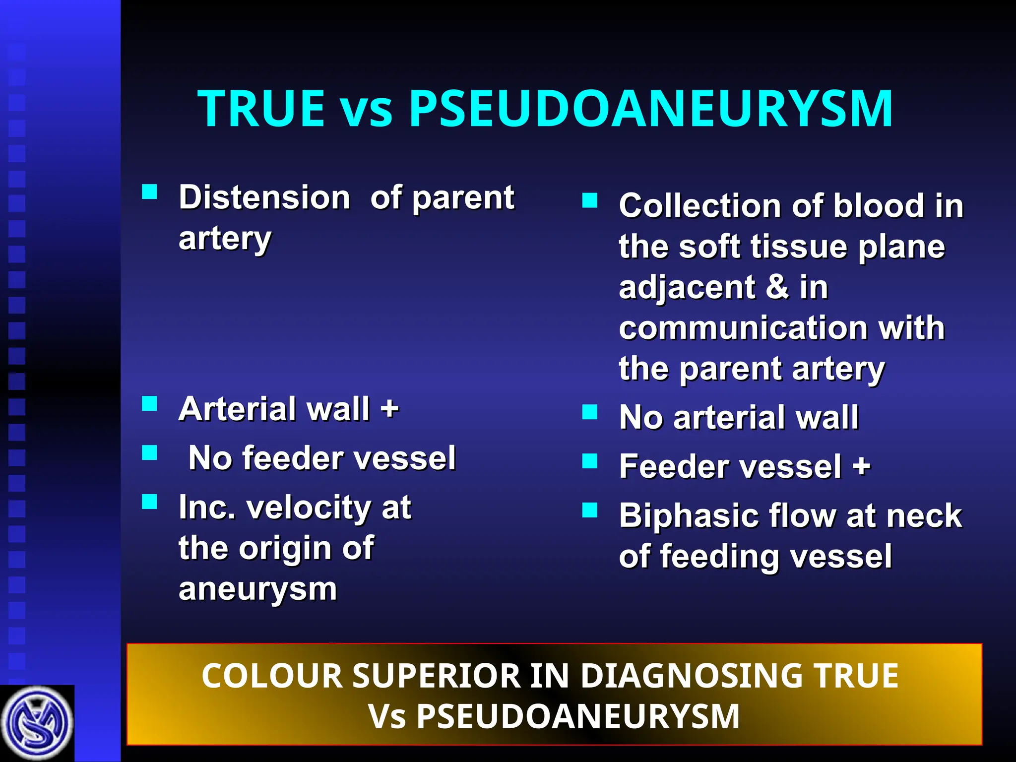
TRUE vs PSEUDOANEURYSM
Distension of parent
Distension of parent
artery
artery
Arterial wall +
Arterial wall +
No feeder vessel
No feeder vessel
Inc. velocity at
Inc. velocity at
the origin of
the origin of
aneurysm
aneurysm
Collection of blood in
Collection of blood in
the soft tissue plane
the soft tissue plane
adjacent & in
adjacent & in
communication with
communication with
the parent artery
the parent artery
No arterial wall
No arterial wall
Feeder vessel +
Feeder vessel +
Biphasic flow at neck
Biphasic flow at neck
of feeding vessel
of feeding vessel
COLOUR SUPERIOR IN DIAGNOSING TRUE
Vs PSEUDOANEURYSM
76.
Pseudo aneurysm
Therapy
Therapy
Feedingchannel can be occluded
Feeding channel can be occluded
with transducer pressure
with transducer pressure
Thrombosis
Thrombosis
77.
Pseudo aneurysm
Criteria
Criteria
Pseudoaneurysm < 1 month
Pseudo aneurysm < 1 month
Length of the neck (> 5mm)
Length of the neck (> 5mm)
Duration of compression
Duration of compression
varies
varies
79.
50 days afterdetection
Occluded
45 days after occlusion
-Refilling
80.
A-V FISTULA
Congenital
Congenital
Traumatic
Traumatic
Iatrogenic
Iatrogenic
High velocity arterialised flow in vein
High velocity arterialised flow in vein
Marked increase in systolic flow in
Marked increase in systolic flow in
artery
artery
Congenital
Clinical –skin discolouration
Clinical – skin discolouration
USG – Limited role
USG – Limited role
Helpful in patients who had
Helpful in patients who had
undergone sclerotherapy
undergone sclerotherapy
Thoracic outlet
Recordingthe pressures on the Left
Recording the pressures on the Left
90
90 0
0
abduction & external rotation
abduction & external rotation
Adson maneuver
Adson maneuver
Post exercise
Duration in
Durationin
(min.)
(min.)
Right
Right Left
Left
2
2 128
128 56
56
4
4 124
124 78
78
6
6 122
122 84
84
8
8 122
122 90
90
10
10 122
122 90
90
12
12 124
124 90
90
Patient had unbearable pain in left leg after 3mnts.
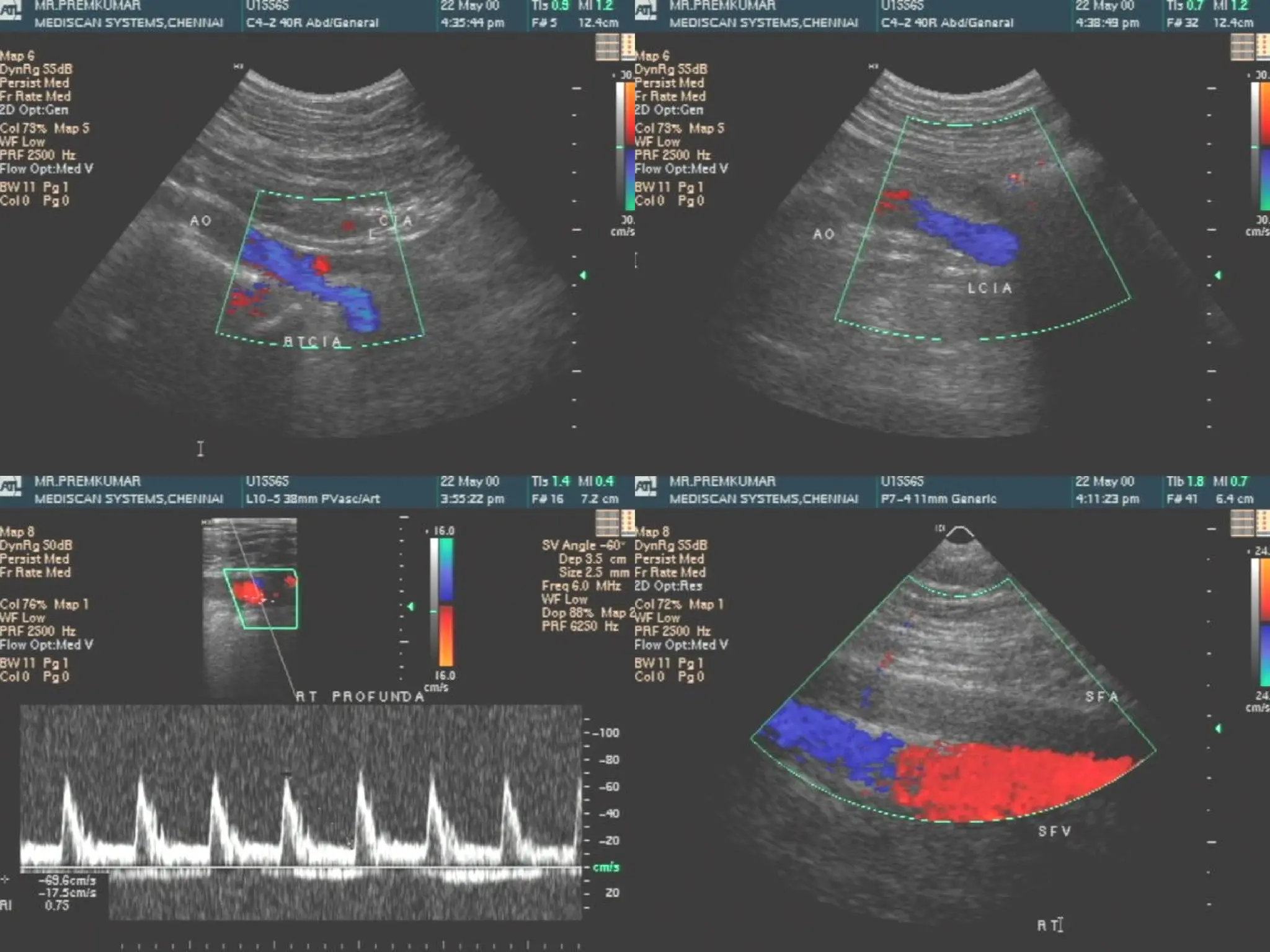
102.
USG FINDINGS
Totalocclusion of left common iliac artery.
Total occlusion of left common iliac artery.
Left external iliac artery was fed by the left
Left external iliac artery was fed by the left
internal iliac artery.
internal iliac artery.
OCCLUSIVE ARTERIAL DISEASE.
OCCLUSIVE ARTERIAL DISEASE.
TOTAL OCCLUSION OF LEFT COMMON
TOTAL OCCLUSION OF LEFT COMMON
ILIAC ARTERY.
ILIAC ARTERY.
REFORMATION OF LEFT EXTERNAL ILIAC
REFORMATION OF LEFT EXTERNAL ILIAC
ARTERY SEEN THROUGH LEFT INTERNAL
ARTERY SEEN THROUGH LEFT INTERNAL
ILIAC ARTERY.
ILIAC ARTERY.
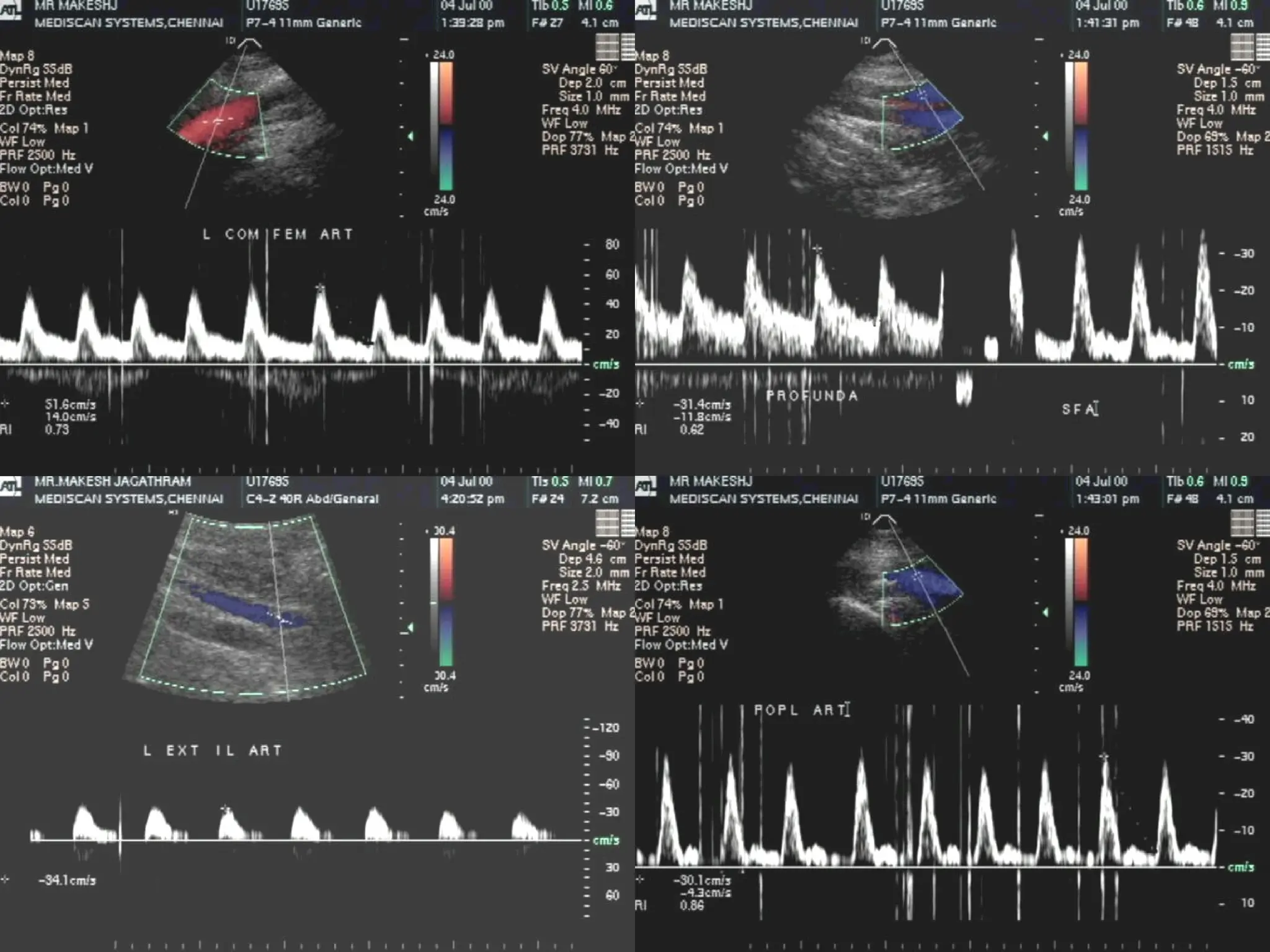
103.
Case II
Mr.PM – 48 Yrs
Mr. PM – 48 Yrs
Smoker– 10 Yrs
Smoker– 10 Yrs
Pain in left foot since 3 weeks
Pain in left foot since 3 weeks
USG findings
Rightside : -
Right side : -
Total occlusion of right superficial femoral
Total occlusion of right superficial femoral
artery.
artery.
Collateral seen joining the popliteal artery with
Collateral seen joining the popliteal artery with
reduced forward flow distally.
reduced forward flow distally.
Left side : -
Left side : -
Total occlusion of external iliac artery and lower
Total occlusion of external iliac artery and lower
limb arteries.
limb arteries.
Normal flow seen in aorta, both common iliac
Normal flow seen in aorta, both common iliac
artery and external iliac artery on right side.
artery and external iliac artery on right side.
Normal flow seen in right common femoral artery
Normal flow seen in right common femoral artery
and profunda femoris.
and profunda femoris.


![Vibe Coding vs. Spec-Driven Development [Free Meetup]](https://cdn.slidesharecdn.com/ss_thumbnails/vibecodingvsspecdrivendevelopment-251209105622-43f455e7-thumbnail.jpg?width=640&height=640&fit=bounds)


