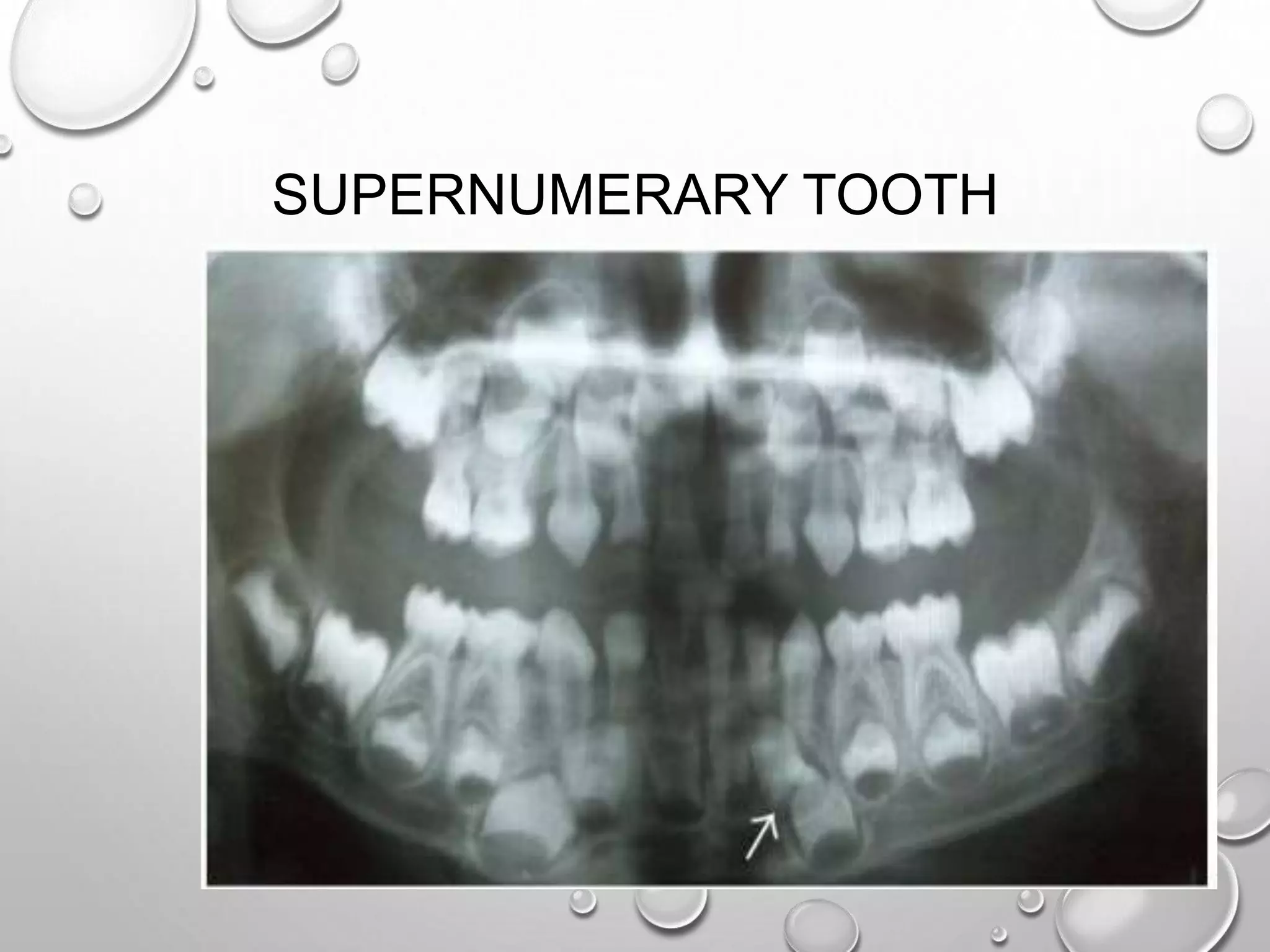
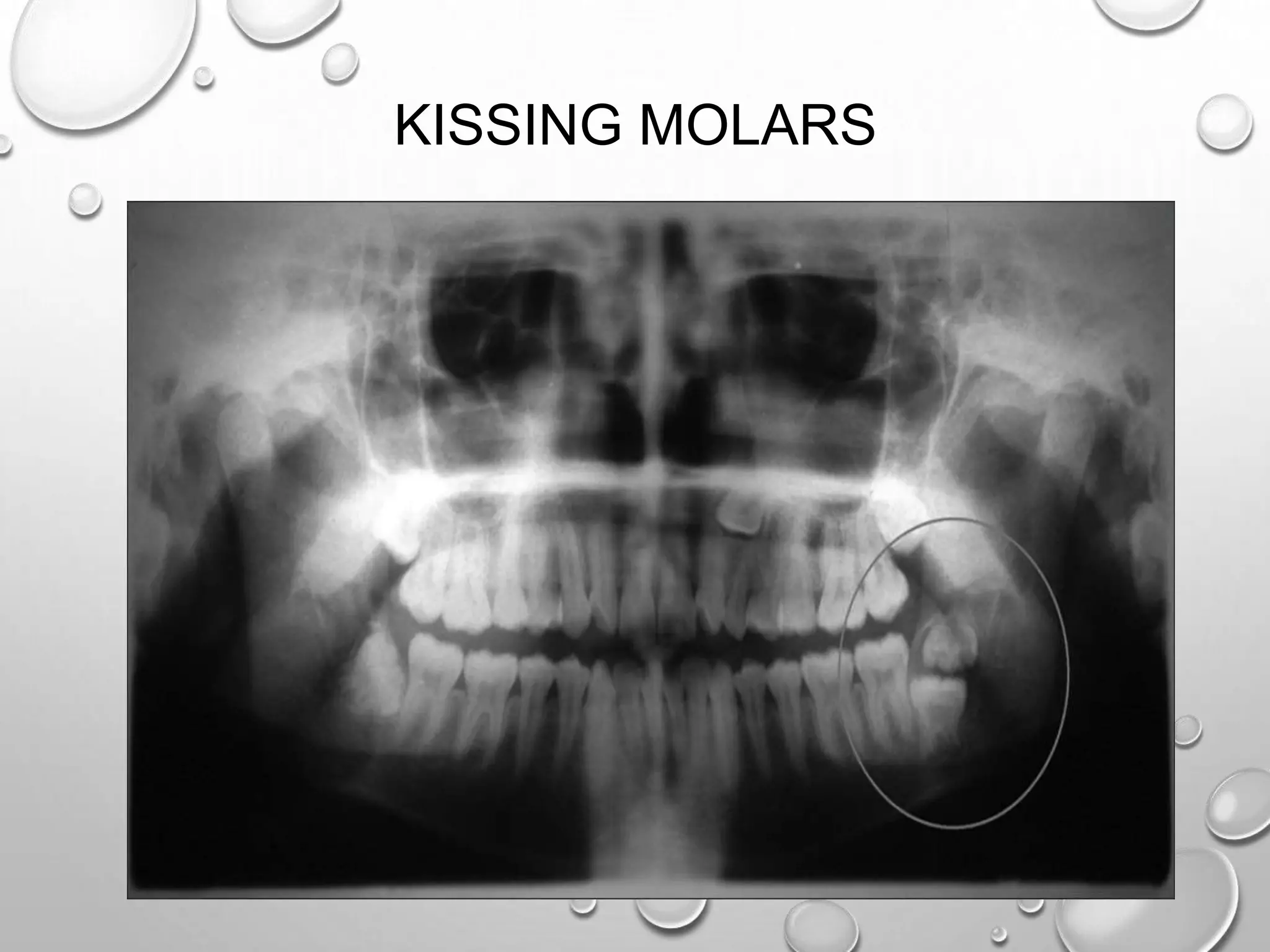
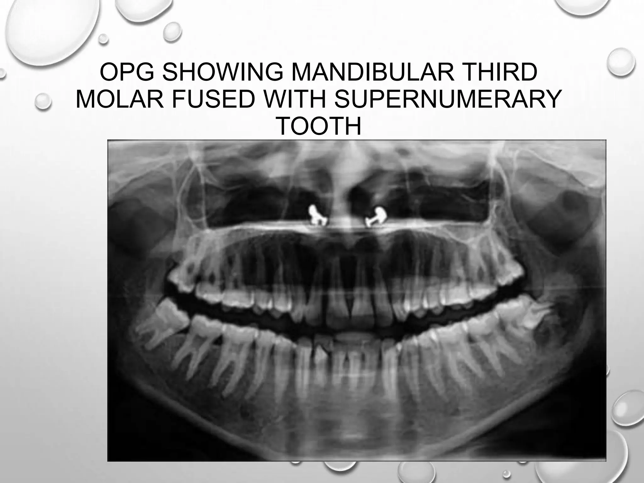
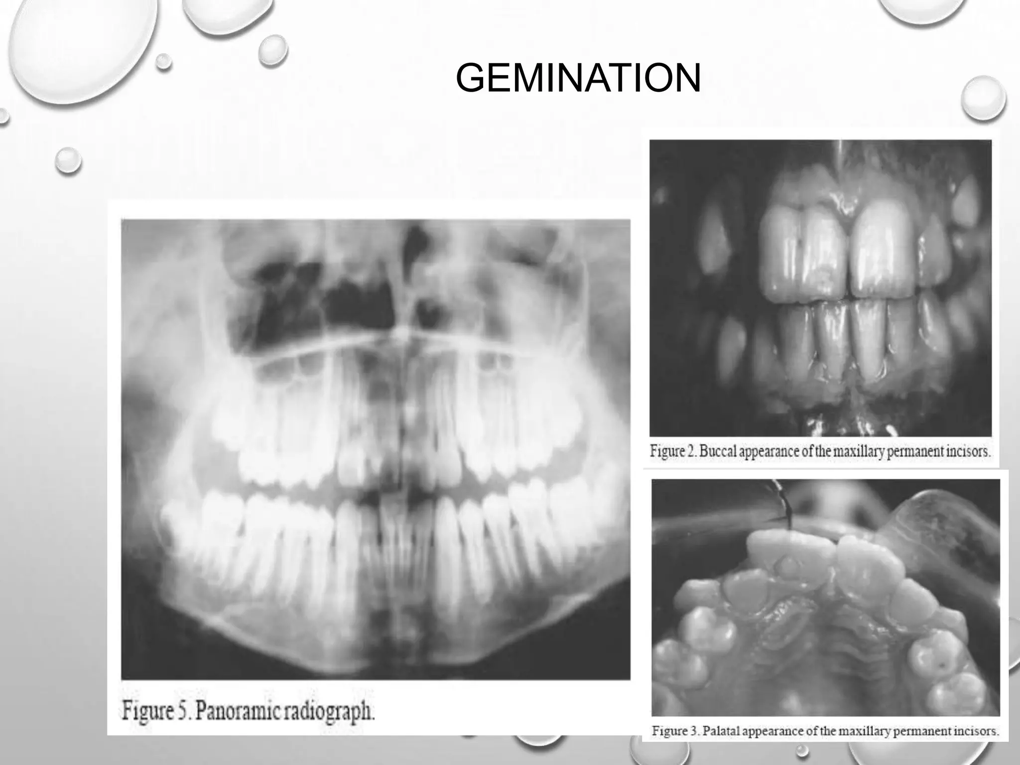
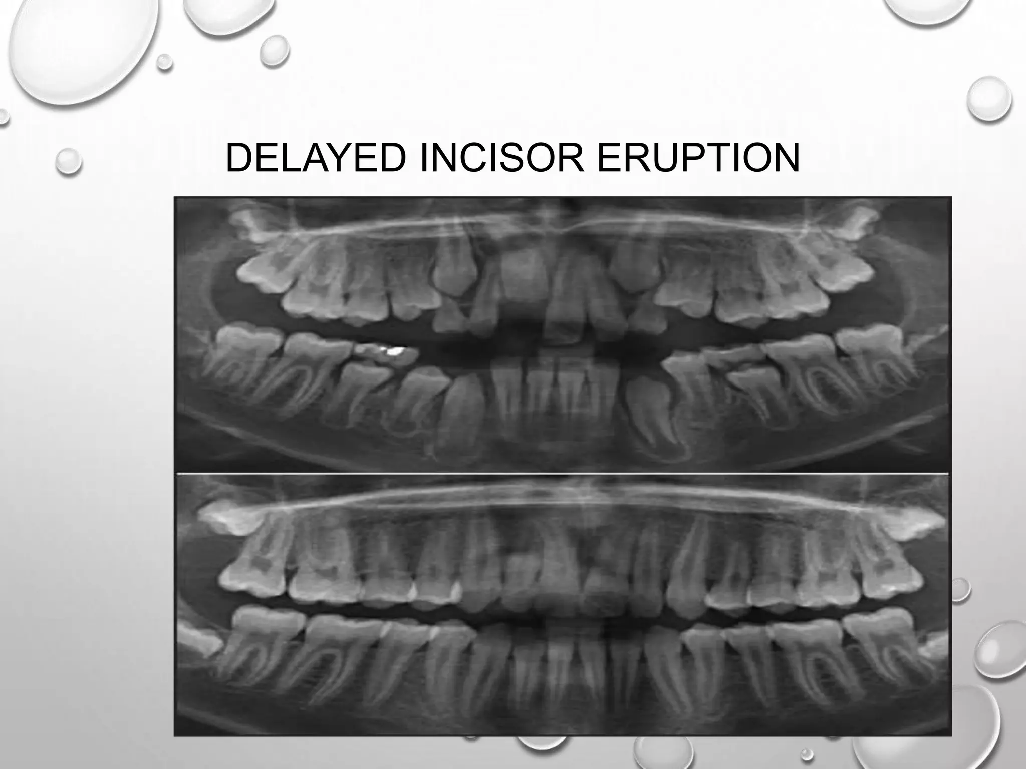
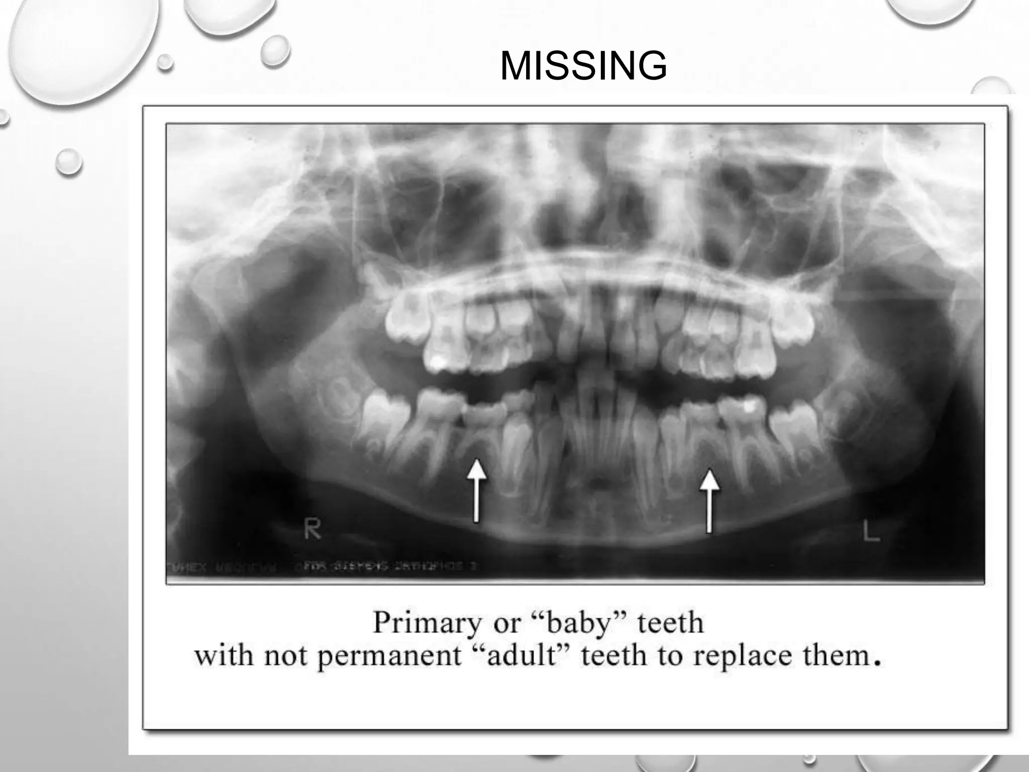
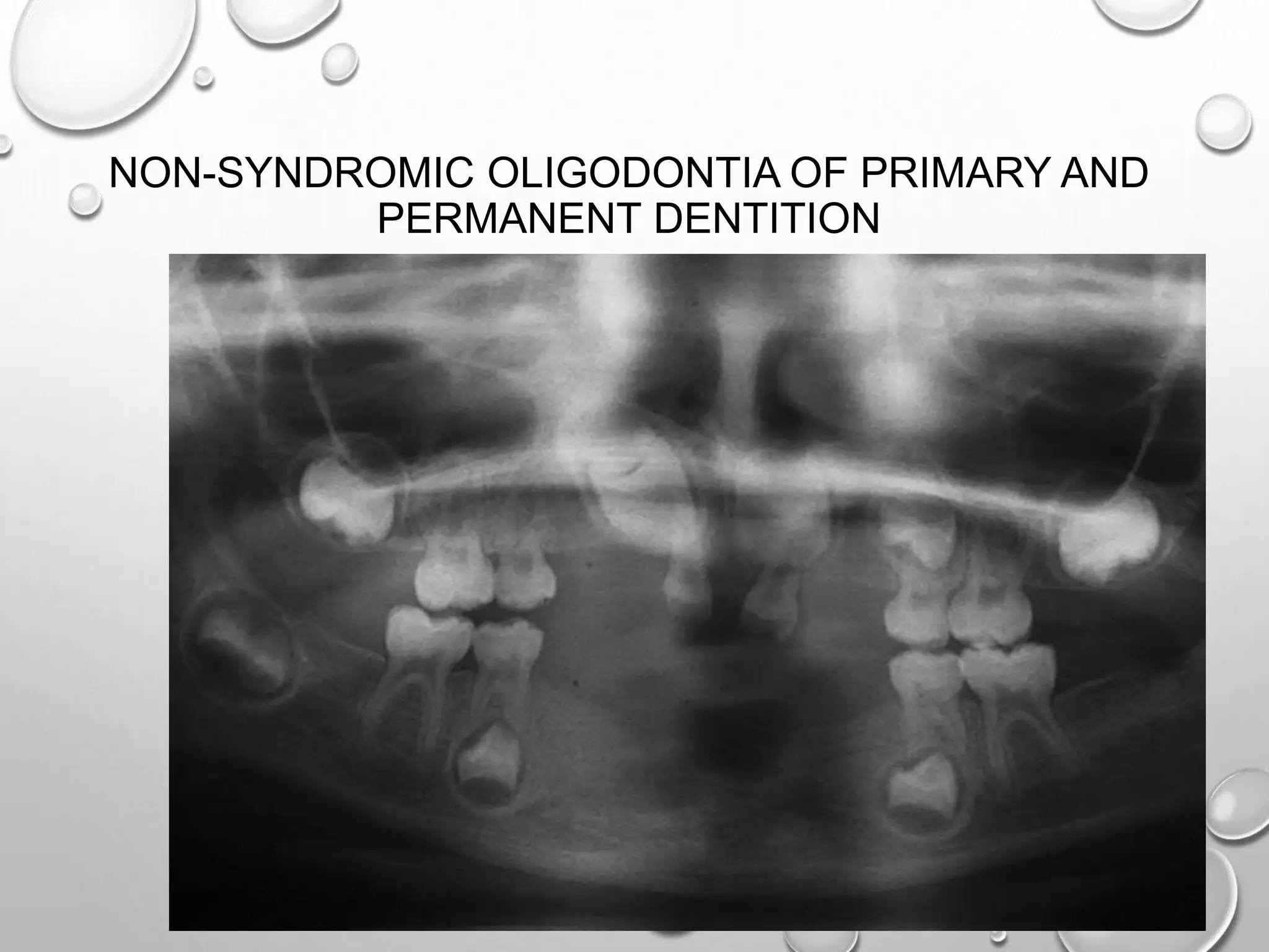
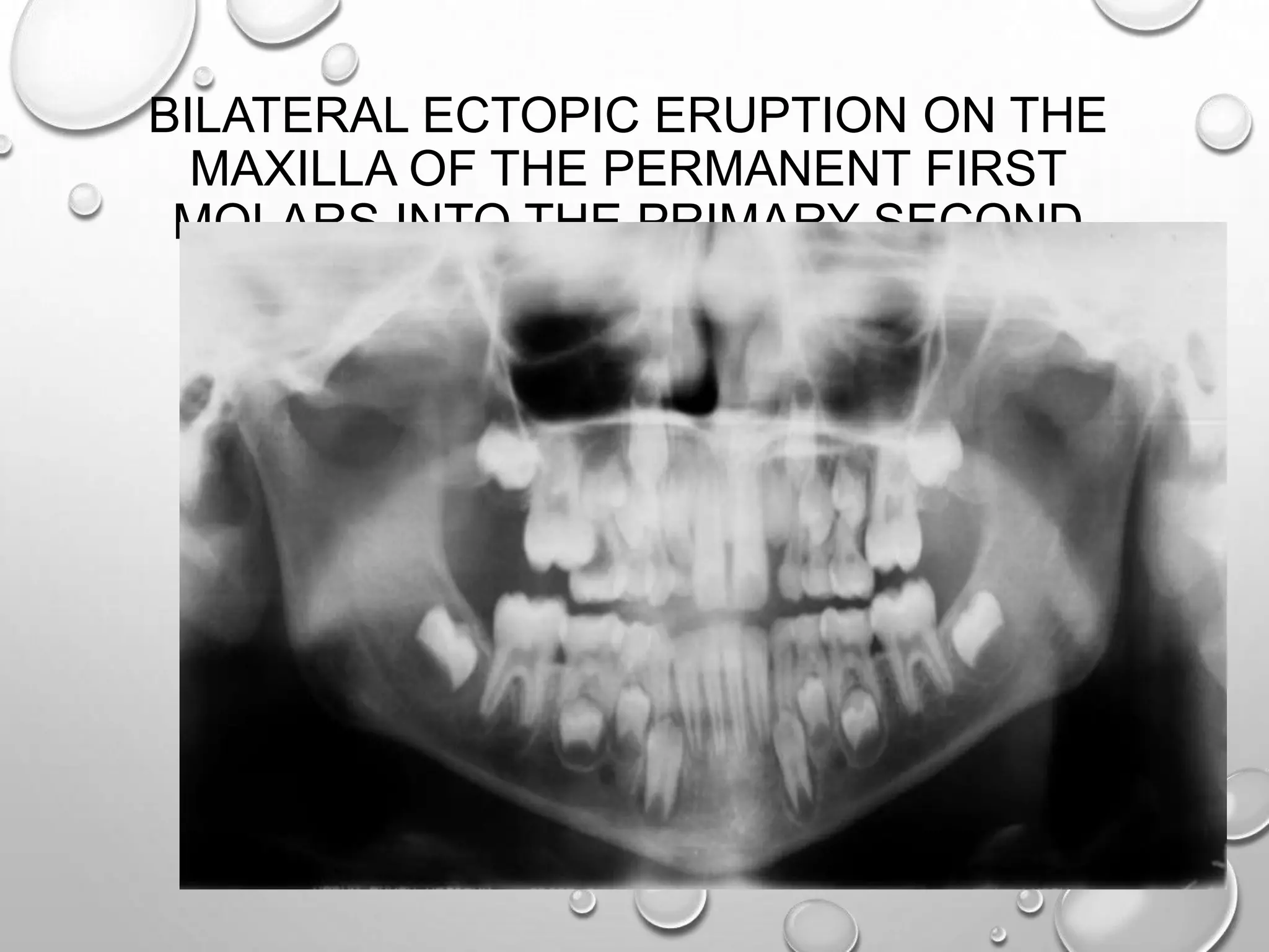
This document discusses the application of panoramic radiography (OPG) in orthodontics. It provides examples of what can be seen on panoramic x-rays including tooth eruption patterns, missing or extra teeth, tooth fractures, and root resorption. Normal anatomy of the jaws is also displayed with labels pointing out various bony landmarks visible on panoramic radiographs. A variety of dental anomalies and orthodontic problems that can be identified and monitored with OPG images are presented.