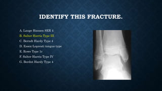
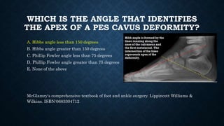
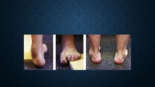
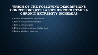
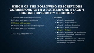
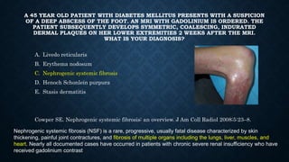
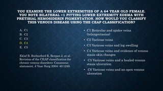
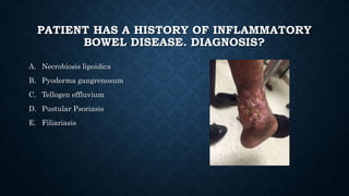
APMLE2CK PRACTICE
A PATIENT SUSTAINS AN ANKLE FRACTURE
WITH 1MM OF TALAR DISPLACEMENT. BY
WHAT PERCENTAGE IS THE CONTACT AREA
WITH THE TIBIAL PLAFOND DIMINISHED?
• 10%
• 22%
• 30%
• 42%
• 50%
A PATIENT SUSTAINS AN ANKLE FRACTURE
WITH 1MM OF TALAR DISPLACEMENT. BY
WHAT PERCENTAGE IS THE CONTACT AREA
WITH THE TIBIAL PLAFOND DIMINISHED?
• 10%
• 22%
• 30%
• 42%
• 50%
• JBJS 1976, Foot and Ankle International Oct 2006
WHAT IS TRUE REGARDING BLOOD-FILLED
FRACTURE BLISTERS AND CLEAR FLUID
FRACTURE BLISTERS?
• A. Blood-filled fracture blisters have been shown histologically to be complete separation
of the dermis from the epidermis. Clear fluid-filled fracture blisters represent a partial
epidermal separation of the epidermis from the underlying dermis, with a few scattered
areas of epithelial cells retained on the dermis.
• B. Clear fluid-filled fracture blisters have been shown histologically to be complete
separation of the dermis from the epidermis. Blood-filled fracture blisters represent a
partial epidermal separation of the epidermis from the underlying dermis, with a few
scattered areas of epithelial cells retained on the dermis.
• C. It is advisable to place incisions through blood-filled fracture blisters in early operative
treatment.
• D. It is advisable to place incisions through clear fluid-filled fracture blisters in early
operative treatment.
WHAT IS TRUE REGARDING BLOOD-FILLED
FRACTURE BLISTERS AND CLEAR FLUID
FRACTURE BLISTERS?
• A. Blood-filled fracture blisters have been shown histologically to be complete separation of the
dermis from the epidermis. Clear fluid-filled fracture blisters represent a partial epidermal
separation of the epidermis from the underlying dermis, with a few scattered areas of
epithelial cells retained on the dermis.
• B. Clear fluid-filled fracture blisters have been shown histologically to be complete separation
of the dermis from the epidermis. Blood-filled fracture blisters represent a partial epidermal
separation of the epidermis from the underlying dermis, with a few scattered areas of
epithelial cells retained on the dermis.
• C. It is advisable to place incisions through blood-filled fracture blisters in early operative
treatment.
• D. It is advisable to place incisions through clear fluid-filled fracture blisters in early operative
treatment.
• Giordano CP J Trauma 1995
IDENTIFY THIS FRACTURE.
A. Lauge Hansen SER 4
B. Salter Harris Type III
C. Berndt Hardy Type 3
D. Essex-Lopresti tongue type
E. Rowe Type 1c
F. Salter Harris Type IV
G. Berdnt Hardy Type 4
IDENTIFY THIS FRACTURE.
A. Lauge Hansen SER 4
B. Salter Harris Type III
C. Berndt Hardy Type 4
D. Essex-Lopresti tongue type
E. Rowe Type 1c
F. Salter Harris Type IV
G. Berdnt Hardy Type 4
WHAT IS THE MOST COMMON CAUSE OF
HYPERCALCEMIA AND WHAT IS THE BEST
TREATMENT FOR SYMPTOMATIC DISEASE?
A. Vitamin D deficiency, replacement with 2000 units vitamin D3 per day
B. Primary Hyperparathyroidism, parathyroidectomy
C. Malignancy, chemotherapy and radiation
D. Chronic kidney disease, dialysis
C Marcocci, F Cetani NEJM 365: 25
WHICH OF THE FOLLOWING IS TRUE
REGARDING OSTEOMALACIA VS
OSTEOPOROSIS?
A. Osteomalacia is soft bone while osteoporosis is porous, brittle bone
B. Osteomalacia can be diagnosed by using moderate force to press the thumb on the
anterior tibia to elicit bone pain which is not true for osteoporosis
C. With osteomalacia, not osteoporosis, mineral composition is decreased relative to
collagen
D. With osteoporosis, not osteomalacia, mineral to collagen ratio is good
E. All of the above are true
Holick MF, NEJM 357: 3
WHAT IS A SKEWFOOT?
A. Metatarsus adductus with a pathologic rearfoot valgus component
B. Metatarsus adductus with a pathologic rearfoot varus component
C. Metatarsus adductus with a pathologic forefoot valgus component
D. Metatarsus adductus with a pathologic forefoot varus component
WHAT IS A SKEWFOOT?
A. Metatarsus adductus with a pathologic rearfoot valgus component
B. Metatarsus adductus with a pathologic rearfoot varus component
C. Metatarsus adductus with a pathologic forefoot valgus component
D. Metatarsus adductus with a pathologic forefoot varus component
Kite H, JAMA 141: 449, 1949
PROXIMAL COMPENSATION AT THE KNEE FOR
EQUINUS DEFORMITY WOULD INCLUDE:
A. Genu valgum
B. Genu varum
C. Genu recurvatum
D. Genu callosum
PROXIMAL COMPENSATION AT THE KNEE FOR
EQUINUS DEFORMITY WOULD INCLUDE:
A. Genu valgum
B. Genu varum
C. Genu recurvatum
D. Genu callosum
Young R, Nix S, Wholohan A, et al. Interventions for increasing ankle joint dorsiflexion: a
systematic review and meta-analysis. J Foot Ankle Res. 2013;6(1):46.
WHICH IS THE ANGLE THAT IDENTIFIES
THE APEX OF A PES CAVUS DEFORMITY?
A. Hibbs angle less than 150 degrees
B. Hibbs angle greater than 150 degrees
C. Phillip Fowler angle less than 75 degrees
D. Phillip Fowler angle greater than 75 degrees
E. None of the above
WHICH IS THE ANGLE THAT IDENTIFIES
THE APEX OF A PES CAVUS DEFORMITY?
A. Hibbs angle less than 150 degrees
B. Hibbs angle greater than 150 degrees
C. Phillip Fowler angle less than 75 degrees
D. Phillip Fowler angle greater than 75 degrees
E. None of the above
McGlamry's comprehensive textbook of foot and ankle surgery. Lippincott Williams &
Wilkins. ISBN:0683304712
A PATIENT WITH PES CAVUS IS SEEN IN THE CLINIC. HIS RIGHT FOOT IS PLACED
ON A WOOD BLOCK WITH THE HEEL AND LATERAL BORDER OF THE FOOT
BEARING FULL WEIGHT AND 1ST METATARSAL ALLOWED TO HANG FREELY. THE
PATIENT’S HEEL VARUS CORRECTS TO NEUTRAL. WHAT IS THE SIGNIFICANCE OF
THIS FINDING?
• A. The hindfoot is considered flexible, surgical procedures can focus on the forefoot. This
is known as the Japas block test.
• B. The hindfoot is considered rigid, surgical procedures must focus on the forefoot and
hindfoot. This is known as the Japas block test.
• C. The hindfoot is considered rigid, surgical procedures must focus on the forefoot and
hindfoot. This is known as the Coleman block test.
• D. The hindfoot is considered flexible, surgical procedures can focus on the forefoot. This
is known as the Coleman block test.
• Clin Orthop Relat Res. 1977 Mar-Apr;(123):60-2.
WHICH IS TRUE REGARDING SYNOVIAL
SARCOMAS?
A. These tumors arise from synovial tissue
B. The most common sites for metastasis are the lungs and lymph nodes
C. These tumors have a fair to good prognosis.
D. The most common presentation is a painful, slow growing mass
E. MRI shows high signal on T1 weighted images and low signal on T2 weighted
images.
WHICH IS TRUE REGARDING SYNOVIAL
SARCOMAS?
A. These tumors arise from synovial tissue
B. The most common sites for metastasis are the lungs and lymph nodes
C. These tumors have a fair to good prognosis.
D. The most common presentation is a painful, slow growing mass
E. MRI shows high signal on T1 weighted images and low signal on T2 weighted images.
JAPMA Vol 106 No 4 July/August 2016.
WHICH OF THE FOLLOWING DESCRIPTIONS
CORRESPOND WITH A RUTHERFORD STAGE 4
CHRONIC EXTREMITY ISCHEMIA?
A. Patient with moderate claudication
B. Patient with severe claudication
C. Patient with rest pain
D. Patient with ischemic non-healing ulcer
E. Patient with focal gangrene
WHICH OF THE FOLLOWING DESCRIPTIONS
CORRESPOND WITH A RUTHERFORD STAGE 4
CHRONIC EXTREMITY ISCHEMIA?
A. Patient with moderate claudication
B. Patient with severe claudication
C. Patient with rest pain
D. Patient with ischemic non-healing ulcer
E. Patient with focal gangrene
J Vasc Surg. 1997;26(3):517.
• Rutherford
• ●Stage 0 – Asymptomatic
• ●Stage 1 – Mild claudication
• ●Stage 2 – Moderate claudication
• ●Stage 3 – Severe claudication
• ●Stage 4 – Rest pain
• ●Stage 5 – Minor tissue loss with ischemic
non-healing ulcer or focal gangrene with
diffuse pedal ischemia
• ●Stage 6 – Major tissue loss – extending
above transmetatarsal level, functional
foot no longer salvageable
WHICH OF THE FOLLOWING DESCRIPTIONS
CORRESPOND WITH A FONTAINE STAGE 3
CHRONIC EXTREMITY ISCHEMIA?
A. Asymptomatic
B. Intermittent claudication, short distances
C. Intermittent claudication, long distances
D. Rest pain
E. Gangrene
WHICH OF THE FOLLOWING DESCRIPTIONS
CORRESPOND WITH A FONTAINE STAGE 3
CHRONIC EXTREMITY ISCHEMIA?
A. Asymptomatic
B. Intermittent claudication, short
distances
C. Intermittent claudication, long
distances
D. Rest pain
E. Gangrene
Helv Chir Acta. 1954;21(5-6):499.
• Fontaine
• ●Stage 1 – No symptoms
• ●Stage 2 – Intermittent claudication
subdivided into:
• ●Stage 2a – without pain on resting,
but with claudication at a distance of
greater than 650 feet (200 meters)
• ●Stage 2b – without pain on resting,
but with a claudication distance of less
than 650 feet (200 meters)
• ●Stage 3 – Nocturnal and/or rest pain
• ●Stage 4 – Necrosis (death of
tissue) and/or gangrene in the limb
WHICH OF THE FOLLOWING MEDICATIONS IS A
COMMON CAUSE OF LOWER EXTREMITY
EDEMA?
A. Amlodipine
B. Cephalexin
C. Dantrolene
D. Lisinopril
E. Rosuvastatin
WHICH OF THE FOLLOWING MEDICATIONS IS A
COMMON CAUSE OF LOWER EXTREMITY
EDEMA?
A. Amlodipine
B. Cephalexin
C. Dantrolene
D. Lisinopril
E. Rosuvastatin
Arch Surg 2003 138: 152-61
WHICH OF THE FOLLOWING IS NOT A
CAUSE OF LOWER EXTREMITY EDEMA?
• A. Congestive heart Failure
• B. NSAIDs
• C. Pregnancy
• D. Sleep apnea
• E. All of the above are causes of lower extremity edema
WHICH OF THE FOLLOWING IS NOT A
CAUSE OF LOWER EXTREMITY EDEMA?
• A. Congestive heart Failure
• B. NSAIDs
• C. Pregnancy
• D. Sleep apnea
• E. All of the above are causes of lower extremity edema
• Am JMed 1998 105: 192. Pulmonary HTN commonly results from sleep apnea and is an
underrecognized cause of lower extremity edema.
• What test do you order R/O pulmonary HTN?
ALL ARE SIGNS/SYMPTOMS CONSISTENT WITH
A DIAGNOSIS OF LYMPHEDEMA EXCEPT:
A. Absence of pitting
B. Brawny induration
C. Positive Kaposi-Stemmer sign
D. Papillomatosis
E. Spares dorsum of the foot
ALL ARE SIGNS/SYMPTOMS CONSISTENT WITH
A DIAGNOSIS OF LYMPHEDEMA EXCEPT:
A. Absence of pitting
B. Brawny induration
C. Positive Kaposi-Stemmer sign
D. Papillomatosis
E. Spares dorsum of the foot
Geriatrics 1993: 48-34-40, Arch Surg 2003: 138 – 152
WHICH OF THE FOLLOWING IS THE DRUG
OF CHOICE FOR IDIOPATHIC EDEMA?
A. Atorvastatin
B. Furosemide
C. Griseofulvin
D. Omeprazole
E. Spironolactone
WHICH OF THE FOLLOWING IS THE DRUG
OF CHOICE FOR IDIOPATHIC EDEMA?
A. Atorvastatin
B. Furosemide
C. Griseofulvin
D. Omeprazole
E. Spironolactone
• Endocrinol Metab Clin North Am 1995; 24: 531-47.
WHICH OF THE FOLLOWING NATURAL
SUBSTANCES HAS EVIDENCE FOR THE
TREATMENT OF CHRONIC VENOUS
INSUFFICIENCY?
A.St. John’s Wort
B.Ginseng
C.Horse chestnut seed extract
D.Yohimibine
E.Turmeric
WHICH OF THE FOLLOWING NATURAL
SUBSTANCES HAS EVIDENCE FOR THE
TREATMENT OF CHRONIC VENOUS
INSUFFICIENCY?
A. St. John’s Wort
B. Ginseng
C. Horse chestnut seed extract
D. Yohimibine
E. Turmeric
Pittler MH, Ernst E. Horse chestnut seed extract for chronic venous insufficiency. Cochrane
Database Syst Rev 2002 (1):CD003230; Pittler MH, Ernst E. Horse-chestnut seed extract
for chronic venous insufficiency. A criteria-based systematic review. Arch Dermatol 1998;
134: 1356-60; Diehm C, Trampisch HJ, Lange S, Schmidt C. Comparison of leg compression
stocking and oral horse-chestnut seed extract therapy in patients with chronic venous
insufficiency. Lancet 1996; 347: 292-4.
WHICH OF THE FOLLOWING IS NOT A FORM
OF LYMPHEDEMA?
A. Caisson disease
B. Milroy’s disease
C. Meige disease
D. Lymphedema praecox
E. Lymphedema tarda
WHICH OF THE FOLLOWING IS NOT A FORM
OF LYMPHEDEMA?
A. Caisson disease
B. Milroy’s disease
C. Meige disease
D. Lymphedema praecox
E. Lymphedema tarda
Szuba A, Rockson SG. Lymphedema: classification, diagnosis, and therapy. Vasc Med 1998;
3: 145-6.
WHICH OF THE FOLLOWING IS A STRONG
RELATIVE CONTRAINDICATION FOR A LOWER
EXTREMITY MRI?
A. Vascular stainless steel or nickel titanium stents in peripheral vessels
B. Implanted IVC filter
C. Prosthetic heart valve
D. Implantable cardioverter defibrillator
E. Tattoo
WHICH OF THE FOLLOWING IS A STRONG
RELATIVE CONTRAINDICATION FOR A LOWER
EXTREMITY MRI?
A. Vascular stainless steel or nickel titanium stents in peripheral vessels
B. Implanted IVC filter
C. Prosthetic heart valve
D. Implantable cardioverter defibrillator
E. Tattoo
Dill: Contraindications to magnetic resonance imaging. Heart 2008;94;943-948
WHICH OF THE FOLLOWING IS A STRONG
RELATIVE CONTRAINDICATION FOR A LOWER
EXTREMITY MRI?
A. Cochlear implant
B. IUD in a 1.5 tesla MRI field (metal-containing IUDs and the ESSURE sterilization have
not yet been fully evaluated in the 3 T field, which is why they represent
a contraindication to MRI)
C. Bare metal coronary artery stent
D. Drug eluting coronary artery stent
E. Most aortic stent grafts
WHICH OF THE FOLLOWING IS A STRONG
RELATIVE CONTRAINDICATION FOR A LOWER
EXTREMITY MRI?
A. Cochlear implant
B. IUD in a 1.5 tesla MRI field
C. Bare metal coronary artery stent
D. Drug eluting coronary artery stent
E. Most aortic stent grafts
Dill: Contraindications to magnetic resonance imaging. Heart 2008;94;943-948
A 45 YEAR OLD PATIENT WITH DIABETES MELLITUS PRESENTS WITH A SUSPICION
OF A DEEP ABSCESS OF THE FOOT. AN MRI WITH GADOLINIUM IS ORDERED. THE
PATIENT SUBSEQUENTLY DEVELOPS SYMMETRIC, COALESCING, INDURATED
DERMAL PLAQUES ON HER LOWER EXTREMITIES 2 WEEKS AFTER THE MRI.
WHAT IS YOUR DIAGNOSIS?
A. Livedo reticularis
B. Erythema nodosum
C. Nephrogenic systemic fibrosis
D. Henoch Schonlein purpura
E. Stasis dermatitis
Cowper SE. Nephrogenic systemic fibrosis: an overview. J Am Coll Radiol 2008;5:23–8.
Nephrogenic systemic fibrosis (NSF) is a rare, progressive, usually fatal disease characterized by skin
thickening, painful joint contractures, and fibrosis of multiple organs including the lungs, liver, muscles, and
heart. Nearly all documented cases have occurred in patients with chronic severe renal insufficiency who have
received gadolinium contrast
YOU EXAMINE THE LOWER EXTREMITIES OF A 64 YEAR OLD FEMALE.
YOU NOTE BILATERAL +1 PITTING LOWER EXTREMITY EDEMA WITH
PRETIBIAL HEMOSIDERIN PIGMENTATION. HOW WOULD YOU CLASSIFY
THIS VENOUS DISEASE USING THE CEAP CLASSIFICATION?
A. C1
B. C2
C. C3
D. C4
E. C5
YOU EXAMINE THE LOWER EXTREMITIES OF A 64 YEAR OLD FEMALE.
YOU NOTE BILATERAL +1 PITTING LOWER EXTREMITY EDEMA WITH
PRETIBIAL HEMOSIDERIN PIGMENTATION. HOW WOULD YOU CLASSIFY
THIS VENOUS DISEASE USING THE CEAP CLASSIFICATION?
A. C1
B. C2
C. C3
D. C4
E. C5
Eklof B, Rutherford R, Bergan J, et al.
Revision of the CEAP classification for
chronic venous disorders: Consensus
statement. J Vasc Surg 2004; 40:1248.
• C1 Reticular and spider veins
(telangectasias)
• C2 Varicose veins
• C3 Varicose veins and leg swelling
• C4 Varicose veins and evidence of venous
stasis skin changes
• C5 Varicose veins and a healed venous
stasis ulceration
• C6 Varicose veins and an open venous
ulceration
A 54 YEAR OLD PATIENT WITH TYPE 2 DIABETES AND STAGE 3 CHRONIC
KIDNEY DISEASE DEVELOPS HETEROTOPIC OSSIFICATION STATUS POST
TRANSMETATARSAL AMPUTATION. WHICH OF THE FOLLOWING IS THE
BEST CHOICE FOR THE PREVENTION OF THIS OCCURRENCE?
A. Calcitonin
B. NSAID
C. Prednisone
D. Radiation
E. Tumor necrosis factor inhibitor
The Journal of Foot and Ankle Surgery
Volume 55, Issue 4, July-August 2016,
Pages 714-719
• Radiation preferred over NSAIDs in
patients with renal disease.
PATIENT HAS A HISTORY OF INFLAMMATORY
BOWEL DISEASE. DIAGNOSIS?
A. Necrobiosis lipoidica
B. Pyoderma gangrenosum
C. Tellogen effluvium
D. Pustular Psoriasis
E. Filiariasis
PATIENT HAS HISTORY OF INFLAMMATORY
BOWEL DISEASE. DIAGNOSIS?
A. Necrobiosis lipoidica
B. Pyoderma gangrenosum
C. Tellogen effluvium
D. Pustular Psoriasis
E. Filiariasis
BMJ. 2006 Jul 22; 333(7560): 181–184.

APMLE2CK practice 2016 NEW.pptx

  • 1.
  • 2.
    A PATIENT SUSTAINSAN ANKLE FRACTURE WITH 1MM OF TALAR DISPLACEMENT. BY WHAT PERCENTAGE IS THE CONTACT AREA WITH THE TIBIAL PLAFOND DIMINISHED? • 10% • 22% • 30% • 42% • 50%
  • 3.
    A PATIENT SUSTAINSAN ANKLE FRACTURE WITH 1MM OF TALAR DISPLACEMENT. BY WHAT PERCENTAGE IS THE CONTACT AREA WITH THE TIBIAL PLAFOND DIMINISHED? • 10% • 22% • 30% • 42% • 50% • JBJS 1976, Foot and Ankle International Oct 2006
  • 4.
    WHAT IS TRUEREGARDING BLOOD-FILLED FRACTURE BLISTERS AND CLEAR FLUID FRACTURE BLISTERS? • A. Blood-filled fracture blisters have been shown histologically to be complete separation of the dermis from the epidermis. Clear fluid-filled fracture blisters represent a partial epidermal separation of the epidermis from the underlying dermis, with a few scattered areas of epithelial cells retained on the dermis. • B. Clear fluid-filled fracture blisters have been shown histologically to be complete separation of the dermis from the epidermis. Blood-filled fracture blisters represent a partial epidermal separation of the epidermis from the underlying dermis, with a few scattered areas of epithelial cells retained on the dermis. • C. It is advisable to place incisions through blood-filled fracture blisters in early operative treatment. • D. It is advisable to place incisions through clear fluid-filled fracture blisters in early operative treatment.
  • 5.
    WHAT IS TRUEREGARDING BLOOD-FILLED FRACTURE BLISTERS AND CLEAR FLUID FRACTURE BLISTERS? • A. Blood-filled fracture blisters have been shown histologically to be complete separation of the dermis from the epidermis. Clear fluid-filled fracture blisters represent a partial epidermal separation of the epidermis from the underlying dermis, with a few scattered areas of epithelial cells retained on the dermis. • B. Clear fluid-filled fracture blisters have been shown histologically to be complete separation of the dermis from the epidermis. Blood-filled fracture blisters represent a partial epidermal separation of the epidermis from the underlying dermis, with a few scattered areas of epithelial cells retained on the dermis. • C. It is advisable to place incisions through blood-filled fracture blisters in early operative treatment. • D. It is advisable to place incisions through clear fluid-filled fracture blisters in early operative treatment. • Giordano CP J Trauma 1995
  • 6.
    IDENTIFY THIS FRACTURE. A.Lauge Hansen SER 4 B. Salter Harris Type III C. Berndt Hardy Type 3 D. Essex-Lopresti tongue type E. Rowe Type 1c F. Salter Harris Type IV G. Berdnt Hardy Type 4
  • 7.
    IDENTIFY THIS FRACTURE. A.Lauge Hansen SER 4 B. Salter Harris Type III C. Berndt Hardy Type 4 D. Essex-Lopresti tongue type E. Rowe Type 1c F. Salter Harris Type IV G. Berdnt Hardy Type 4
  • 8.
    WHAT IS THEMOST COMMON CAUSE OF HYPERCALCEMIA AND WHAT IS THE BEST TREATMENT FOR SYMPTOMATIC DISEASE? A. Vitamin D deficiency, replacement with 2000 units vitamin D3 per day B. Primary Hyperparathyroidism, parathyroidectomy C. Malignancy, chemotherapy and radiation D. Chronic kidney disease, dialysis
  • 9.
    WHAT IS THEMOST COMMON CAUSE OF HYPERCALCEMIA AND WHAT IS THE BEST TREATMENT FOR SYMPTOMATIC DISEASE? A. Vitamin D deficiency, replacement with 2000 units vitamin D3 per day B. Primary Hyperparathyroidism, parathyroidectomy C. Malignancy, chemotherapy and radiation D. Chronic kidney disease, dialysis C Marcocci, F Cetani NEJM 365: 25
  • 10.
    WHICH OF THEFOLLOWING IS TRUE REGARDING OSTEOMALACIA VS OSTEOPOROSIS? A. Osteomalacia is soft bone while osteoporosis is porous, brittle bone B. Osteomalacia can be diagnosed by using moderate force to press the thumb on the anterior tibia to elicit bone pain which is not true for osteoporosis C. With osteomalacia, not osteoporosis, mineral composition is decreased relative to collagen D. With osteoporosis, not osteomalacia, mineral to collagen ratio is good E. All of the above are true
  • 11.
    WHICH OF THEFOLLOWING IS TRUE REGARDING OSTEOMALACIA VS OSTEOPOROSIS? A. Osteomalacia is soft bone while osteoporosis is porous, brittle bone B. Osteomalacia can be diagnosed by using moderate force to press the thumb on the anterior tibia to elicit bone pain which is not true for osteoporosis C. With osteomalacia, not osteoporosis, mineral composition is decreased relative to collagen D. With osteoporosis, not osteomalacia, mineral to collagen ratio is good E. All of the above are true Holick MF, NEJM 357: 3
  • 12.
    WHAT IS ASKEWFOOT? A. Metatarsus adductus with a pathologic rearfoot valgus component B. Metatarsus adductus with a pathologic rearfoot varus component C. Metatarsus adductus with a pathologic forefoot valgus component D. Metatarsus adductus with a pathologic forefoot varus component
  • 13.
    WHAT IS ASKEWFOOT? A. Metatarsus adductus with a pathologic rearfoot valgus component B. Metatarsus adductus with a pathologic rearfoot varus component C. Metatarsus adductus with a pathologic forefoot valgus component D. Metatarsus adductus with a pathologic forefoot varus component Kite H, JAMA 141: 449, 1949
  • 14.
    PROXIMAL COMPENSATION ATTHE KNEE FOR EQUINUS DEFORMITY WOULD INCLUDE: A. Genu valgum B. Genu varum C. Genu recurvatum D. Genu callosum
  • 15.
    PROXIMAL COMPENSATION ATTHE KNEE FOR EQUINUS DEFORMITY WOULD INCLUDE: A. Genu valgum B. Genu varum C. Genu recurvatum D. Genu callosum Young R, Nix S, Wholohan A, et al. Interventions for increasing ankle joint dorsiflexion: a systematic review and meta-analysis. J Foot Ankle Res. 2013;6(1):46.
  • 16.
    WHICH IS THEANGLE THAT IDENTIFIES THE APEX OF A PES CAVUS DEFORMITY? A. Hibbs angle less than 150 degrees B. Hibbs angle greater than 150 degrees C. Phillip Fowler angle less than 75 degrees D. Phillip Fowler angle greater than 75 degrees E. None of the above
  • 17.
    WHICH IS THEANGLE THAT IDENTIFIES THE APEX OF A PES CAVUS DEFORMITY? A. Hibbs angle less than 150 degrees B. Hibbs angle greater than 150 degrees C. Phillip Fowler angle less than 75 degrees D. Phillip Fowler angle greater than 75 degrees E. None of the above McGlamry's comprehensive textbook of foot and ankle surgery. Lippincott Williams & Wilkins. ISBN:0683304712
  • 18.
    A PATIENT WITHPES CAVUS IS SEEN IN THE CLINIC. HIS RIGHT FOOT IS PLACED ON A WOOD BLOCK WITH THE HEEL AND LATERAL BORDER OF THE FOOT BEARING FULL WEIGHT AND 1ST METATARSAL ALLOWED TO HANG FREELY. THE PATIENT’S HEEL VARUS CORRECTS TO NEUTRAL. WHAT IS THE SIGNIFICANCE OF THIS FINDING? • A. The hindfoot is considered flexible, surgical procedures can focus on the forefoot. This is known as the Japas block test. • B. The hindfoot is considered rigid, surgical procedures must focus on the forefoot and hindfoot. This is known as the Japas block test. • C. The hindfoot is considered rigid, surgical procedures must focus on the forefoot and hindfoot. This is known as the Coleman block test. • D. The hindfoot is considered flexible, surgical procedures can focus on the forefoot. This is known as the Coleman block test.
  • 19.
    A PATIENT WITHPES CAVUS IS SEEN IN THE CLINIC. HIS RIGHT FOOT IS PLACED ON A WOOD BLOCK WITH THE HEEL AND LATERAL BORDER OF THE FOOT BEARING FULL WEIGHT AND 1ST METATARSAL ALLOWED TO HANG FREELY. THE PATIENT’S HEEL VARUS CORRECTS TO NEUTRAL. WHAT IS THE SIGNIFICANCE OF THIS FINDING? • A. The hindfoot is considered flexible, surgical procedures can focus on the forefoot. This is known as the Japas block test. • B. The hindfoot is considered rigid, surgical procedures must focus on the forefoot and hindfoot. This is known as the Japas block test. • C. The hindfoot is considered rigid, surgical procedures must focus on the forefoot and hindfoot. This is known as the Coleman block test. • D. The hindfoot is considered flexible, surgical procedures can focus on the forefoot. This is known as the Coleman block test. • Clin Orthop Relat Res. 1977 Mar-Apr;(123):60-2.
  • 21.
    WHICH IS TRUEREGARDING SYNOVIAL SARCOMAS? A. These tumors arise from synovial tissue B. The most common sites for metastasis are the lungs and lymph nodes C. These tumors have a fair to good prognosis. D. The most common presentation is a painful, slow growing mass E. MRI shows high signal on T1 weighted images and low signal on T2 weighted images.
  • 22.
    WHICH IS TRUEREGARDING SYNOVIAL SARCOMAS? A. These tumors arise from synovial tissue B. The most common sites for metastasis are the lungs and lymph nodes C. These tumors have a fair to good prognosis. D. The most common presentation is a painful, slow growing mass E. MRI shows high signal on T1 weighted images and low signal on T2 weighted images. JAPMA Vol 106 No 4 July/August 2016.
  • 23.
    WHICH OF THEFOLLOWING DESCRIPTIONS CORRESPOND WITH A RUTHERFORD STAGE 4 CHRONIC EXTREMITY ISCHEMIA? A. Patient with moderate claudication B. Patient with severe claudication C. Patient with rest pain D. Patient with ischemic non-healing ulcer E. Patient with focal gangrene
  • 24.
    WHICH OF THEFOLLOWING DESCRIPTIONS CORRESPOND WITH A RUTHERFORD STAGE 4 CHRONIC EXTREMITY ISCHEMIA? A. Patient with moderate claudication B. Patient with severe claudication C. Patient with rest pain D. Patient with ischemic non-healing ulcer E. Patient with focal gangrene J Vasc Surg. 1997;26(3):517. • Rutherford • ●Stage 0 – Asymptomatic • ●Stage 1 – Mild claudication • ●Stage 2 – Moderate claudication • ●Stage 3 – Severe claudication • ●Stage 4 – Rest pain • ●Stage 5 – Minor tissue loss with ischemic non-healing ulcer or focal gangrene with diffuse pedal ischemia • ●Stage 6 – Major tissue loss – extending above transmetatarsal level, functional foot no longer salvageable
  • 25.
    WHICH OF THEFOLLOWING DESCRIPTIONS CORRESPOND WITH A FONTAINE STAGE 3 CHRONIC EXTREMITY ISCHEMIA? A. Asymptomatic B. Intermittent claudication, short distances C. Intermittent claudication, long distances D. Rest pain E. Gangrene
  • 26.
    WHICH OF THEFOLLOWING DESCRIPTIONS CORRESPOND WITH A FONTAINE STAGE 3 CHRONIC EXTREMITY ISCHEMIA? A. Asymptomatic B. Intermittent claudication, short distances C. Intermittent claudication, long distances D. Rest pain E. Gangrene Helv Chir Acta. 1954;21(5-6):499. • Fontaine • ●Stage 1 – No symptoms • ●Stage 2 – Intermittent claudication subdivided into: • ●Stage 2a – without pain on resting, but with claudication at a distance of greater than 650 feet (200 meters) • ●Stage 2b – without pain on resting, but with a claudication distance of less than 650 feet (200 meters) • ●Stage 3 – Nocturnal and/or rest pain • ●Stage 4 – Necrosis (death of tissue) and/or gangrene in the limb
  • 27.
    WHICH OF THEFOLLOWING MEDICATIONS IS A COMMON CAUSE OF LOWER EXTREMITY EDEMA? A. Amlodipine B. Cephalexin C. Dantrolene D. Lisinopril E. Rosuvastatin
  • 28.
    WHICH OF THEFOLLOWING MEDICATIONS IS A COMMON CAUSE OF LOWER EXTREMITY EDEMA? A. Amlodipine B. Cephalexin C. Dantrolene D. Lisinopril E. Rosuvastatin Arch Surg 2003 138: 152-61
  • 29.
    WHICH OF THEFOLLOWING IS NOT A CAUSE OF LOWER EXTREMITY EDEMA? • A. Congestive heart Failure • B. NSAIDs • C. Pregnancy • D. Sleep apnea • E. All of the above are causes of lower extremity edema
  • 30.
    WHICH OF THEFOLLOWING IS NOT A CAUSE OF LOWER EXTREMITY EDEMA? • A. Congestive heart Failure • B. NSAIDs • C. Pregnancy • D. Sleep apnea • E. All of the above are causes of lower extremity edema • Am JMed 1998 105: 192. Pulmonary HTN commonly results from sleep apnea and is an underrecognized cause of lower extremity edema. • What test do you order R/O pulmonary HTN?
  • 31.
    ALL ARE SIGNS/SYMPTOMSCONSISTENT WITH A DIAGNOSIS OF LYMPHEDEMA EXCEPT: A. Absence of pitting B. Brawny induration C. Positive Kaposi-Stemmer sign D. Papillomatosis E. Spares dorsum of the foot
  • 32.
    ALL ARE SIGNS/SYMPTOMSCONSISTENT WITH A DIAGNOSIS OF LYMPHEDEMA EXCEPT: A. Absence of pitting B. Brawny induration C. Positive Kaposi-Stemmer sign D. Papillomatosis E. Spares dorsum of the foot Geriatrics 1993: 48-34-40, Arch Surg 2003: 138 – 152
  • 33.
    WHICH OF THEFOLLOWING IS THE DRUG OF CHOICE FOR IDIOPATHIC EDEMA? A. Atorvastatin B. Furosemide C. Griseofulvin D. Omeprazole E. Spironolactone
  • 34.
    WHICH OF THEFOLLOWING IS THE DRUG OF CHOICE FOR IDIOPATHIC EDEMA? A. Atorvastatin B. Furosemide C. Griseofulvin D. Omeprazole E. Spironolactone • Endocrinol Metab Clin North Am 1995; 24: 531-47.
  • 35.
    WHICH OF THEFOLLOWING NATURAL SUBSTANCES HAS EVIDENCE FOR THE TREATMENT OF CHRONIC VENOUS INSUFFICIENCY? A.St. John’s Wort B.Ginseng C.Horse chestnut seed extract D.Yohimibine E.Turmeric
  • 36.
    WHICH OF THEFOLLOWING NATURAL SUBSTANCES HAS EVIDENCE FOR THE TREATMENT OF CHRONIC VENOUS INSUFFICIENCY? A. St. John’s Wort B. Ginseng C. Horse chestnut seed extract D. Yohimibine E. Turmeric Pittler MH, Ernst E. Horse chestnut seed extract for chronic venous insufficiency. Cochrane Database Syst Rev 2002 (1):CD003230; Pittler MH, Ernst E. Horse-chestnut seed extract for chronic venous insufficiency. A criteria-based systematic review. Arch Dermatol 1998; 134: 1356-60; Diehm C, Trampisch HJ, Lange S, Schmidt C. Comparison of leg compression stocking and oral horse-chestnut seed extract therapy in patients with chronic venous insufficiency. Lancet 1996; 347: 292-4.
  • 37.
    WHICH OF THEFOLLOWING IS NOT A FORM OF LYMPHEDEMA? A. Caisson disease B. Milroy’s disease C. Meige disease D. Lymphedema praecox E. Lymphedema tarda
  • 38.
    WHICH OF THEFOLLOWING IS NOT A FORM OF LYMPHEDEMA? A. Caisson disease B. Milroy’s disease C. Meige disease D. Lymphedema praecox E. Lymphedema tarda Szuba A, Rockson SG. Lymphedema: classification, diagnosis, and therapy. Vasc Med 1998; 3: 145-6.
  • 39.
    WHICH OF THEFOLLOWING IS A STRONG RELATIVE CONTRAINDICATION FOR A LOWER EXTREMITY MRI? A. Vascular stainless steel or nickel titanium stents in peripheral vessels B. Implanted IVC filter C. Prosthetic heart valve D. Implantable cardioverter defibrillator E. Tattoo
  • 40.
    WHICH OF THEFOLLOWING IS A STRONG RELATIVE CONTRAINDICATION FOR A LOWER EXTREMITY MRI? A. Vascular stainless steel or nickel titanium stents in peripheral vessels B. Implanted IVC filter C. Prosthetic heart valve D. Implantable cardioverter defibrillator E. Tattoo Dill: Contraindications to magnetic resonance imaging. Heart 2008;94;943-948
  • 41.
    WHICH OF THEFOLLOWING IS A STRONG RELATIVE CONTRAINDICATION FOR A LOWER EXTREMITY MRI? A. Cochlear implant B. IUD in a 1.5 tesla MRI field (metal-containing IUDs and the ESSURE sterilization have not yet been fully evaluated in the 3 T field, which is why they represent a contraindication to MRI) C. Bare metal coronary artery stent D. Drug eluting coronary artery stent E. Most aortic stent grafts
  • 42.
    WHICH OF THEFOLLOWING IS A STRONG RELATIVE CONTRAINDICATION FOR A LOWER EXTREMITY MRI? A. Cochlear implant B. IUD in a 1.5 tesla MRI field C. Bare metal coronary artery stent D. Drug eluting coronary artery stent E. Most aortic stent grafts Dill: Contraindications to magnetic resonance imaging. Heart 2008;94;943-948
  • 43.
    A 45 YEAROLD PATIENT WITH DIABETES MELLITUS PRESENTS WITH A SUSPICION OF A DEEP ABSCESS OF THE FOOT. AN MRI WITH GADOLINIUM IS ORDERED. THE PATIENT SUBSEQUENTLY DEVELOPS SYMMETRIC, COALESCING, INDURATED DERMAL PLAQUES ON HER LOWER EXTREMITIES 2 WEEKS AFTER THE MRI. WHAT IS YOUR DIAGNOSIS? A. Livedo reticularis B. Erythema nodosum C. Nephrogenic systemic fibrosis D. Henoch Schonlein purpura E. Stasis dermatitis
  • 44.
    A 45 YEAROLD PATIENT WITH DIABETES MELLITUS PRESENTS WITH A SUSPICION OF A DEEP ABSCESS OF THE FOOT. AN MRI WITH GADOLINIUM IS ORDERED. THE PATIENT SUBSEQUENTLY DEVELOPS SYMMETRIC, COALESCING, INDURATED DERMAL PLAQUES ON HER LOWER EXTREMITIES 2 WEEKS AFTER THE MRI. WHAT IS YOUR DIAGNOSIS? A. Livedo reticularis B. Erythema nodosum C. Nephrogenic systemic fibrosis D. Henoch Schonlein purpura E. Stasis dermatitis Cowper SE. Nephrogenic systemic fibrosis: an overview. J Am Coll Radiol 2008;5:23–8. Nephrogenic systemic fibrosis (NSF) is a rare, progressive, usually fatal disease characterized by skin thickening, painful joint contractures, and fibrosis of multiple organs including the lungs, liver, muscles, and heart. Nearly all documented cases have occurred in patients with chronic severe renal insufficiency who have received gadolinium contrast
  • 45.
    YOU EXAMINE THELOWER EXTREMITIES OF A 64 YEAR OLD FEMALE. YOU NOTE BILATERAL +1 PITTING LOWER EXTREMITY EDEMA WITH PRETIBIAL HEMOSIDERIN PIGMENTATION. HOW WOULD YOU CLASSIFY THIS VENOUS DISEASE USING THE CEAP CLASSIFICATION? A. C1 B. C2 C. C3 D. C4 E. C5
  • 46.
    YOU EXAMINE THELOWER EXTREMITIES OF A 64 YEAR OLD FEMALE. YOU NOTE BILATERAL +1 PITTING LOWER EXTREMITY EDEMA WITH PRETIBIAL HEMOSIDERIN PIGMENTATION. HOW WOULD YOU CLASSIFY THIS VENOUS DISEASE USING THE CEAP CLASSIFICATION? A. C1 B. C2 C. C3 D. C4 E. C5 Eklof B, Rutherford R, Bergan J, et al. Revision of the CEAP classification for chronic venous disorders: Consensus statement. J Vasc Surg 2004; 40:1248. • C1 Reticular and spider veins (telangectasias) • C2 Varicose veins • C3 Varicose veins and leg swelling • C4 Varicose veins and evidence of venous stasis skin changes • C5 Varicose veins and a healed venous stasis ulceration • C6 Varicose veins and an open venous ulceration
  • 47.
    A 54 YEAROLD PATIENT WITH TYPE 2 DIABETES AND STAGE 3 CHRONIC KIDNEY DISEASE DEVELOPS HETEROTOPIC OSSIFICATION STATUS POST TRANSMETATARSAL AMPUTATION. WHICH OF THE FOLLOWING IS THE BEST CHOICE FOR THE PREVENTION OF THIS OCCURRENCE? A. Calcitonin B. NSAID C. Prednisone D. Radiation E. Tumor necrosis factor inhibitor
  • 48.
    A 54 YEAROLD PATIENT WITH TYPE 2 DIABETES AND STAGE 3 CHRONIC KIDNEY DISEASE DEVELOPS HETEROTOPIC OSSIFICATION STATUS POST TRANSMETATARSAL AMPUTATION. WHICH OF THE FOLLOWING IS THE BEST CHOICE FOR THE PREVENTION OF THIS OCCURRENCE? A. Calcitonin B. NSAID C. Prednisone D. Radiation E. Tumor necrosis factor inhibitor The Journal of Foot and Ankle Surgery Volume 55, Issue 4, July-August 2016, Pages 714-719 • Radiation preferred over NSAIDs in patients with renal disease.
  • 49.
    PATIENT HAS AHISTORY OF INFLAMMATORY BOWEL DISEASE. DIAGNOSIS? A. Necrobiosis lipoidica B. Pyoderma gangrenosum C. Tellogen effluvium D. Pustular Psoriasis E. Filiariasis
  • 50.
    PATIENT HAS HISTORYOF INFLAMMATORY BOWEL DISEASE. DIAGNOSIS? A. Necrobiosis lipoidica B. Pyoderma gangrenosum C. Tellogen effluvium D. Pustular Psoriasis E. Filiariasis BMJ. 2006 Jul 22; 333(7560): 181–184.

Editor's Notes

  • #21 The Coleman block test is performed by placing a block under the lateral foot and allowing the first metatarsal to come to the ground. A and B, In these photographs, the foot is quite rigid, and no correction of the heel varus took place. C, The patient was an adolescent. The heel corrected into neutral with the test, suggesting more of a forefoot-driven varus deformity.
  • #31 Echo