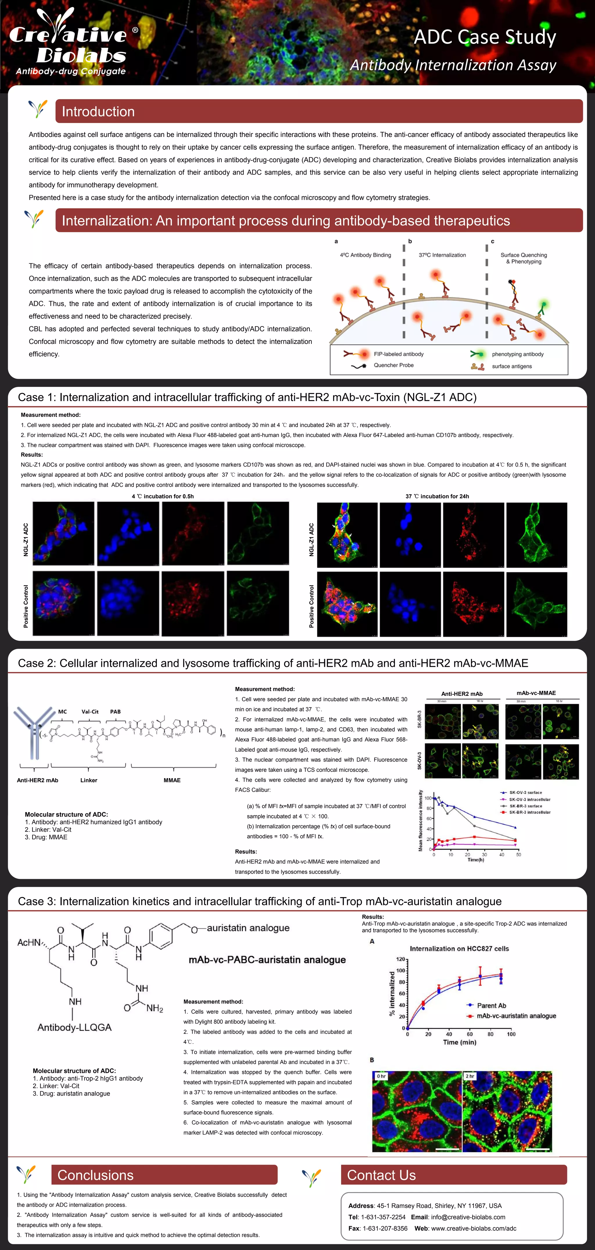
1. Creative Biolabs provides an antibody internalization assay service using confocal microscopy and flow cytometry to detect the rate and extent of antibody and antibody-drug conjugate (ADC) internalization.
2. Three case studies are presented demonstrating the successful use of this service to detect internalization of various ADCs into lysosomes over time, including anti-HER2 and anti-Trop-2 ADCs.
3. The internalization assay allows clients to verify that their antibodies and ADCs are efficiently internalized, an important factor for therapeutic effectiveness.
Kolkata Call Girls Services 9907093804 @24x7 High Class Babes Here Call Now
Antibody Internalization Assay Case Studies
1. ADC Case Study
Antibody Internalization Assay
Introduction
Internalization: An important process during antibody-based therapeutics
The efficacy of certain antibody-based therapeutics depends on internalization process.
Once internalization, such as the ADC molecules are transported to subsequent intracellular
compartments where the toxic payload drug is released to accomplish the cytotoxicity of the
ADC. Thus, the rate and extent of antibody internalization is of crucial importance to its
effectiveness and need to be characterized precisely.
CBL has adopted and perfected several techniques to study antibody/ADC internalization.
Confocal microscopy and flow cytometry are suitable methods to detect the internalization
efficiency.
Conclusions Contact Us
Antibodies against cell surface antigens can be internalized through their specific interactions with these proteins. The anti-cancer efficacy of antibody associated therapeutics like
antibody-drug conjugates is thought to rely on their uptake by cancer cells expressing the surface antigen. Therefore, the measurement of internalization efficacy of an antibody is
critical for its curative effect. Based on years of experiences in antibody-drug-conjugate (ADC) developing and characterization, Creative Biolabs provides internalization analysis
service to help clients verify the internalization of their antibody and ADC samples, and this service can be also very useful in helping clients select appropriate internalizing
antibody for immunotherapy development.
Presented here is a case study for the antibody internalization detection via the confocal microscopy and flow cytometry strategies.
Case 1: Internalization and intracellular trafficking of anti-HER2 mAb-vc-Toxin (NGL-Z1 ADC)
Measurement method:
1. Cell were seeded per plate and incubated with NGL-Z1 ADC and positive control antibody 30 min at 4 ℃ and incubated 24h at 37 ℃, respectively.
2. For internalized NGL-Z1 ADC, the cells were incubated with Alexa Fluor 488-labeled goat anti-human IgG, then incubated with Alexa Fluor 647-Labeled anti-human CD107b antibody, respectively.
3. The nuclear compartment was stained with DAPI. Fluorescence images were taken using confocal microscope.
PositiveControl
NGL-Z1ADCPositiveControl
NGL-Z1ADC
37 ℃ incubation for 24h
1. Using the "Antibody Internalization Assay" custom analysis service, Creative Biolabs successfully detect
the antibody or ADC internalization process.
2. "Antibody Internalization Assay" custom service is well-suited for all kinds of antibody-associated
therapeutics with only a few steps.
3. The internalization assay is intuitive and quick method to achieve the optimal detection results.
Address: 45-1 Ramsey Road, Shirley, NY 11967, USA
Tel: 1-631-357-2254 Email: info@creative-biolabs.com
Fax: 1-631-207-8356 Web: www.creative-biolabs.com/adc
Results:
NGL-Z1 ADCs or positive control antibody was shown as green, and lysosome markers CD107b was shown as red, and DAPI-stained nuclei was shown in blue. Compared to incubation at 4℃ for 0.5 h, the significant
yellow signal appeared at both ADC and positive control antibody groups after 37 ℃ incubation for 24h,and the yellow signal refers to the co-localization of signals for ADC or positive antibody (green)with lysosome
markers (red), which indicating that ADC and positive control antibody were internalized and transported to the lysosomes successfully.
Case 2: Cellular internalized and lysosome trafficking of anti-HER2 mAb and anti-HER2 mAb-vc-MMAE
Molecular structure of ADC:
1. Antibody: anti-HER2 humanized IgG1 antibody
2. Linker: Val-Cit
3. Drug: MMAE
Measurement method:
1. Cell were seeded per plate and incubated with mAb-vc-MMAE 30
min on ice and incubated at 37 ℃.
2. For internalized mAb-vc-MMAE, the cells were incubated with
mouse anti-human lamp-1, lamp-2, and CD63, then incubated with
Alexa Fluor 488-labeled goat anti-human IgG and Alexa Fluor 568-
Labeled goat anti-mouse IgG, respectively.
3. The nuclear compartment was stained with DAPI. Fluorescence
images were taken using a TCS confocal microscope.
4. The cells were collected and analyzed by flow cytometry using
FACS Calibur:
Results:
Anti-HER2 mAb and mAb-vc-MMAE were internalized and
transported to the lysosomes successfully.
(a) % of MFI tx=MFI of sample incubated at 37 ℃/MFI of control
sample incubated at 4 ℃ × 100.
(b) Internalization percentage (% tx) of cell surface-bound
antibodies = 100 - % of MFI tx.
Anti-HER2 mAb Linker MMAE
Anti-HER2 mAb mAb-vc-MMAE
Molecular structure of ADC:
1. Antibody: anti-Trop-2 hIgG1 antibody
2. Linker: Val-Cit
3. Drug: auristatin analogue
Case 3: Internalization kinetics and intracellular trafficking of anti-Trop mAb-vc-auristatin analogue
Measurement method:
1. Cells were cultured, harvested, primary antibody was labeled
with Dylight 800 antibody labeling kit.
2. The labeled antibody was added to the cells and incubated at
4℃.
3. To initiate internalization, cells were pre-warmed binding buffer
supplemented with unlabeled parental Ab and incubated in a 37℃.
4. Internalization was stopped by the quench buffer. Cells were
treated with trypsin-EDTA supplemented with papain and incubated
in a 37℃ to remove un-internalized antibodies on the surface.
5. Samples were collected to measure the maximal amount of
surface-bound fluorescence signals.
6. Co-localization of mAb-vc-auristatin analogue with lysosomal
marker LAMP-2 was detected with confocal microscopy.
Results:
Anti-Trop mAb-vc-auristatin analogue , a site-specific Trop-2 ADC was internalized
and transported to the lysosomes successfully.
4 ℃ incubation for 0.5h