The document discusses the essentials of equipment used in anaesthesia, critical care, and peri-operative medicine, highlighting various medical devices and safety practices. It provides a comprehensive overview of gas supply systems, anaesthetic machines, breathing systems, monitoring equipment, and point-of-care testing, alongside updates and new additions pertinent to medical practice. The fifth edition aims to simplify these concepts, making it exam-focused for trainees and practitioners, and includes access to supplemental online resources.


![1
8 Medical gas supply
machine end should be permanently fixed using a nut
and liner union where the thread is gas specific and
non-interchangeable (a non-interchangeable screw
thread [NIST] is the British Standard).
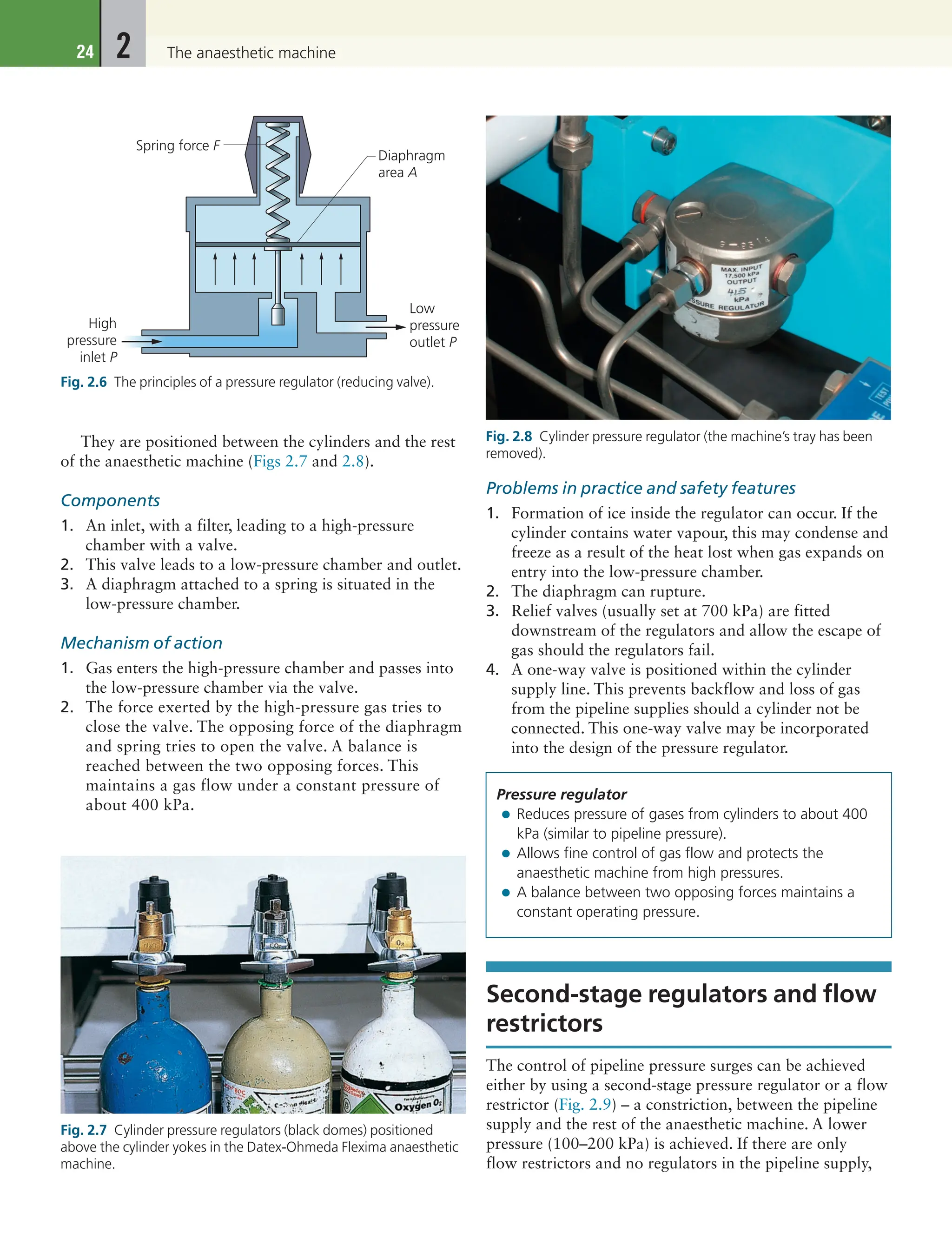
7. Isolating valves behind break-glass covers are positioned
at strategic points throughout the pipeline network.
They are also known as area valve service units (AVSUs)
(Fig. 1.16). They can be accessed to isolate the supply to
an area in cases of fire or other emergency.
Problems in practice and safety features
1. A reserve bank of cylinders is available should the
primary supply fail. Low-pressure alarms detect gas
supply failure (Fig. 1.17).
2. A single-hose test is performed to detect
cross-connection.
3. A tug test is performed to detect misconnection.
4. Regulations for PMGV installation, repair and
modification are enforced.
5. Anaesthetists are responsible for the gases supplied
from the terminal outlet through to the anaesthetic
machine. Pharmacy, supplies and engineering
departments share the responsibility for the gas
pipelines ‘behind the wall’.
6. There is a risk of fire from worn or damaged hoses
that are designed to carry gases under pressure from
Fig. 1.14 Outlet sockets mounted in a retractable ceiling unit.
(Courtesy Penlon Ltd, Abingdon, UK [http://www.penlon.com].)
Fig. 1.15 Colour-coded hoses with NIST fittings attached to an
anaesthetic machine.
Fig. 1.16 An area valve service unit (AVSU).
Fig. 1.17 Medical gas alarm panel. (Courtesy Penlon Ltd,
Abingdon, UK [http://www.penlon.com].)](https://image.slidesharecdn.com/anesthesiabooks2019essentialsofe-240617194721-1da90945/75/Anesthesia_Books-2019-Essentials-of-E-pdf-22-2048.jpg)


![11
Oxygen concentrators
The information obtained is sent to the hospital alarm
system. When required, fresh supplies of liquid oxygen
are pumped from a tanker into the vessel.
4. The cold oxygen gas is warmed once outside the vessel
in a coil of copper tubing. The increase in temperature
causes an increase in pressure.
5. At a temperature of 15°C and atmospheric pressure,
liquid oxygen can give 842 times its volume as gas.
Problems in practice and safety features
1. Reserve banks of cylinders are kept in case of supply
failure.
2. A liquid oxygen storage vessel should be housed away
from main buildings due to the fire hazard. The risk of
fire is increased in cases of liquid spillage.
3. Spillage of cryogenic liquid can cause cold burns,
frostbite and hypothermia.
Oxygen concentrators
Oxygen concentrators, also known as pressure swing
adsorption systems, extract oxygen from air by differential
adsorption. These devices may be small, designed to
supply oxygen to a single patient (Fig. 1.22), supply
oxygen to an anaesthetic machine (Fig. 1.23) or they
can be large enough to supply oxygen for a medical gas
pipeline system (Fig. 1.24).
Components
A zeolite molecular sieve is used. Zeolites are hydrated
aluminium silicates of the alkaline earth metals in a
powder or granular form in a lattice structure. Many
zeolite columns are used.
Mechanism of action (Fig. 1.25)
1. Ambient air is filtered and pressurized to about
137kPa by a compressor.
2. Air is exposed to a zeolite molecular sieve column,
forming a very large surface area, at a certain pressure.
3. The sieve selectively retains nitrogen, water vapour
and other unwanted components of air. These are
released into the atmosphere after heating the column
and applying a vacuum.
4. The sequential changeover between the columns is
made by a time switch, typically cycles of around 20
seconds, allowing for a continuous supply of oxygen.
5. The maximum oxygen concentration achieved is 95%
by volume. Argon is the main remaining constituent
with a concentration of approximately 5%. Argon is
concentrated from air in a similar fashion to oxygen.
6. The life of the zeolite crystal can be expected to be at
least 20000hours (which is about 10 years of use).
Routine maintenance consists of changing filters at
regular intervals.
Problems in practice and safety features
Although the oxygen concentration achieved is sufficient
for the vast majority of clinical applications, its use with
the circle system leads to argon accumulation. To avoid
this, higher fresh gas flows are required.
Exam tip: For the exam, make sure you understand
how the VIE works. It is very important to memorize
the relevant temperatures and pressures of liquid and
gaseous oxygen.
You also need to be able to explain what happens
when there is an overdemand or an underdemand for
the oxygen supply.
Fig. 1.22 The portable Eclipse 3 home oxygen concentrator. (Courtesy
Chart BioMedical Ltd, Wokingham, UK [www.sequal.com].)
Source of supply
• Cylinder manifold: banks of large cylinders, usually size
J, are used.
• Liquid oxygen: a thermally insulated vessel at a
temperature of −150°C to −170°C and at a pressure of
500–1000kPa is used.
• Oxygen concentrator: a zeolite molecular sieve is used.](https://image.slidesharecdn.com/anesthesiabooks2019essentialsofe-240617194721-1da90945/75/Anesthesia_Books-2019-Essentials-of-E-pdf-25-2048.jpg)
![1
12 Medical gas supply
Entonox
This is a compressed gas mixture containing 50% oxygen
and 50% nitrous oxide by volume. It is commonly
used in the A&E and labour ward and pre-hospital
care settings to provide analgesia. A two-stage pressure
demand regulator is attached to the Entonox cylinder
when in use (Fig. 1.26). As the patient inspires through
the mask or mouth piece, gas flow is allowed to occur.
Gas flow ceases at the end of an inspiratory effort.
Entonox is compressed into cylinders to a pressure of
13700 kPa. Entonox cylinders should be stored at 10°C
for 24 hours before use.
If the temperature of the Entonox cylinder is decreased
to below −5.5°C, liquefaction and separation of the two
components occur (Poynting effect). This results in:
• a liquid mixture containing mostly nitrous oxide with
about 20% oxygen dissolved in it
• above the liquid, a gas mixture of high oxygen
concentration.
This means that when used at a constant flow rate,
a gas with a high concentration of oxygen is supplied
first. This is followed by a gas of decreasing oxygen
concentration as the liquid evaporates. This may lead
to the supply of hypoxic mixtures, with less than 20%
oxygen, as the cylinder is nearly empty.
Rewarming and mixing of both the cylinder and its
contents reverses the separation and liquefaction.
Fig. 1.24 The RA40/D/M hospital oxygen concentrator. It produces
80L/min of oxygen. (Courtesy Rimer Alco Ltd, Thetford, UK
[www.i4innovation.co.uk].)
95% Oxygen
Tank
Zeolite towers
Nitrogen vent
Switch valve
Compressor
Room air
21% oxygen
To anaesthetic machine
and patient
Fig. 1.25 Mechanism of action of a concentrator.
Fig. 1.23 The universal anaesthetic machine (UAM) which has a
built-in oxygen concentrator (see Ch. 2 for more details). (Courtesy
Gradian Health Systems, New York, NY, USA [www.uamglobal.org].)](https://image.slidesharecdn.com/anesthesiabooks2019essentialsofe-240617194721-1da90945/75/Anesthesia_Books-2019-Essentials-of-E-pdf-26-2048.jpg)
![13
Problems in practice and safety features
Liquefaction and separation of the components can be
prevented by:
1. Cylinders being stored horizontally for about 24
hours at temperatures of or above 5°C before
use. The horizontal position increases the area for
diffusion. If the contents are well mixed by repeated
inversion, cylinders can be used earlier than 24 hours.
2. Large cylinders are equipped with a dip tube with
its tip ending in the liquid phase. This results in the
liquid being used first, preventing the delivery of an
oxygen concentration of less than 20%. Prolonged use
of Entonox should be avoided because of the effect
of nitrous oxide on the bone marrow especially in the
critically ill patient. Adequate facilities for scavenging
should be provided to protect hospital staff.
Compressed air
Medical air is supplied in a hospital for clinical uses or
to drive power tools. The former is supplied at a pressure
of 400kPa and the latter at 700 kPa. The anaesthetic
machines and most intensive care ventilator blenders
accept a 400kPa supply. The terminal outlets for the two
pressures are different, each with its own Schrader valve,
to prevent misconnection.
Air may be supplied from cylinder manifolds, or
more economically from a compressor plant with duty
and back-up compressors (Fig. 1.27). Each compressor
is capable to meeting the hospital’s expected demands.
The temperature of air increases as it is compressed
(Gay-Lussac’s law, also known as the third gas law). The
air is then cooled and the condensation is captured in
special traps. Oil-free medical air is cleaned by filters and
separators then dried and pressure regulated before use.
Compressor air intake should be carefully situated to
prevent contamination with regular inspection.
Centralized vacuum or suction
system (Fig. 1.28)
Suction devices play a crucial part in the care of patients
in the operating theatre, intensive care unit and other parts
of the hospital.
Components
1. A pump or a power source that is capable of
continuously generating a negative pressure of
−500mmHg.
Fig. 1.26 Entonox cylinder showing the integrated valve, Schroder
valve, hose and the patient’s demand valve. (Courtesy BOC
Healthcare, Manchester, UK [http://www.bochealthcare.co.uk].)
Entonox
• A compressed gas mixture with 50% oxygen and 50%
nitrous oxide by volume at a pressure of 13700kPa.
• A two-stage pressure demand regulator is usually used.
• Liquification and separation occurs at temperatures
below −5.5°C (Poynting effect). This can potentially
lead to hypoxic mixtures. Large cylinders are designed to
prevent this. Fig. 1.27 Compressed medical air plant. (Courtesy Penlon Ltd,
Abingdon, UK [http://www.penlon.com].)
Centralized vacuum or suction system](https://image.slidesharecdn.com/anesthesiabooks2019essentialsofe-240617194721-1da90945/75/Anesthesia_Books-2019-Essentials-of-E-pdf-27-2048.jpg)
![1
14 Medical gas supply
2. A suction controller with a filter.
3. A receiver or a collection vessel.
4. A suction tubing and suction nozzle (e.g. a Yankauer
sucker) or catheter.
Mechanism of action
1. This is a high-pressure, low-flow system (compared
with scavenging systems which are low-pressure, high-
flow; see Chapter 3).
2. Negative pressure is generated by an electric motor
and pneumatic-driven pumps using the Venturi
principle.
3. The amount of vacuum generated can be manually
adjusted by the suction controller. This device has
a variable orifice with a float assembly, a back-up
filter to prevent liquid entering the system and ports
to connect to a collection vessel or reservoir through
flexible tubing.
4. The reservoir must have sufficient capacity to receive
the aspirated material. Too large a capacity will make
the system cumbersome and will take a long time to
generate adequate negative pressure.
5. The suction tubing should be flexible and firm to
prevent collapse. Also it should be transparent so
that the contents aspirated can be visualized, and of
sufficient internal diameter and length for optimal
suction.
6. The negative pressure (or degree of suctioning) can
be adjusted to suit its use; e.g. a lesser degree of
suctioning is required to clear oral secretions in a child
than in an adult.
7. Bacterial filters are used to prevent spread of infectious
bacteria, with a removal of 99.999% of bacteria.
Filters are also used to prevent fluids, condensate and
smoke from contaminating the system.
8. It is recommended that there are at least two vacuum
outlets in each operating theatre, one per anaesthetic
room and one per recovery or intensive care unit bed.
Problems in practice and safety features
To prevent trauma to the tissues during suction, the
nozzles should taper, be smooth and have multiple
holes, so that if one is blocked the others will continue
suction.
SUGGESTED FURTHER READING
Crombie, N., 2009. Confusing and ambiguous labelling
of an oxygen cylinder. Anaesthesia 64 (1), 98.
Available from: http://onlinelibrary.wiley.com/
doi/10.1111/j.1365-2044.2008.05805.x/full.
Department of Health, 2006. Health Technical
Memorandum 02-01. Medical gas pipeline systems.
Part A: Design, Installation, Validation and
Verification. The Stationery Office, London.
Department of Health, 2006. Health Technical
Memorandum 02-01. Medical gas pipeline systems.
Part B: Operational Management. The Stationery
Office, London.
Department of Health, 2010. Unsecured medical gas
cylinders, including cylinders on trolleys. Available
from: https://www.gov.uk/government/publications/
unsecured-medical-gas-cylinders-including-cylinders-
on-trolleys.
Fig. 1.28 Medical vacuum plant. (Courtesy Penlon Ltd, Abingdon,
UK [http://www.penlon.com].)
To determine the efficiency of central-piped vacuum
systems
• A negative pressure of at least −53kPa (−400mmHg)
should be maintained at the outlet.
• Each central-piped vacuum outlet should be able to
withstand a flow of free air of at least 40L/min.
• A unit should take no longer than 10 seconds to
generate a vacuum (500mmHg) with a displacement of
air of 25L/min.
Centralized vacuum or suction system
• Consists of a power source, a suction controller, a
receiver, a suction tubing and suction nozzle.
• Efficiency of the system should be tested before use.
• The amount of negative pressure should be adjusted
according to its use.
• Trauma to tissues can be caused by the suction.](https://image.slidesharecdn.com/anesthesiabooks2019essentialsofe-240617194721-1da90945/75/Anesthesia_Books-2019-Essentials-of-E-pdf-28-2048.jpg)


![2
28 The anaesthetic machine
Vaporizers can be classified according to location:
1. Inside the breathing system. Gases pass through a very
low resistance, draw-over vaporizer due to the patient’s
respiratory efforts (e.g. Goldman, Oxford Miniature
Vaporizer [OMV]). Such vaporizers are simple in
design, lightweight, agent non-specific, i.e. allowing the
use of any volatile agent, small and inexpensive. For
these reasons, they are used in the ‘field’ or in otherwise
difficult environments. However, they are not as
efficient as the plenum vaporizers as their performance
is affected as the temperature of the anaesthetic agent
decreases due to loss of latent heat during vaporization.
2. Outside the breathing system. Gases are driven
through a plenum (high resistance, unidirectional and
agent specific) vaporizer due to gas supply pressure.
These vaporizers are reliable and easy to use.
PLENUM VAPORIZER (FIG. 2.17)
Components
1. The case with the filling level indicator and a port for
the filling device.
2. Percentage control dial on top of the case.
3. The bypass channel and the vaporization chamber. The
latter has Teflon wicks or baffles, cowls or nebulizers
to increase the surface area available for vaporization
(Fig. 2.18).
4. The splitting ratio is controlled by a temperature-sensitive
valve utilizing a bimetallic strip (Fig. 2.19). The latter is
made of two strips of metal with different coefficients
of thermal expansion bonded together. It is positioned
inside the vaporization chamber in the Tec Mk 2 whereas
in Tec Mk 3, 4 and 5, it is outside the vaporization
chamber. An ether-filled bellows is the temperature
compensating device in the M&IE Vapamasta Vaporizer
5 and 6 (Key Health Solutions Ltd, Leicestershire, UK).
The bellows contracts as the temperature of the vaporizer
decreases.
5. The vaporizers are mounted on the back bar
(Fig. 2.20) using the interlocking Selectatec system
(Fig. 2.21). The percentage control dial cannot be
moved unless the locking lever of the system is
engaged (in Mk 4 and 5). The interlocking extension
rods prevent more than one vaporizer being used at
any one time, preventing contamination of the one
Fig. 2.17 A plenum vaporizer mounted on the back bar of an
anaesthetic machine. (Courtesy Philips Healthcare, Guildford, UK.)
Bypass
Bypass
Bypass
Fresh
gas in
Dial
Fresh
and
vapour
out
Vaporizing
chamber
Flow
split
Bimetallic
strip
Temperature
compensating
valve
Outlet
port
Percentage
control valve
Fig. 2.18 A schematic diagram of the Tec Mk 5, an example of a
plenum vaporizer.
Fig. 2.19 Mechanism of action of a bimetallic strip.](https://image.slidesharecdn.com/anesthesiabooks2019essentialsofe-240617194721-1da90945/75/Anesthesia_Books-2019-Essentials-of-E-pdf-42-2048.jpg)


![2
36 The anaesthetic machine
3. A second length of tubing leading from the self-
inflating bag to two OMVs.
4. An oxygen cylinder can be connected upstream of the
vaporizers. A third length of tubing acts as an oxygen
reservoir during expiration.
Mechanism of action
1. The Triservice apparatus can be used for both
spontaneous and controlled ventilation.
a. The patient can draw air through the vaporizers.
The exhaled gases are vented out via the non-
rebreathing valve.
b. The self-inflating bag can be used for controlled or
assisted ventilation.
2. The OMV is a variable bypass, low resistance draw-
over vaporizer with a capacity for 50mL of anaesthetic
agent. The wick is made of metal with no temperature
compensation features. However there is an ethylene
glycol jacket acting as a heat sink to help to stabilize
the vaporizer temperature. The calibration scale on the
Oxygen
concentrator
Oxygen analyser
and apnoea alarm
NPRV
air inlet
REGS
CYL/pipeline
pin-index yoke
+ N20 option
O2/N2O/air
flow control
O2 inlet
Emergency oxygen
inlet on the back
of the UAM
2 Litre reservoir
bag on back bar
Oxygen analyser
sampling port
Silicone
bellows
Bellows
block +
2 valves
FGF inlet
Y-piece outlet
Balloon
occluder
Balloon actuator
pipe
Scavenger
EXP
INSP
To patient
DOV for
HAL/ISO
PPRV
Fig. 2.35 Diagrammatic representation of UAM. (Courtesy Gradian Health Systems, New York, NY, USA [www.uamglobal.org].)
Fig. 2.36 The Triservice apparatus.
Inflating valve Inlet valve
0.1
Patient
Contents
gauge
Oxygen
Pressure
reducing
valve
Reservoir
Inflating bag
Vaporizers
1 L/min. 4 L/min.
Fig. 2.37 Mechanism of action of the Triservice apparatus.](https://image.slidesharecdn.com/anesthesiabooks2019essentialsofe-240617194721-1da90945/75/Anesthesia_Books-2019-Essentials-of-E-pdf-50-2048.jpg)


![47
Charcoal canisters (Cardiff Aldasorber)
receiving and disposal components. A bacterial filter
situated downstream and a visual flow indicator
positioned between the receiving and disposal
systems can be used. A reservoir bag with two
spring-loaded safety valves can also be used as a
receiving system.
3. The active disposal system consists of a fan or a pump
used to generate a vacuum (Fig. 3.5).
Mechanism of action
1. The vacuum drives the gases through the system.
Active scavenging systems are able to deal with a wide
range of expiratory flow rates (30–130L/min).
2. A motorized fan, a pump or a Venturi system is used
to generate the vacuum or negative pressure that is
transmitted through pipes.
3. The receiving system is capable of coping with changes
in gas flow rates. Increased demands (or excessive
negative pressure) allows ambient air to be entrained
thereby maintaining the pressure. The opposite occurs
during excessive positive pressure. As a result, a
uniform gas flow is passed to the disposal system.
Problems in practice and safety features
1. The reservoir is designed to prevent excessive negative
or positive pressures being applied to the patient.
Excessive negative pressure leads to the collapse of
the reservoir bag of the breathing system and the risk
of rebreathing. Excessive positive pressure increases
the risk of barotrauma should an obstruction occur
beyond the receiving system.
2. An independent vacuum pump should be used for
scavenging purposes.
3. More recently, the ISO standard 80601-2-13 Anaesthetic
Workstation, states ‘under single fault conditions, the
pressure at the inlet to the anaesthetic gas scavenging
system shall not exceed 20cm H2O with an exhaust flow
rate of 75L/min’. A safety valve (Fig. 3.6) prevents the
build-up of pressure in the breathing system in case the
transfer system/tubing is occluded causing back pressure
to the patient. Gases are released into the atmosphere.
Charcoal canisters (Cardiff
Aldasorber)
The canister is a compact passive scavenging system
(Fig. 3.7).
Danger
415 volts
Fig. 3.5 Anaesthetic gases scavenging system (AGSS) vacuum
pumps used in an active scavenging system. (Courtesy Penlon Ltd,
Abingdon, UK [http://www.penlon.com].)
Scavenging
• Active or passive systems are incorporated to collect gas
waste.
• This method consists of a collecting and transfer system,
a receiving system and a disposal system.
• Both excessive positive and negative pressure variations
in the system are limited.
• Other methods are used to reduce theatre pollution:
theatre ventilation, circle system, total intravenous and
regional anaesthesia.
Exam tip: All the safety features in both the passive
and active scavenging systems need to be understood.
How is the patient protected against excessive positive
or negative pressures within the breathing system?
Fig. 3.6 Anaesthesia gas scavenging pressure relief valve. (Courtesy
Intersurgical, Wokingham, UK.)](https://image.slidesharecdn.com/anesthesiabooks2019essentialsofe-240617194721-1da90945/75/Anesthesia_Books-2019-Essentials-of-E-pdf-61-2048.jpg)


![4
58 Breathing systems
Mechanism of action
1. It has a similar mechanism to the Magill system except
the Lack system is a coaxial version. The fresh gas
flows through the outside tube whereas the exhaled
gases flow through the inside tube.
2. A FGF rate of about 70mL/kg/min is required in
order to prevent rebreathing. This makes it an efficient
breathing system for spontaneous ventilation.
3. Since it is based on the Magill system, it is not suitable
for controlled ventilation.
Instead of the coaxial design, a parallel tubing
version of the system exists (Fig. 4.7B). This has separate
inspiratory and expiratory tubing, and retains the same
flow characteristics as the coaxial version.
Mapleson B and C systems
(Figs 4.1 and 4.8)
Components
1. A reservoir bag. In the B system, corrugated tubing is
attached to the bag and both act as a reservoir.
2. An APL valve at the patient’s end.
3. FGF is added just proximal to the APL.
Mechanism of action
Both systems are not efficient during spontaneous
ventilation. An FGF of more than 1.5–2 times the minute
volume is required to prevent rebreathing.
During controlled ventilation, the B system is more
efficient due to the corrugated tubing acting as a reservoir.
An FGF of more than 50% of the minute ventilation is
still required to prevent rebreathing.
Mapleson C (also known as the Waters circuit) is
lightweight and compact. It is used in resuscitation
situations as an alternative to self-inflating bag. By
adjusting the APL valve, it allows positive end-expiratory
pressure (PEEP) with a visual and tactile ventilation
monitor.
Bain system (Mapleson D)
The Bain system is a coaxial version of the Mapleson D
system (Fig. 4.9). It is lightweight and compact at the
patient end. It is useful where access to the patient is
limited, such as during head and neck surgery.
The Manley ventilator, which has been switched to a
spontaneous ventilation mode, is an example of a non-
coaxial Mapleson D system.
Components
1. A length of coaxial tubing (tube inside a tube). The
usual length is 180cm, but it can be supplied at 270cm
(for dental or ophthalmic surgery) and 540cm (for
magnetic resonance imaging [MRI] scans where the
anaesthetic machine needs to be kept outside the
scanner’s magnetic field). Increasing the length of the
tubing does not affect the physical properties of the
breathing system.
2. The fresh gas flows through the narrow inner tube
(6mm) while the exhaled gases flow through the
outside tube (22mm) (Fig. 4.10). The internal lumen
Lack breathing system
• Coaxial version of Mapleson A, making it efficient for
spontaneous ventilation. FGF rate of about 70mL/kg/
min is required.
• FGF is delivered along the outside tube and the exhaled
gases flow along the inner tube.
• APL valve is at the machine end.
• Not suitable for controlled ventilation.
Fig. 4.8 Intersurgical adult Mapleson C system.
Mapleson B and C breathing systems
• B system has a tubing and bag reservoir.
• Both B and C systems are not efficient for spontaneous
and controlled ventilation.
• B system is more efficient than the A system during
controlled ventilation.
• C system is lightweight and used in resuscitation
situation.
Fig. 4.9 The Bain breathing system.](https://image.slidesharecdn.com/anesthesiabooks2019essentialsofe-240617194721-1da90945/75/Anesthesia_Books-2019-Essentials-of-E-pdf-72-2048.jpg)


![63
The circle breathing system and soda lime
Components
1. A vertically positioned canister contains soda lime. The
canister has two ports, one to deliver inspired gases to
the patient and the other to receive exhaled gases from
the patient.
2. Inspiratory and expiratory tubings are connected to
the canister. Each port incorporates a unidirectional
valve. Corrugated tubings are used to prevent
kinking.
3. FGF from the anaesthetic machine is positioned
distal to the soda lime canister, but proximal to the
inspiratory valve; i.e. on the inspiratory limb.
4. An APL valve is positioned between the expiratory
valve and canister; i.e. on the expiratory limb. It is
connected to a 2-L reservoir bag.
5. A vaporizer is mounted on the anaesthetic machine
back bar (vaporizer outside circle [VOC]) or a
vaporizer positioned on the expiratory limb within the
system (vaporizer inside circle [VIC]).
6. Soda lime consists of 94% calcium hydroxide and
5% sodium hydroxide with a small amount of
potassium hydroxide (less than 0.1%). It has a pH
of 13.5 and a moisture content of 14–19%. Some
modern types of soda lime have no potassium
hydroxide. Soda lime granules are prone to powder
formation, especially during transport. Disintegrated
granules increase resistance to breathing. Because of
this, silica (0.2%) is added to harden the absorbents
and reduce powder formation. A dye or colour
indicator is added to change the granules’ colour
when the soda lime is exhausted. Colour changes
can be from white to violet/purple (ethyl violet
dye), from pink to white (titan yellow dye) or from
green to violet. Colour changes occur when the pH
is less than 10. Newer types of soda lime have a
low concentration of a zeolite added. This helps to
maintain the pH at a high level for longer and retains
moisture so improving carbon dioxide absorption
and reducing the formation of carbon monoxide and
compound A.
7. The size of soda lime granules is 4–8 mesh.
Strainers with 4–8 mesh have four and eight
openings/in, respectively. Therefore, the higher
the mesh number, the smaller the particles are.
Recently produced soda lime made to a uniform
shape of 3–4-mm spheres allows a more even flow
of gases and a reduction in channelling. This results
in a longer life with lower dust content and lower
resistance to flow: 1 kg can absorb more than 120 L
of CO2.
8. Barylime, which consists of barium hydroxide (80%)
and calcium hydroxide (20%), is widely used in
the United States. Another absorber is Amsorb that
consists of CaCl2 and Ca(OH)2.
Mechanism of action
1. High FGF of several L/min is needed in the initial
period to denitrogenate the circle system and the
FRC. This is important to avoid the build-up of
unacceptable levels of nitrogen in the system. In closed
circle anaesthesia, a high FGF is needed for up to 15
minutes. In low-flow anaesthesia, a high FGF of up to
6 minutes is required. The FGF can be later reduced
to 0.5–1L/min. If no N2O is used during anaesthesia
(i.e. an oxygen/air mix is used), it is not necessary to
eliminate nitrogen because air contains nitrogen. A
short period of high flow is needed to prime the system
and the patient with the inhalational agent.
2. Exhaled gases are circled back to the canister, where
carbon dioxide absorption takes place and water and
heat (exothermic reaction) are produced. The warmed
and humidified gas joins the FGF to be delivered to the
patient (Fig. 4.19).
3. Chemical sequences for the absorption of carbon
dioxide by soda lime:
a. Note how both NaOH and KOH are regenerated at
the expense of Ca(OH)2. This explains soda lime’s
mix – only a little Na(OH) and K(OH) and a lot of
Ca(OH)2:
H O CO H CO
2 2 2 3
+ ®
then
H CO KOH K CO H O
2 3 2 3 2
2 2
+ ® +
then
K CO Ca OH CaCO KOH
2 3 2 3 2
+ ( ) ® +
or
CO NaOH Na CO H O heat
2 2 3 2
2
+ ® + +
then
Na CO Ca OH NaOH CaCO
2 3 2 3
2
+ ( ) ® +
Connection to bag
in bottle ventilator
Soda lime
canister
Unidirectional
valve
Unidirectional
valve
Sampled gas from monitor
returned to circle
APL
Reservoir bag
To patient
FGF
Vent/bag
switch
From patient
Fig. 4.19 Mechanism of action of the circle breathing system. APL,
Adjustable pressure limiting; FGF, fresh gas flow.](https://image.slidesharecdn.com/anesthesiabooks2019essentialsofe-240617194721-1da90945/75/Anesthesia_Books-2019-Essentials-of-E-pdf-77-2048.jpg)


![5
88 Tracheal tubes, tracheostomy tubes and airways
Components
1. The rounded curved body of the nasopharyngeal
airway.
2. The bevel is left-facing.
3. The proximal end has a flange. A ‘safety pin’ is
provided to prevent the airway from migrating into
the nose.
Mechanism of action
1. It is an alternative to the oropharyngeal airway when
the mouth cannot be opened or an oral airway does
not relieve the obstruction.
2. Nasotracheal suction can be performed using a
catheter passed through the nasal airway.
3. It is better tolerated by semi-awake patients than the
oral airway.
4. A lubricant is used to help in its insertion.
5. The size inserted can be estimated as size 6 for an average
height female and size 7 for an average height male.
6. Once lubricated, it can be inserted through either
nares, although the left-facing bevel is designed to ease
insertion into the right nostril. On insertion, it should
be passed backwards through the nasopharynx, such
that its distal end lies beyond the pharyngeal border of
the soft palate but not beyond the epiglottis.
Problems in practice and safety features
1. Its use is not recommended when the patient has
a bleeding disorder, is on anticoagulants, has nasal
deformities or sepsis.
2. Excess force should not be used during insertion as a
false passage may be created. There is a potential risk of
intracranial placement in cases of basal skull fracture.
3. An airway that is too large can result in pressure
necrosis of the nasal mucosa, whereas an airway that
is too small may be ineffective at relieving airway
obstruction.
Supraglottic airway devices
The introduction of the laryngeal mask airway heralded
an era of hands-free airway maintenance without the need
for tracheal intubation. Many other airway devices (well
over 100 designs) that lie outside the trachea and attempt
to provide a leak-free seal for spontaneous ventilation,
while some provide an adequate seal for positive pressure
ventilation under normal conditions, have been used. These
devices are collectively known as supraglottic airways
devices (SADs). Worldwide, millions of such devices are
used each year. SADs can be divided into:
• First-generation SADs
These fit the description ‘simple airway devices’,
including the original LMA Classic with only one
ventilation channel. They may or may not protect
against aspiration in the event of regurgitation, but have
no specific design features that lessen this risk.
• Second-generation SADs
SADs that have design features to reduce the risk of
aspiration with a separation of ventilatory and gastric
access channels; e.g. LMA ProSeal (Teleflex, Dublin,
Ireland), LMA Supreme (Teleflex, Dublin, Ireland),
AuraGain (Ambu, St Ives, Cambridgeshire, UK) and i-gel
(Intersurgical, Wokingham, UK) (see later). Some designs
have a ‘bite block’ to prevent the patient biting the tube
and occluding it during emergence from anaesthesia.
SADs, in general, provide the following:
1. The ability to be placed without direct visualization
of the larynx.
2. Increased speed and ease of placement when
compared with tracheal intubation, both by
experienced and less experienced operators.
3. Increased cardiovascular stability on insertion and
emergence.
4. During emergence, improved oxygen saturation and
lower frequency of coughing.
5. Minimal rise in intraocular pressure on insertion.
6. When the device is properly placed, it can act as
a conduit for oral tracheal intubation due to the
anatomical alignment of its aperture with the glottic
opening.
7. In the ‘can’t intubate, can’t ventilate’ scenario, the
decision to use such devices should be made early
to gain time while attempts are made to secure a
definite airway.
8. First-generation SADs normally provide little or
no protection against aspiration of refluxed gastric
contents, and are therefore contraindicated in patients
with full stomachs or prone to reflux. However,
second-generation devices (e.g. LMA ProSeal, LMA
Supreme, AuraGain and i-gel [see later]) offer many
improvements such as high cuff seal, second seal,
gastric access and drain tube. These allow for rapid
drainage of gastric fluids or secretions and reduce
the risk of gastric gas insufflation during ventilation.
Future indications might even be in emergency
medicine, where gastric vacuity is unknown, and in
cases of increased risk of regurgitation.
Nasopharyngeal airway
• Inserted through the nose into the nasopharynx.
• A useful alternative to the oropharyngeal airway.
• Not recommended in coagulopathy, nasal sepsis and
deformities.](https://image.slidesharecdn.com/anesthesiabooks2019essentialsofe-240617194721-1da90945/75/Anesthesia_Books-2019-Essentials-of-E-pdf-102-2048.jpg)


![125
Characteristics of the ideal ventilator
to this pressure gradient as well as the elastic recoil
of the lungs and chest wall.
b. Negative pressure:
i. Tank ventilator or ‘iron lung’: gas is pumped
out of the airtight tank to generate a vacuum
around the body, decreasing intrapulmonary
pressure and leading to chest expansion. As the
vacuum is released, elastic recoil of the chest
leads to expiration.
ii. Cuirass ventilator: an upper body shell or
cuirass generates negative pressure only around
the chest. This technique has been improved by
the ability to generate two pressures, biphasic
cuirass ventilation. This allows control of
expiration as well as inspiration, and therefore
the I:E ratio and respiratory rate.
3. Control system:
a. Closed and open loops systems: in closed loop
systems, microprocessors allow feedback loops
between the control variable (such as tidal volume)
as set by the operator and the measured control
variable (exhaled tidal volume). If the two differ,
for example due to a leak, the ventilator can adjust
to achieve the desired expired tidal volume by
increasing the volume delivered. Open loop systems
deliver ventilation as set by the operator but do not
measure or adjust.
b. Pneumatic circuit: the means of delivering gas flow
from a high-pressure gas source to the patient can
be either:
i. Internal
• Single: the gas from the high pressure source
flows directly to the patient (e.g. modern
intensive care unit [ICU] ventilators)
• Double: the power source causes gas flow to
compress a chamber such as bellows or ‘bag-
in-a-chamber’. The gas in the chamber is then
delivered to the patient.
ii. External: tubing goes from the ventilator to the
patient.
4. Suitability for use in theatre and/or intensive care.
5. Suitability for paediatric practice.
6. Drive mechanism: the internal hardware that converts
electrical power or gas pressure into a breath to the
patient.
a. Flow devices: compressors, driven by pistons, rotating
blades, diaphragms or bellows, move atmospheric
pressure gas into a higher pressure storage chamber
which is then delivered as a breath. Blowers generate
high flows of gas as the direct ventilator output.
b. Volume displacement devices: the volume of gas to
be delivered to the patient is displaced by a moving
part such as a piston or spring loaded bellows.
7. Output control mechanism: valves regulate gas flow to
and from the patient.
a. Proportional solenoid valve: opens in very small
increments dependent upon flow required.
b. Digital on/off valves: a collection of valves, whereby
each one is either fully open or closed. Each valve
produces a certain flow by controlling the opening/
closing of a specifically sized orifice.
Characteristics of the ideal
ventilator
1. The ventilator should be simple, portable, robust and
economical to purchase and use. If compressed gas is
used to drive the ventilator, a significant wastage of
the compressed gas is expected. Some ventilators use
a Venturi to drive the bellows, to reduce the use of
compressed oxygen.
2. It should be versatile and supply tidal volumes up
to 1500mL with a respiratory rate of up to 60
breaths/min and variable I:E ratio. It can be used
Power source Electrically powered
Pneumatically powered
Pneumatically powered
microprocessor-controlled
Pressure generation Positive pressure
Negative pressure
Control system Open and closed loops
systems
Pneumatic circuit:
Internal: single or double
External
Suitability for use Operating theatre
Intensive care unit
Both
Paediatric practice Yes/no
Drive mechanisms Flow devices
Volume displacement devices
Output control
mechanism
Proportional solenoid valve
Digital on/off valves
Table 8.1 Summary of the methods used in
classifying ventilators
Exam tip: A basic understanding of the classification
of ventilators is important.](https://image.slidesharecdn.com/anesthesiabooks2019essentialsofe-240617194721-1da90945/75/Anesthesia_Books-2019-Essentials-of-E-pdf-139-2048.jpg)

![127
Bag in bottle ventilator
used with different breathing systems (Fig. 8.2). It is a
volume-preset, time-cycled, flow generator in adult use.
In paediatric use, it is a pressure-preset, time-cycled, flow
generator.
Components
1. The control module consists of an airway pressure
gauge (cm H2O), inspiratory and expiratory time dials
(seconds), inspiratory flow rate dial (L/s) and an on/
off switch. Underneath the control module there are
connections for the driving gas supply and the valve
block. Tubing connects the valve block to the airway
pressure gauge.
2. The valve block has three ports:
a. a port for tubing to connect to the breathing system
reservoir bag mount
b. an exhaust port which can be connected to the
scavenging system
c. a pressure relief valve which opens at 60cm H2O.
3. The valve block can be changed to a paediatric
(Newton) valve.
Mechanism of action
1. The ventilator is powered by a driving gas
independent from the FGF. The commonly used
driving gas is oxygen (at about 400 kPa) supplied
from the compressed oxygen outlets on the
anaesthetic machine. The driving gas should not reach
the patient as it dilutes the FGF, lightening the depth
of anaesthesia.
2. It can be used with different breathing systems such
as Bain, Humphrey ADE, T-piece and the circle. In the
Bain and circle systems, the reservoir bag is replaced
by the tubing delivering the driving gas from the
ventilator. The APL valve of the breathing system must
be fully closed during ventilation.
3. The inspiratory and expiratory times can be adjusted
to the desired I:E ratio. The tidal volume is determined
by adjusting the inspiratory time and inspiratory flow
rate controls. The inflation pressure is adjusted by the
inspiratory flow rate control.
4. With its standard valve, the ventilator acts as a
time-cycled flow generator to deliver a minimal tidal
volume of 50 mL. When the valve is changed to a
paediatric (Newton) valve, the ventilator changes
to a time-cycled pressure generator capable of
delivering tidal volumes between 10 and 300 mL.
This makes it capable of ventilating premature
babies and neonates. It is recommended that the
Newton valve is used for children of less than 20 kg
body weight.
5. A PEEP valve may be fitted to the exhaust port.
Problems in practice and safety features
1. The ventilator continues to cycle despite breathing
system disconnection without an alarm.
2. Requires high flows of driving gas.
Bag in bottle ventilator
Modern anaesthetic machines often incorporate a bag in
bottle ventilator.
Components
1. A driving unit consisting of:
a. a chamber (Fig. 8.3) with a tidal volume range of
0–1500mL (a paediatric version with a range of
0–400mL exists)
b. an ascending bellows accommodating the FGF.
2. A control unit with a variety of controls, displays
and alarms: the tidal volume, respiratory rate (6–40
breaths/min), I:E ratio, airway pressure and power
supply (Figs 8.3 and 8.4).
Mechanism of action
1. It is a time-cycled ventilator that is pneumatically
powered and electronically controlled.
2. The fresh gas is accommodated in the bellows.
Fig. 8.2 The Penlon Nuffield 200 ventilator. (Courtesy Penlon Ltd,
Abingdon, UK [http://www.penlon.com].)
Penlon Nuffield Anaesthesia Ventilator Series 200
• An intermittent blower with a pressure gauge,
inspiratory and expiratory time and flow controls.
• Powered by a driving gas.
• Can be used for both adults and paediatric patients.
• Can be used with different breathing systems.](https://image.slidesharecdn.com/anesthesiabooks2019essentialsofe-240617194721-1da90945/75/Anesthesia_Books-2019-Essentials-of-E-pdf-141-2048.jpg)
![8
128 Ventilators
3. Compressed air is used as the driving gas (Fig. 8.5).
On entering the chamber, the compressed air forces
the bellows down, delivering the fresh gas to the
patient.
4. The driving gas and the fresh gas remain separate.
5. The volume of the driving gas reaching the chamber is
equal to the tidal volume.
6. Some designs feature a descending bellows instead.
Problems in practice and safety features
1. Positive pressure in the standing bellows causes a PEEP
of 2–4cm H2O.
2. The ascending bellows collapses to an empty
position and remains stationary in cases of
disconnection or leak.
3. The descending bellows hangs down to a fully
expanded position in a case of disconnection and
may continue to move almost normally in a case of
leakage.
SERVO-U ventilator
The SERVO-U is a versatile intensive care ventilator,
capable of being used for paediatric and adult patients. It
is fully transportable, utilizing at least two 12V batteries
when mains electricity is not available. It is not intended
for use with inhalational anaesthetics; however, it can be
used with intravenous anaesthetics in the theatre setting
if required. It can also be used to ventilate patients with
a tight-fitting nasal mask or prong, face mask or hood,
instead of a standard endotracheal tube or tracheostomy.
This is termed non-invasive ventilation (NIV). Leaks are
compensated for during NIV.
The most modern versions have advanced tools to
safely perform lung recruitment utilizing software that
regulates PEEP and aims to maintain lung compliance.
Neurally adjusted ventilatory assist (NAVA) uses a
specially adapted nasogastric tube that detects the phrenic
Fig. 8.3 Bag in bottle AV800 ventilator. (Courtesy Penlon Ltd,
Abingdon, UK [http://www.penlon.com].)
Fig. 8.4 Control panel of the Datex-Ohmeda 7900 ventilator.
Driving
gas
Fresh gas
100
200
300
400
500
600
700
800
900
1000
1100
1200
1300
1400
1500
Fig. 8.5 Mechanism of action of the bag in bottle ventilator.
Bag in bottle ventilator
• It is a time-cycled ventilator.
• Consists of driving and control units.
• Fresh gas is within the bellows whereas the driving gas
is within the chamber.
Exam tip: The bag in bottle ventilator is the most
commonly used type of ventilator in the operating
theatre. It is important to have good understanding of
its components and functions.](https://image.slidesharecdn.com/anesthesiabooks2019essentialsofe-240617194721-1da90945/75/Anesthesia_Books-2019-Essentials-of-E-pdf-142-2048.jpg)


![133
Self-inflating bag and mask
Components
1. A high-pressure oxygen source functions at about
400kPa (from the anaesthetic machine or direct from
a pipeline).
2. It has an on/off trigger.
3. Connection tubing can withstand high pressures.
4. A needle is of suitable gauge, which allows good air
entrainment without creating excessive airway pressures.
Mechanism of action
1. The high-pressure oxygen is injected intermittently
through the needle placed at the proximal end of the
bronchoscope or a cricothyroid needle.
2. This creates a Venturi effect, entraining atmospheric
air and inflating the lungs with oxygen-enriched air.
3. Oxygenation and carbon dioxide elimination are
achieved with airway pressures of 25–30cm H2O.
Problems in practice and safety features
1. Barotrauma is possible. Airway pressure monitoring is
not available. As exhalation is passive, it is essential to
allow full exhalation before delivering the next breath
to avoid air stacking and barotrauma.
2. Gastric distension can occur should ventilation
commence before the distal end of the bronchoscope is
beyond the larynx.
Self-inflating bag and mask
This is a means of providing manual IPPV. It is portable
and is used during resuscitation, transport and short-term
ventilation (Fig. 8.13). It can be used with or without a
pressurized gas supply.
Components
1. Self-inflating bag with a connection for added oxygen.
The bag is usually made of clear silicone.
2. A one-way valve with three ports:
a. inspiratory inlet allowing the entry of fresh gas
during inspiration
b. expiratory outlet allowing the exit of exhaled gas
c. connection to the face mask or tracheal tube, and
marked ‘patient’.
3. A reservoir for oxygen to increase the FiO2 delivered
to the patient.
Mechanism of action
1. The non-rebreathing valve (Ambu valve; artificial
manual breathing unit) incorporates a silicone
rubber membrane (Fig. 8.14). It has a small dead
space and low resistance to flow. At a flow of 25 L/
min, an inspiratory resistance of 0.4 cm H2O and an
expiratory resistance of 0.6 cm H2O are achieved.
The valve can easily be dismantled for cleaning and
sterilization.
2. The valve acts as a spill-over valve allowing
excess inspiratory gas to be channelled
directly to the expiratory outlet, bypassing
the patient port.
3. The valve is suitable for both IPPV and spontaneous
ventilation.
Venturi injector device
• Manually controlled Venturi used during rigid
bronchoscopy and rescue ventilation.
• High-pressure oxygen injected through a needle
entraining air.
Fig. 8.13 A range of self-inflating resuscitation bags with oxygen
reservoirs.
Valve housing
Inspiratory
connector
Expiratory
connector Patient connector
Valve
membrane
Fig. 8.14 An Ambu valve disassembled. (Courtesy Ambu
International [UK] Ltd.)](https://image.slidesharecdn.com/anesthesiabooks2019essentialsofe-240617194721-1da90945/75/Anesthesia_Books-2019-Essentials-of-E-pdf-147-2048.jpg)


![10
154 Non-invasive monitoring
Mechanism of action
1. The oxygen saturation is estimated by measuring the
transmission of light, through a pulsatile vascular
tissue bed (e.g. finger). This is based on Beer’s law
(the relation between the light absorbed and the
concentration of solute in the solution) and Lambert’s
law (relation between absorption of light and the
thickness of the absorbing layer).
2. The amount of light transmitted depends on many
factors. The light absorbed by non-pulsatile tissues (e.g.
skin, soft tissues, bone and venous blood) is constant
(direct current [DC]). The non-constant absorption (AC)
is the result of arterial blood pulsations (Fig. 10.14). The
sensitive photodetector generates a voltage proportional
to the transmitted light. The AC component of the wave
is about 1–5% of the total signal.
3. The high frequency of the LEDs allows the absorption
to be sampled many times during each pulse beat. This
is used to enable running averages of saturation to be
calculated many times per second. This decreases the
‘noise’ (e.g. movement) effect on the signal.
4. The microprocessor is programmed to mathematically
analyse both the DC and AC components at 660 and
940nm calculating the ratio of absorption at these two
frequencies (R:IR ratio). The result is related to the
arterial saturation. The absorption of oxyhaemoglobin
and deoxyhaemoglobin at these two wavelengths is
very different. This allows these two wavelengths to
provide good sensitivity. 805nm is one of the isobestic
points of oxyhaemoglobin and deoxyhaemoglobin.
The OFF part allows a baseline measurement for any
changes in ambient light.
5. A more recent design uses multiple wavelengths to
eradicate false readings from carboxyhaemoglobin
and methaemoglobinaemia. Advanced oximeters use
more than seven light wavelengths. This has enabled
the measurement of haemoglobin value, oxygen
content, carboxyhaemoglobin and methaemoglobin
concentrations.
6. A variable pitch beep provides an audible signal of
changes in saturation.
Problems in practice and safety features
1. It is accurate (±2%) in the 70–100% range. Below
the saturation of 70%, readings are extrapolated.
2. The absolute measurement of oxygen saturation may
vary from one probe to another but with accurate
trends. This is due to the variability of the centre
wavelength of the LEDs.
3. Carbon monoxide poisoning (including smoking),
coloured nail varnish, intravenous injections of certain
dyes (e.g. methylene blue, indocyanine green) and drugs
responsible for the production of methaemoglobinaemia
are all sources of error (Table 10.3).
Display
Photodetector
LED sequence
Microprocessor
Sequence repeated many times per second
On Off
Off On
Off Off
940
nm
660
nm
Fig. 10.13 Working principles of the pulse oximeter. The LEDs
operate in sequence and when both are off the photodetector
measures the background level of ambient light.
Arterial
Venous
Skin
Tissue
Bone
Time
Absorbance
Fig. 10.14 Schematic representation of the contribution of various
body components to the absorbance of light.
HbF No significant clinical change
(absorption spectrum is similar
to the adult Hb over the range of
wavelengths used)
MetHb False low reading
CoHb False high reading
SulphHb Not a clinical problem
Bilirubin Not a clinical problem
Dark skin No effect
Methylene blue False low reading
Indocyanine
green
False low reading
Nail varnish May cause false low reading
Table 10.3 Sources of error in pulse oximetry](https://image.slidesharecdn.com/anesthesiabooks2019essentialsofe-240617194721-1da90945/75/Anesthesia_Books-2019-Essentials-of-E-pdf-168-2048.jpg)

![10
156 Non-invasive monitoring
less than arterial CO2. This difference is reduced if
the lungs are ventilated with large tidal volumes. The
Greek root kapnos, meaning ‘smoke’, give us the term
capnography (CO2 can be thought as the ‘smoke’ of
cellular metabolism).
End tidalCO alveolarCO aCO
-
2 2 2
P
In reality, the devices used cannot determine the
different phases of respiration but simply report the
minimum and maximum CO2 concentrations during each
respiratory cycle.
Components
1. The sampling chamber can either be positioned
within the patient’s gas stream (main-stream version,
Fig. 10.16) or connected to the distal end of the
breathing system via a sampling tube (side-stream
version, Fig. 10.17).
2. A photodetector measures light reaching it from a light
source at the correct infrared wavelength (using optical
filters) after passing through two chambers. One acts
as a reference whereas the other one is the sampling
chamber (Fig. 10.18).
Mechanism of action
1. Carbon dioxide absorbs the infrared radiation
particularly at a wavelength of 4.3μm.
2. The amount of infrared radiation absorbed is
proportional to the number of carbon dioxide
molecules (partial pressure of carbon dioxide) present
in the chamber.
3. The remaining infrared radiation falls on the
thermopile detector, which in turn produces heat.
The heat is measured by a temperature sensor and is
proportional to the partial pressure of carbon dioxide
gas present in the mixture in the sample chamber. This
produces an electrical output. This means that the
amount of gas present is inversely proportional to the
amount of infrared light present at the detector in the
sample chamber (Fig 10.19).
4. In the same way, a beam of light passes through the
reference chamber which contains room air. The
absorption detected from the sample chamber is
compared to that in the reference chamber. This allows
the calculation of carbon dioxide values.
5. The inspired and exhaled carbon dioxide forms a
square wave, with a zero baseline unless there is
rebreathing (Fig. 10.20A).
6. A microprocessor-controlled infrared lamp is
used. This produces a stable infrared source with
Fig. 10.16 A main-stream end-tidal carbon dioxide analyser.
Fig. 10.17 The Penlon PM9000 Express which measures end-tidal
CO2, oximetry and inhalational agent concentration using a side-
stream method. (Courtesy Penlon Ltd, Abingdon, UK [http://www.
penlon.com].)
Sample gas
Sample chamber Multigas filter
Light source
Detector
Fig. 10.18 Components of a gas analyser using an infrared light
source suitable for end-tidal carbon dioxide measurement. The
reference chamber has been omitted for the sake of clarity.](https://image.slidesharecdn.com/anesthesiabooks2019essentialsofe-240617194721-1da90945/75/Anesthesia_Books-2019-Essentials-of-E-pdf-170-2048.jpg)


![167
Peripheral nerve stimulators and neuromuscular blockade monitoring
Peripheral nerve stimulators
and neuromuscular blockade
monitoring
These devices are used to monitor transmission across
the neuromuscular junction. The depth, adequate reversal
and type of neuromuscular blockade can be established
(Fig. 10.36).
Components
1. Two surface electrodes (small ECG electrodes) are
positioned over the nerve and connected via the leads
to the nerve stimulator.
2. Alternatively skin contact can be made via ball electrodes
which are mounted on the nerve stimulator casing.
3. The case consists of an on/off switch, facility to deliver
a twitch, train-of-four (at 2Hz) and tetanus (50Hz).
The stimulator is battery operated.
Mechanism of action
1. A supramaximal stimulus is used to stimulate a
peripheral nerve. This ensures that all the motor fibres
of the nerve are depolarized. The response of the
muscle(s) supplied by the nerve is observed. A current
of 15–40mA is used for the ulnar nerve (a current of
50–60mA may have to be used in obese patients).
2. This device should be battery powered and capable
of delivering a constant current. It is the current
magnitude that determines whether the nerve
depolarizes or not, so delivering a constant current is
more important than delivering a constant voltage as
the skin resistance is variable (Ohm’s law).
3. The muscle contraction can be observed visually, palpated,
measured using a force transducer, or the electrical activity
can be measured (electromyogram [EMG]).
4. The duration of the stimulus is less than 0.2–0.3ms.
The stimulus should have a monophasic square wave
shape to avoid repetitive nerve firing.
5. Superficial, accessible peripheral nerves are most
commonly used for monitoring purposes, e.g. ulnar
nerve at the wrist, common peroneal nerve at the neck
of the fibula, posterior tibial nerve at the ankle and the
facial nerve.
6. The negative electrode is positioned directly over
the most superficial part of the nerve. The positive
electrode is positioned along the proximal course of
the nerve to avoid direct muscle stimulation.
7. Consider the ulnar nerve at the wrist. Two electrodes
are positioned over the nerve, with the negative
electrode placed distally and the positive electrode
positioned about 2cm proximally. Successful ulnar
nerve stimulation causes the contraction of the
adductor pollicis brevis muscle.
More advanced devices offer continuous monitoring
of the transmission across the neuromuscular junction
(Fig. 10.37). A graphical and numerical display of the
train-of-four (Fig. 10.38) and the trend provide optimal
monitoring. Skin electrodes are used. A reference
Fig. 10.36 The RS7 peripheral nerve stimulator. (Courtesy Blue Box
Medical Ltd/G Rutter Ltd, Southampton, UK.) Fig. 10.37 Continuous neuromuscular transmission.](https://image.slidesharecdn.com/anesthesiabooks2019essentialsofe-240617194721-1da90945/75/Anesthesia_Books-2019-Essentials-of-E-pdf-181-2048.jpg)


![10
172 Non-invasive monitoring
to a very ‘deep’ level of anaesthesia and values close to
100 correspond to the awake patient. Like BIS, values
between 40 and 60 represent clinically desirable depths of
anaesthesia. At this level, the SE and RE indexes should be
similar if not identical.
As the patient awakens, an increase in the difference
between the SE and RE values is seen due to a diminishing
effect of drugs on the central nervous system (CNS) and
an increasing contribution from frontalis EMG.
SUGGESTED FURTHER READING
Kodali, B.S., 2013. Capnography outside the operating
rooms. Anesthesiology 118 (1), 192–201. Available
from: http://anesthesiology.pubs.asahq.org/article.
aspx?articleid=2034665resultClick=3.
McGrath, C.D., Hunter, J.M., 2006. Monitoring of
neuromuscular block. Contin Educ Anaesth Crit
Care Pain 6 (1), 7–12. Available from: http://ceaccp.
oxfordjournals.org/content/6/1/7.extract.
Medicines and Healthcare Products Regulatory Agency
(MHRA), 2012. Top tips for pulse oximetry. Available
from: http://webarchive.nationalarchives.gov.
uk/20141205150130/http://www.mhra.gov.uk/home/
groups/dts-iac/documents/publication/con103021.pdf.
National Institute of Health and Care Excellence, 2012.
Depth of anaesthesia monitors – Bispectral Index
(BIS), E-Entropy and Narcotrend-Compact M. NICE
diagnostics guidance [DG6]. Available from: http://
www.nice.org.uk/guidance/dg6.
Pandit, J.J., Cook, T.M. (Eds.), 2014. Accidental awareness
during general anaesthesia in the United Kingdom
and Ireland. National Audit Project 5. Available from:
http://www.nationalauditprojects.org.uk/NAP5report.
Patel, S., Souter, M., 2008. Equipment-related
electrocardiographic artifacts: causes, characteristics,
consequences, and correction. Anesthesiology 108,
138–148. Available from: http://anesthesiology.pubs.
asahq.org/article.aspx?articleid=1931969.
Entropy of EEG
1. The ‘regularity’ or the amount of disorder of the
EEG signal is used to measure the depth of sedation/
anaesthesia.
2. During anaesthesia, low levels correlate with deep
unconsciousness.
3. State entropy (SE) index corresponds predominantly to
EEG activity.
4. Response entropy (RE) index includes EMG activity from
frontalis muscle.](https://image.slidesharecdn.com/anesthesiabooks2019essentialsofe-240617194721-1da90945/75/Anesthesia_Books-2019-Essentials-of-E-pdf-186-2048.jpg)


![181
Invasive arterial pressure monitoring
Mechanism of action
1. The saline column moves back and forth with the
arterial pulsation causing the diaphragm to move.
This causes changes in the resistance and current flow
through the wires of the transducer.
2. The transducer is connected to a Wheatstone bridge
circuit (Fig. 11.6). This is an electrical circuit for the
precise comparison of resistors. It uses a null-deflection
system consisting of a very sensitive galvanometer and
four resistors in two parallel branches: two constant
resistors, a variable resistor and the unknown resistor.
Changes in resistance and current are measured,
electronically converted and displayed as systolic,
diastolic and mean arterial pressures. The Wheatstone
bridge circuit is ideal for measuring the small changes
in resistance found in strain gauges. Most pressure
transducers contain four strain gauges that form the
four resistors of the Wheatstone bridge.
3. The flushing device allows 3–4mL/h of saline (or
heparinized saline) to flush the cannula. This is to prevent
clotting and backflow through the catheter. Manual
flushing of the system is also possible when indicated.
4. The radial artery is the most commonly used artery
because the ulnar artery is the dominant artery in the
hand. The ulnar artery is connected to the radial artery
through the palmar arch in 95% of patients. The
brachial, femoral, ulnar or dorsalis pedis arteries are
used occasionally.
5. The information gained from invasive arterial pressure
monitoring includes heart rate, pulse pressure, the
presence of a respiratory swing, left ventricular (LV)
contractility, vascular tone (systematic vascular
resistance [SVR]) and stroke volume.
The arterial pressure waveform (Fig. 11.7)
1. This can be characterized as a complex sine wave
that is the summation of a series of simple sine waves
of different amplitudes and frequencies.
2. The fundamental frequency (or first harmonic) is equal
to the heart rate, so a heart rate of 60 beats/min=1
beat/s or 1 cycle/s or 1Hz. The first 10 harmonics of the
fundamental frequency contribute to the waveform.
3. The system used to measure arterial blood pressure
should be capable of responding to a frequency
range of 0.5–40Hz in order to display the arterial
waveform correctly.
4. The dicrotic notch in the arterial pressure waveform
represents changes in pressure because of vibrations
caused by the closure of the aortic valve.
5. The rate of rise of the upstroke part of the wave (dP/
dt) reflects the myocardial contractility. A slow rise
upstroke might indicate a need for inotropic support.
A positive response to the inotropic support will
show a steeper upstroke. The maximum upward
slope of the arterial waveform during systole is
related to the speed of ventricular ejection.
6. The position of the dicrotic notch on the downstroke
of the wave reflects the peripheral vascular resistance.
In vasodilated patients, e.g. following an epidural
block or in septic patients, the dicrotic notch is
positioned lower on the curve. The notch is higher in
vasoconstricted patients.
7. The downstroke slope indicates resistance to outflow.
A slow fall is seen in vasoconstriction.
8. The stroke volume can be estimated by measuring
the area from the beginning of the upstroke to the
dicrotic notch. Multiply that by the heart rate and
the cardiac output can be estimated.
9. Systolic time indicates the myocardial oxygen demand.
Diastolic time indicates myocardial oxygen supply.
10. Mean blood pressure is the average pressure
throughout the cardiac cycle. As systole is shorter
than diastole, the mean arterial pressure (MAP)
is slightly less than the value half way between
systolic and diastolic pressures. An estimate of
MAP can be obtained by adding a third of the pulse
pressure (systolic−diastolic pressure) to the diastolic
pressure. MAP can also be determined by integrating
a pressure signal over the duration of one cycle,
divided by time.
R4
Unknown
resistance
R3
Variable
resistance
R2
R1
Fig. 11.6 The Wheatstone bridge circuit where null deflection of
the galvanometer implies R1/R2=R3/R4.
120/70
(91)
ABP
0
150
ABP
5v
160
90
Fig. 11.7 Arterial pressure waveform showing systolic, diastolic and
mean pressures. (Courtesy Philips.)](https://image.slidesharecdn.com/anesthesiabooks2019essentialsofe-240617194721-1da90945/75/Anesthesia_Books-2019-Essentials-of-E-pdf-195-2048.jpg)


![185
Central venous catheterization and pressure
This filter must stay dry to maintain direct connection
with the atmosphere.
3. The vertical manometer is filled to about the 20-cm
mark. By opening the three-way tap to the patient, a
swing of the column should be seen with respiration.
The CVP is read in cm H2O when the fluid level
stabilizes.
4. The manometer uses a balance of forces: downward
pressure of the fluid (determined by density and
height) against pressure of the central venous system
(caused by hydrostatic and recoil forces).
In both techniques, the monitoring system has to be
zeroed at the level of the right atrium (usually at the
midaxillary line). This eliminates the effect of hydrostatic
pressure on the CVP value.
CATHETERS
Different types of catheters are used for central venous
cannulation and CVP measurement. They differ in their
lumen size, length, number of lumens, the presence or
absence of a subcutaneous cuff and the material they are
made of. The vast majority of catheters are designed to be
inserted using the Seldinger technique although some are
designed as ‘long’ intravenous cannulae (cannula over a
needle) (Fig. 11.14).
Antimicrobial-coated catheters have been designed
to reduce the incidence of catheter-related bloodstream
infection. These can be either antiseptic coated (e.g.
chlorhexidine/silver sulfadiazine, benzalkonium chloride,
platinum/silver) or antibiotic coated (e.g. minocycline/
rifampin) on either the internal or external surface or
both. The antibiotic-coated central lines are thought to
be more effective in reducing the incidence of infection
(Fig. 11.15).
Multilumen catheter
1. The catheter has two or more lumens of different sizes,
e.g. 16G and 18G (Fig. 11.16). Paediatric sizes also
exist (Fig. 11.17).
2. The different lumens should be flushed with
heparinized saline before insertion.
3. Single and double lumen versions exist.
4. Simultaneous administration of drugs and CVP
monitoring is possible. It does not allow the insertion
of a pulmonary artery (PA) catheter.
5. These catheters are made of polyurethane. This
provides good tensile strength, allowing larger lumens
for smaller internal diameter.
Long central catheters/peripherally inserted
central catheters (PICC)
1. These catheters, 60cm in length, are designed to
be inserted through an introducing cannula via
an antecubital fossa vein, usually the basilic vein
(Fig. 11.18).
Fig. 11.14 Argon cannula over a needle central line. (Courtesy
Argon Medical Devices.)
Fig. 11.15 Smiths Medex silver impregnated triple lumen
central venous catheter. Both the inside and outside surfaces are
impregnated with silver.
Fig. 11.16 An adult triple lumen catheter. (Courtesy Vygon [UK]
Ltd. © Vygon [UK] Ltd.)](https://image.slidesharecdn.com/anesthesiabooks2019essentialsofe-240617194721-1da90945/75/Anesthesia_Books-2019-Essentials-of-E-pdf-199-2048.jpg)
![11
186 Invasive monitoring
2. They are used when a central catheter is required in
situations when it is undesirable to gain access via the
internal jugular or the subclavian veins, for example during
head and neck surgery or prolonged antibiotic therapy.
They are made of soft flexible polyurethane or silicone.
Hickman catheters
1. These central catheters are made of polyurethane
or silicone and are usually inserted into the internal
jugular or subclavian vein. The catheter can have one,
two or three lumens (Fig. 11.19).
2. The proximal end is tunnelled under the skin for a
distance of about 10cm.
3. A Dacron cuff is positioned 3–4cm from the site
of entry into the vein under the skin. It induces a
fibroblastic reaction to anchor the catheter in place
(Fig. 11.20). The cuff also reduces the risk of infection
as it stops the spread of infection from the site of
entry to the skin. Some catheters also have a silver
impregnated cuff that acts as an antimicrobial barrier.
4. They are used for long-term chemotherapy, parenteral
nutrition, blood sampling or as a readily available
venous access especially in children requiring frequent
anaesthetics during cancer treatment.
5. These lines are designed to remain in situ for several
months unless they become infected but require some
degree of daily maintenance.
Dialysis catheters
These are large-calibre catheters designed to allow high
flow rates of at least 300 mL/min. They are made of
silicone or polyurethane. Most of them are dual lumen
with staggered end and side holes to prevent admixture
Fig. 11.18 Double lumen PICC line. (Courtesy Teleflex Medical.)
Fig. 11.19 A double lumen long-term Hickman catheter. Note the
Dacron cuff. (Courtesy Vygon [UK] Ltd. © Vygon [UK] Ltd.)
Fig. 11.20 Final position of a tunnelled Hickman catheter.
(Reproduced with permission from Viggo-Spectramed, a division of
BOC Healthcare, Manchester, UK.)
Fig. 11.17 A paediatric triple lumen catheter. (Courtesy Vygon
[UK] Ltd. © Vygon [UK] Ltd.)](https://image.slidesharecdn.com/anesthesiabooks2019essentialsofe-240617194721-1da90945/75/Anesthesia_Books-2019-Essentials-of-E-pdf-200-2048.jpg)


![189
Balloon-tipped flow-guided PA catheter
2. The proximal lumen should ideally open in the right
atrium, being positioned about 30cm from the tip of
the catheter. It can be used to continuously monitor
the CVP, to administer the injectate, to measure the
cardiac output (by thermodilution) or to infuse fluids.
Depending on the design, a second proximal lumen
may be present which is usually dedicated to infusions
of drugs.
3. Another lumen contains two insulated wires leading
to a thermistor that is about 3.7 cm from the
catheter tip. Proximally it is connected to a cardiac
output computer.
4. The balloon inflation lumen is used to inflate the
balloon which is situated at the catheter tip.
Up to 1.5mL of air is needed. When the balloon is
inflated, the catheter floats with the blood flow into a
pulmonary artery branch (Fig. 11.23).
Mechanism of action
1. Before insertion, flush all the lines and test the balloon
with 1–1.5mL of air.
2. The distal lumen of the catheter is connected to a
transducer pressure measuring system for continuous
monitoring as the catheter is advanced. As the catheter
passes via the superior vena cava to the right atrium,
low pressure waves (mean of 3–8mmHg normally) are
displayed (Fig. 11.24). The distance from the internal
jugular or the subclavian vein to the right atrium is
about 15–20cm.
3. The balloon is partly inflated, enabling the blood
flow to carry the catheter tip through the tricuspid
valve into the right ventricle. Tall pressure waves
(15–25mmHg systolic and 0–10mmHg diastolic) are
displayed.
4. As the balloon tip floats through the pulmonary valve
into the PA, the pressure waveform changes with
higher diastolic pressure (10–20mmHg), but similar
systolic pressures. The dicrotic notch, caused by the
closure of the pulmonary valve, can be noted. The
distance from the right ventricle to the pulmonary
artery should be less than 10cm, unless there is
cardiomegaly.
5. The balloon is fully inflated enabling the blood flow
to carry the tip of the catheter into a pulmonary artery
branch, where it wedges. This is shown as a damped
pressure waveform (pulmonary capillary wedge
pressure [PCWP], mean pressure of 4–12mmHg).
This reflects the left atrial filling pressure. The balloon
should then be deflated so the catheter floats back
Fig. 11.22 Argon balloon-tipped flow-guided pulmonary artery
catheter with five lumens. (Courtesy Argon Medical Devices.)
Proximal
lumen
Fig. 11.23 Position of the pulmonary artery catheter tip in a
pulmonary artery branch. The desired position of the proximal
lumen in the right atrium is indicated by an arrow.
Normal
values
(mmHg)
Pulmonary
artery wedge
Pulmonary
artery
Right
ventricle
Right
atrium
4–12
25/10
25/0
3–8
25
20
15
10
5
0
Fig. 11.24 Diagrammatic representation of the pressure waveforms
seen as a pulmonary artery flotation catheter is advanced until it
wedges in a branch of the pulmonary artery.](https://image.slidesharecdn.com/anesthesiabooks2019essentialsofe-240617194721-1da90945/75/Anesthesia_Books-2019-Essentials-of-E-pdf-203-2048.jpg)


![13
234 Additional equipment used in anaesthesia and intensive care
Single best answers (SBA)
8. CPAP respiratory support
a. Should be administered by a loose-fitting mask.
b. Can only be used in type 2 respiratory failure.
c. Can be used with high inspired oxygen
concentrations.
d. Typically uses an expiratory valve between
20–30cm H2O.
e. Has no effects on the cardiovascular system.
9. The blood gas analyser
a. Can use either heparinized or unheparinized
samples.
b. All the results are individually measured by the
machine.
c. The pH is related to the [H+
].
d. Its results are unaffected by temperature.
e. Only blood samples can be analysed.](https://image.slidesharecdn.com/anesthesiabooks2019essentialsofe-240617194721-1da90945/75/Anesthesia_Books-2019-Essentials-of-E-pdf-248-2048.jpg)


![239
Original TEG/ROTEM
3. The test cuvette contains the following activation reagents:
a. low range: Celite, potato dextrin, stabilizers and
buffers
b. high range: Silica, kaolin, phospholipid, stabilizers
and buffers.
Mechanism of action
1. The cuvette is warmed to 37°C.
2. A single drop of blood is needed to be added to the
warmed cuvette.
3. The result is available in minutes.
Problems in practice and safety features
1. Memory function to store results.
2. Printer option available.
3. Quality control testing is necessary at regular intervals.
4. The ACT reading is prolonged by heparin, hypothermia,
haemodilution, low platetlet count, warfarin, clotting
factor deficiencies, hypofibrinogenaemia, qualitative
platelet abnormalities, aprotinin.
Original TEG/ROTEM
BACKGROUND
Speed to clot formation and the firmness (quality) of the
clot formed at the point of care can be assessed by two
similar technologies. They differ in essence via the simple
principles of testing the firmness of a clot as it forms. One
spins a cup around a fixed pin (thromboelastography
[TEG]). The other spins a pin in a fixed cup (rotational
thromboelastometry [ROTEM]).
Prothrombin time (PT), activated partial
thromboplastin time (APTT) and thrombin time (TT) are
traditional coagulation laboratory-based tests, with the
associated time delay to results being available.
Because of the speed of access to results with
TEG/ROTEM, these point-of-care machines are
rapidly becoming the gold standard for the treatment
of bleeding in major trauma, cardiac and liver
transplantation surgery and massive haemorrhage
resulting from other causes.
A small volume of whole blood is added to a cup
containing a clot activator assay. Other cups that contain
heparinase (an enzyme that neutralizes heparin) with the
clot activator, and other reagents are also available.
How it works
The cup is placed on the machine, and a sensor pin is
inserted into the blood. Rotational movement occurs
between the pin and the cup in one direction, followed by a
short pause. Rotation then occurs in the opposite direction.
This back and forth rotation continues throughout the
analysis, and is intended to represent sluggish venous
blood flow. The whole system is kept at 37°C. As the clot
begins to form, the blood becomes more viscous and exerts
more resistance to rotation. This resistance is detected and
plotted on a graph of clot strength against time.
TEG 6s
This is the most modern and truly portable point-of-
care device at the time of publication (Fig. 14.3). This
device uses a resonance method instead of a cup and pin
(as discussed previously) to produce the clot formation
measurements.
Components
1. A compact, portable unit that has an interactive screen
to display and control the functions of the TEG 6s.
2. The unit accepts a single microfluidic cartridge that
is primed with fresh whole blood from the patient.
Each cartridge can have an array of tests depending on
preference.
Fig. 14.3 TEG 6s with cartridge.](https://image.slidesharecdn.com/anesthesiabooks2019essentialsofe-240617194721-1da90945/75/Anesthesia_Books-2019-Essentials-of-E-pdf-253-2048.jpg)


![15
246 Electrical safety
The electrical equipment used in the operating theatre and
intensive care unit is designed to improve patient care and
safety. At the same time, however, there is the potential
of exposing both the patient and staff to an increased
risk of electric shock. It is essential for the anaesthetist to
have a thorough understanding of the basic principles of
electricity, even though these devices include specific safety
features.
In the UK, mains power source of electricity is supplied
as an alternating current with a frequency of 50Hz. The
current travels from the power station to the substation
where it is converted to main power voltage by a
transformer. From the substation, the current travels in
two conductors, the live and neutral wires. The live wire
is at a potential of 240V (or more accurately 240 RMS
[root mean square]). The neutral is connected to the earth
at the substation so keeping its potential approximately
the same as earth. The live wire carries the potential to the
equipment whereas the neutral wire returns the current
back to the source, so completing the circuit.
Principles of electricity (Fig. 15.1)
Electric current (I)
An electric current is the flow of electrons through a
conductor past a given point per unit of time, propelled
by the driving force, i.e. the voltage (potential difference).
The current is measured in amperes (A). One ampere
represents a flow of 6.24×10¹⁸
electrons (one coulomb of
charge) past a specific point in 1 second.
1. Direct current (DC): the current flows in one direction
(e.g. flow from a battery).
2. Alternating current (AC): the flow of electrons
reverses direction at regular intervals (e.g. main power
supply); in the UK, the frequency of AC is 50 cycles
per second (Hz).
Potential difference or voltage (V)
It is the electrical force that drives the electric current.
When a current of 1 A is carried along a conductor, such
that 1 watt (W) of power is dissipated between two points,
the potential difference between those points is 1 volt (V).
Electrical resistance (R)
Electrical resistance is the resistance along a conductor to
the flow of electrical current. It is not dependent on the
frequency of the current. Electrical resistance is measured
in ohms (Ω).
Impedance (Z)
Impedance is the sum of the forces that oppose the
movement of electrons in an AC circuit. The unit for
impedance is the ohm (Ω). The term impedance covers
resistors, capacitors and inductors and is dependent on the
frequency of the current. Substances with high impedance
are known as insulators. Substances with low impedance
are known as conductors. The impedance through
capacitors and inductors is related to the frequency (Hz)
at which AC reverses direction. Such an impedance, i.e.
frequency related, is known as reactance (X).
1. Capacitor: impedance ∝ 1/frequency.
2. Inductor: impedance ∝ frequency.
Ohm’s law
Electric potential (volts)=current (amperes)×resistance
(ohms) [E=I×R]
Cell
Switch
Lamp Voltmeter Ammeter
Motor
Battery
Resistor Variable resistor
Alternating current Direct current
Transformer
Capacitor
Inductor
Earth
Fig. 15.1 Symbols of the common electric components.](https://image.slidesharecdn.com/anesthesiabooks2019essentialsofe-240617194721-1da90945/75/Anesthesia_Books-2019-Essentials-of-E-pdf-260-2048.jpg)


