Anatomy related to obstetrics (U G).pptx
•Download as PPTX, PDF•
0 likes•3 views
Anatomy of the reproductive system. Mainly related to obstetrics
Report
Share
Report
Share

Recommended
CBL Management of labour.pdf
This document provides an overview of the management of normal labor in 3 stages:
1) Preadmission - The patient is not admitted until cervical dilation is at least 4-5cm unless membranes have ruptured. Fetal presentation and vital signs are checked.
2) Admission - IV access is started and monitoring begun. The patient is allowed to move freely and pushing begins in the second stage with an episiotomy if needed.
3) Recovery - The patient is observed for 2 hours for bleeding or preeclampsia before discharge. Proper management of labor focuses on fetal monitoring, cervical checks, amniotomy if needed and analgesia to assist a natural delivery.
The obstetric examination ppt
This document provides information on examining a pregnant patient and summarizing the stages of labor. It begins with guidelines for conducting the abdominal examination, including obtaining consent and maintaining privacy. It then describes assessing the abdomen through inspection, palpation, and auscultation to determine fetal position, presentation, and other details. Finally, it divides labor into three stages - the first stage from onset to full dilation, the second from full dilation to delivery, and the third being delivery of the placenta. Key points on assessing cervical dilation and the definition and typical durations of each stage are also provided.
ANATOMICAL CHANGES IN PREGNANCY.pptx
During pregnancy, there are progressive anatomical changes not only to the reproductive organs but all body systems as part of maternal adaptation to the growing fetus. The uterus enlarges dramatically, increasing in weight, length and volume. The cervix softens, endometrium develops into decidua, and the breasts enlarge in preparation for lactation. Unless the extensive physiological adaptations of normal pregnancy are well understood, they could be misinterpreted as pathological.
Labor, PUERPERIUM, Infant, Прочан_b5e062ae4f105da975e276044c4cea06.pdf
1. The document discusses the physiological processes involved in labor and delivery.
2. It describes how contractions increase in the weeks before birth, initially as irregular Braxton Hicks contractions but becoming more frequent and rhythmic.
3. Labor involves three stages - cervical dilation, fetal expulsion, and placental delivery. Key events in each stage are outlined.
Clinic of the labor obstetric
The document summarizes key aspects of labor and delivery:
1. The myometrium consists of 4 layers of smooth muscle cells that contract during labor, driven by hormones like oxytocin and prostaglandins, to expel the fetus.
2. Labor progresses through three stages - early labor involving cervical changes, active labor of rapid cervical dilation, and third stage of delivering the placenta.
3. Multiple signs and assessments are used to monitor labor including cervical exams, fetal monitoring, and assessing contractions.
physiological changes during pregnancy
This document summarizes physiological changes that occur during pregnancy. It discusses changes to the duration of pregnancy, genital organs, ovaries, uterus, cervix, cardiovascular system, breasts, and skin. Key changes include increased blood volume, heart rate, and uterine and breast growth to support the developing fetus. The cervix softens and produces mucus to protect against infection. Pigmentation changes often occur on the face, breasts, and abdomen. Overall, the document outlines the normal anatomical and physiological adaptations involved in sustaining a healthy pregnancy.
7. Normal-Labor-and-Delivery 07.12.14 lecture.ppt
Normal labor usually begins within 2 weeks (before or after) the estimated delivery date. In a first pregnancy, labor usually lasts 12 to 18 hours on average; subsequent labors are often shorter, averaging 6 to 8 hours. Management of complications during labor requires additional measures (eg, induction of labor.
Physiology of labor and pain pathways
The document discusses the physiology of labor and pain pathways. It describes the theories behind the onset of labor, including progesterone withdrawal and estrogen stimulation. It outlines the stages of labor and differences between true and false labor. The passage of the fetus through the birth canal involves changes in position called cardinal movements. Factors that can affect labor include the passenger (fetus), passageway (maternal pelvis), and powers (uterine contractions and maternal efforts). Pain in labor is transmitted via neural pathways and can stimulate various physiological responses.
Cervical insufficiency
This document discusses cervical insufficiency, including its definition, causes, diagnosis, and treatment. Cervical insufficiency is the inability of the cervix to retain a pregnancy due to structural or functional defects in the absence of labor contractions. It can be congenital or acquired due to conditions like cervical trauma. Diagnosis involves history, examination finding an open cervix, and transvaginal ultrasound to measure cervical length. Treatment options include cervical cerclage surgery to provide structural support to the cervix, progesterone to help maintain the cervix, or a pessary. Cerclage is indicated for women with a history suggestive of cervical insufficiency or a short cervix on ultrasound.
Retroverted uterus
Retroversion is the term used when the long axis of the Corpus or body and cervix are inline and the whole organs backwards in relation to the long axis of birth canal.
Retroflexion signifies bending backwards of the Corpus on the cervix at the level of internal OS.
These two conditions are usually present together and are loosely called retroversion or retro displacement.
It is discussed in briefly.
Retro-version of uterus
This presentation of Retro-version of uterus is very interesting topic and easy to understand and learn.
Abnormal uterine action
This document discusses normal and abnormal uterine action during labor. It defines normal labor as having coordinated contractions that gradually increase in frequency and intensity, associated with cervical dilation of at least 1 cm per hour. Abnormal uterine action is any deviation from this pattern and occurs in about 25% of nulliparous and 10% of multiparous women. Types of abnormal action include over-efficient contractions, inefficient contractions like hypotonic and hypertonic inertia, and cervical dystocia. Management involves identifying the type of abnormality and taking appropriate measures like oxytocics to stimulate contractions or cesarean delivery if needed.
Pelvic organ prolapse Dr H.K.Cheema
Pelvic organ prolapse is caused by weakness of the supporting structures of the uterus and vagina, usually due to trauma from childbirth. The uterus and vagina have three tiers of support - the upper tier includes the endopelvic fascia and ligaments, the middle tier includes the peri-cervical ring, and the lower tier includes the pelvic floor muscles. Prolapse can involve the anterior vaginal wall (cystocele/urethrocele), posterior vaginal wall (enterocele/rectocele), uterus, or vaginal vault after hysterectomy. The condition is usually graded based on the degree of descent and is commonly seen in post-menopausal, multiparous
Managment of labor for undergraduate
undergraduate course lectures in Obstetrics&Gynecology prepared by Dr Manal Behery.Professor of OB&GYNE Faculty of medicine Zagazig University
Breech presentation
This document provides information on breech births, including definitions, types, diagnosis, and management. It begins with an introduction defining breech birth as birth where the baby exits the pelvis feet or buttocks first instead of head first. It then describes the different types of breech presentations (complete, incomplete, frank), discusses diagnosis using clinical exams and ultrasound, and outlines the management of breech births including external cephalic version, vaginal delivery or cesarean section depending on the situation. The conclusion states that breech presentations can be effectively managed with early diagnosis and skillful techniques from obstetricians.
Us in infertility
This document discusses transvaginal ultrasound assessment of the female reproductive system for infertility diagnosis and treatment. It covers evaluation of the uterus, including endometrial thickness, uterine anomalies, fibroids, adenomyosis, and other abnormalities. It also discusses ovarian assessment including volume, antral follicle count, polycystic ovary syndrome, cysts, and diminished ovarian reserve. Key diagnostic features of structures like the corpus luteum are also summarized. The document provides guidance on using ultrasound to evaluate infertility and monitor treatment.
Obstetric physical examination-Ramy.ppt
An obstetric examination involves examining the pregnant woman's abdomen and pelvis to determine fetal presentation, position, and station, as well as cervical dilation during labor. Abdominal examination techniques include inspection, palpation using the Leopold maneuvers to determine fetal lie, attitude, and position. Auscultation is used to assess fetal wellbeing. During labor, cervical dilation, effacement, consistency, and position are evaluated along with uterine contractions and whether membranes have ruptured. Scores like the Bishop score are used to determine labor induction likelihood.
RH 2 LECTURE 1.pptx
This document discusses obstetric emergencies including prolapsed umbilical cord and uterine rupture. It defines a prolapsed cord as occurring when the umbilical cord precedes the presenting fetal part. Risk factors include premature rupture of membranes, multiparity, and malpresentation. Immediate management of a prolapsed cord with pulsation includes relieving pressure on the cord by holding the presenting part away from the cord with fingers in the vagina. Uterine rupture is defined as a full thickness tear through the uterus and can occur in scarred or unscarred uteruses. It is a medical emergency requiring prompt cesarean delivery and potential hysterectomy. Complications include hemorrhage, trauma to the fetus, and
Normal labour by Dr shehr bano
The document provides an overview of normal labour, including definitions, criteria, components, anatomy, onset, stages, monitoring and management. It defines labour and normal labour. The criteria for normal labour includes spontaneous expulsion of a single, full-term fetus presented by vertex within 3-18 hours without complications. The components are the passage (birth canal), passenger (fetus), and power (uterine contractions and abdominal muscles). It describes the anatomy of the female pelvis and fetal skull, as well as the onset, three stages and mechanism of labour. Intrapartum monitoring includes monitoring the mother's temperature, pulse, blood pressure and urine as well as fetal monitoring. Management includes pain relief, hydration, fetal monitoring and managing
Obstetric physical examination
An obstetric physical examination involves a full examination of the pregnant woman, including abdominal and pelvic examinations. The abdominal examination assesses the size, shape, and position of the uterus to determine information like fetal presentation, position, and lie. The pelvic examination allows assessment of cervical dilation, effacement, and fetal station and engagement. Together these examinations provide important information about the fetus and progress of the pregnancy or labor.
labour mech.pptx
The normal mechanism of labour involves a series of passive movements that the fetus undergoes to accommodate itself in the maternal pelvis. These include engagement of the fetal head, descent through the pelvis, flexion, internal rotation, extension, restitution, external rotation, and delivery of the shoulders and body. Engagement occurs when the fetal head is in line with the ischial spine. Descent is the downward passage of the presenting part through the pelvis, aided by uterine contractions and fluid pressure. Flexion allows the smallest diameter of the fetal head to navigate the pelvis. Internal rotation positions the occiput anteriorly under the pubic bone. Extension and external rotation allow delivery of the head and shoulders, followed by the
Normal labour and management
The document discusses the structure and function of the myometrium, the muscular layer of the uterine wall, during labor and delivery. It contains three layers of smooth muscle (longitudinal, circular, and oblique) that contract during labor due to hormones like oxytocin and prostaglandins. Calcium entry into uterine muscle cells allows the interaction of actin and myosin fibers to cause contractions. Synchronized contractions of the myometrium expel the fetus through the birth canal in three stages: cervical dilation and effacement in stage one; fetal expulsion in stage two; and placental separation and delivery in stage three.
01 LABOUR.ppt
This document discusses the process of labor and outlines the female pelvis and fetal skull anatomy. It describes the stages of normal labor and the mechanism of labor. Abnormal labor patterns including protraction disorders and arrest disorders are defined. Risk factors for abnormal labor include older age, diabetes, and prior complications. Dystocia can cause issues for both the mother and neonate. Causes of dystocia are classified as abnormal power, abnormal passage, or abnormal passenger. Management may include supportive care, augmentation, and operative delivery depending on the type of dystocia. The role of the partograph in monitoring labor is also summarized.
Breech presentation
This document discusses breech presentation, which occurs when a baby is positioned bottom or feet first in the uterus instead of head first. It defines the different types of breech positions and discusses risk factors, diagnosis, management options, and complications associated with vaginal breech delivery and cesarean section for breech babies. Management options include external cephalic version, vaginal delivery, or cesarean section depending on the specific situation. Risks and procedures for both vaginal delivery and cesarean section are outlined.
UTERUS.pptx
female reproductive organ, gross anatomy of uterus, its parts,position, internal structure, its attachments, supports of uterus, blood supply and lymphatic drainage.
process of Normal labor (1).ppt
Normal labor typically occurs spontaneously at term and is completed within 18 hours without complications. The first stage of labor involves cervical effacement and dilation and lasts up to 20 hours for first-time mothers. The second stage involves fetal descent and birth of the baby, lasting 1-2 hours. The third stage involves placental delivery, lasting 5-30 minutes. Nursing care focuses on monitoring labor progress, providing comfort measures, and ensuring safety of the mother and baby.
Normal labour
This document discusses labour and its stages. Labour is defined as the process by which uterine contractions bring about cervical dilation and effacement, resulting in delivery of the fetus and placenta. It has three components: the passenger (fetus), the passageway (birth canal), and the power (uterine contractions). Labour normally occurs between 37-42 weeks and has three stages: 1) cervical dilation from 0-10cm, 2) delivery of the fetus, 3) delivery of the placenta. The first stage has latent and active phases, and factors like contractions and fetal position affect dilation. The second stage involves descent and rotation of the fetus for delivery.
Maternal Health.ppt
Maternal health presentation
Role of physiotherapy in Gynaecology and obstetrics
Role of physiotherapy in maternal health
Antenatal and postnatal care
Benner "Expanding Pathways to Publishing Careers"
This presentation was provided by Rebecca Benner, Ph.D., of the American Society of Anesthesiologists, for the second session of NISO's 2024 Training Series "DEIA in the Scholarly Landscape." Session Two: 'Expanding Pathways to Publishing Careers,' was held June 13, 2024.
More Related Content
Similar to Anatomy related to obstetrics (U G).pptx
Cervical insufficiency
This document discusses cervical insufficiency, including its definition, causes, diagnosis, and treatment. Cervical insufficiency is the inability of the cervix to retain a pregnancy due to structural or functional defects in the absence of labor contractions. It can be congenital or acquired due to conditions like cervical trauma. Diagnosis involves history, examination finding an open cervix, and transvaginal ultrasound to measure cervical length. Treatment options include cervical cerclage surgery to provide structural support to the cervix, progesterone to help maintain the cervix, or a pessary. Cerclage is indicated for women with a history suggestive of cervical insufficiency or a short cervix on ultrasound.
Retroverted uterus
Retroversion is the term used when the long axis of the Corpus or body and cervix are inline and the whole organs backwards in relation to the long axis of birth canal.
Retroflexion signifies bending backwards of the Corpus on the cervix at the level of internal OS.
These two conditions are usually present together and are loosely called retroversion or retro displacement.
It is discussed in briefly.
Retro-version of uterus
This presentation of Retro-version of uterus is very interesting topic and easy to understand and learn.
Abnormal uterine action
This document discusses normal and abnormal uterine action during labor. It defines normal labor as having coordinated contractions that gradually increase in frequency and intensity, associated with cervical dilation of at least 1 cm per hour. Abnormal uterine action is any deviation from this pattern and occurs in about 25% of nulliparous and 10% of multiparous women. Types of abnormal action include over-efficient contractions, inefficient contractions like hypotonic and hypertonic inertia, and cervical dystocia. Management involves identifying the type of abnormality and taking appropriate measures like oxytocics to stimulate contractions or cesarean delivery if needed.
Pelvic organ prolapse Dr H.K.Cheema
Pelvic organ prolapse is caused by weakness of the supporting structures of the uterus and vagina, usually due to trauma from childbirth. The uterus and vagina have three tiers of support - the upper tier includes the endopelvic fascia and ligaments, the middle tier includes the peri-cervical ring, and the lower tier includes the pelvic floor muscles. Prolapse can involve the anterior vaginal wall (cystocele/urethrocele), posterior vaginal wall (enterocele/rectocele), uterus, or vaginal vault after hysterectomy. The condition is usually graded based on the degree of descent and is commonly seen in post-menopausal, multiparous
Managment of labor for undergraduate
undergraduate course lectures in Obstetrics&Gynecology prepared by Dr Manal Behery.Professor of OB&GYNE Faculty of medicine Zagazig University
Breech presentation
This document provides information on breech births, including definitions, types, diagnosis, and management. It begins with an introduction defining breech birth as birth where the baby exits the pelvis feet or buttocks first instead of head first. It then describes the different types of breech presentations (complete, incomplete, frank), discusses diagnosis using clinical exams and ultrasound, and outlines the management of breech births including external cephalic version, vaginal delivery or cesarean section depending on the situation. The conclusion states that breech presentations can be effectively managed with early diagnosis and skillful techniques from obstetricians.
Us in infertility
This document discusses transvaginal ultrasound assessment of the female reproductive system for infertility diagnosis and treatment. It covers evaluation of the uterus, including endometrial thickness, uterine anomalies, fibroids, adenomyosis, and other abnormalities. It also discusses ovarian assessment including volume, antral follicle count, polycystic ovary syndrome, cysts, and diminished ovarian reserve. Key diagnostic features of structures like the corpus luteum are also summarized. The document provides guidance on using ultrasound to evaluate infertility and monitor treatment.
Obstetric physical examination-Ramy.ppt
An obstetric examination involves examining the pregnant woman's abdomen and pelvis to determine fetal presentation, position, and station, as well as cervical dilation during labor. Abdominal examination techniques include inspection, palpation using the Leopold maneuvers to determine fetal lie, attitude, and position. Auscultation is used to assess fetal wellbeing. During labor, cervical dilation, effacement, consistency, and position are evaluated along with uterine contractions and whether membranes have ruptured. Scores like the Bishop score are used to determine labor induction likelihood.
RH 2 LECTURE 1.pptx
This document discusses obstetric emergencies including prolapsed umbilical cord and uterine rupture. It defines a prolapsed cord as occurring when the umbilical cord precedes the presenting fetal part. Risk factors include premature rupture of membranes, multiparity, and malpresentation. Immediate management of a prolapsed cord with pulsation includes relieving pressure on the cord by holding the presenting part away from the cord with fingers in the vagina. Uterine rupture is defined as a full thickness tear through the uterus and can occur in scarred or unscarred uteruses. It is a medical emergency requiring prompt cesarean delivery and potential hysterectomy. Complications include hemorrhage, trauma to the fetus, and
Normal labour by Dr shehr bano
The document provides an overview of normal labour, including definitions, criteria, components, anatomy, onset, stages, monitoring and management. It defines labour and normal labour. The criteria for normal labour includes spontaneous expulsion of a single, full-term fetus presented by vertex within 3-18 hours without complications. The components are the passage (birth canal), passenger (fetus), and power (uterine contractions and abdominal muscles). It describes the anatomy of the female pelvis and fetal skull, as well as the onset, three stages and mechanism of labour. Intrapartum monitoring includes monitoring the mother's temperature, pulse, blood pressure and urine as well as fetal monitoring. Management includes pain relief, hydration, fetal monitoring and managing
Obstetric physical examination
An obstetric physical examination involves a full examination of the pregnant woman, including abdominal and pelvic examinations. The abdominal examination assesses the size, shape, and position of the uterus to determine information like fetal presentation, position, and lie. The pelvic examination allows assessment of cervical dilation, effacement, and fetal station and engagement. Together these examinations provide important information about the fetus and progress of the pregnancy or labor.
labour mech.pptx
The normal mechanism of labour involves a series of passive movements that the fetus undergoes to accommodate itself in the maternal pelvis. These include engagement of the fetal head, descent through the pelvis, flexion, internal rotation, extension, restitution, external rotation, and delivery of the shoulders and body. Engagement occurs when the fetal head is in line with the ischial spine. Descent is the downward passage of the presenting part through the pelvis, aided by uterine contractions and fluid pressure. Flexion allows the smallest diameter of the fetal head to navigate the pelvis. Internal rotation positions the occiput anteriorly under the pubic bone. Extension and external rotation allow delivery of the head and shoulders, followed by the
Normal labour and management
The document discusses the structure and function of the myometrium, the muscular layer of the uterine wall, during labor and delivery. It contains three layers of smooth muscle (longitudinal, circular, and oblique) that contract during labor due to hormones like oxytocin and prostaglandins. Calcium entry into uterine muscle cells allows the interaction of actin and myosin fibers to cause contractions. Synchronized contractions of the myometrium expel the fetus through the birth canal in three stages: cervical dilation and effacement in stage one; fetal expulsion in stage two; and placental separation and delivery in stage three.
01 LABOUR.ppt
This document discusses the process of labor and outlines the female pelvis and fetal skull anatomy. It describes the stages of normal labor and the mechanism of labor. Abnormal labor patterns including protraction disorders and arrest disorders are defined. Risk factors for abnormal labor include older age, diabetes, and prior complications. Dystocia can cause issues for both the mother and neonate. Causes of dystocia are classified as abnormal power, abnormal passage, or abnormal passenger. Management may include supportive care, augmentation, and operative delivery depending on the type of dystocia. The role of the partograph in monitoring labor is also summarized.
Breech presentation
This document discusses breech presentation, which occurs when a baby is positioned bottom or feet first in the uterus instead of head first. It defines the different types of breech positions and discusses risk factors, diagnosis, management options, and complications associated with vaginal breech delivery and cesarean section for breech babies. Management options include external cephalic version, vaginal delivery, or cesarean section depending on the specific situation. Risks and procedures for both vaginal delivery and cesarean section are outlined.
UTERUS.pptx
female reproductive organ, gross anatomy of uterus, its parts,position, internal structure, its attachments, supports of uterus, blood supply and lymphatic drainage.
process of Normal labor (1).ppt
Normal labor typically occurs spontaneously at term and is completed within 18 hours without complications. The first stage of labor involves cervical effacement and dilation and lasts up to 20 hours for first-time mothers. The second stage involves fetal descent and birth of the baby, lasting 1-2 hours. The third stage involves placental delivery, lasting 5-30 minutes. Nursing care focuses on monitoring labor progress, providing comfort measures, and ensuring safety of the mother and baby.
Normal labour
This document discusses labour and its stages. Labour is defined as the process by which uterine contractions bring about cervical dilation and effacement, resulting in delivery of the fetus and placenta. It has three components: the passenger (fetus), the passageway (birth canal), and the power (uterine contractions). Labour normally occurs between 37-42 weeks and has three stages: 1) cervical dilation from 0-10cm, 2) delivery of the fetus, 3) delivery of the placenta. The first stage has latent and active phases, and factors like contractions and fetal position affect dilation. The second stage involves descent and rotation of the fetus for delivery.
Maternal Health.ppt
Maternal health presentation
Role of physiotherapy in Gynaecology and obstetrics
Role of physiotherapy in maternal health
Antenatal and postnatal care
Similar to Anatomy related to obstetrics (U G).pptx (20)
Recently uploaded
Benner "Expanding Pathways to Publishing Careers"
This presentation was provided by Rebecca Benner, Ph.D., of the American Society of Anesthesiologists, for the second session of NISO's 2024 Training Series "DEIA in the Scholarly Landscape." Session Two: 'Expanding Pathways to Publishing Careers,' was held June 13, 2024.
skeleton System.pdf (skeleton system wow)
🔥🔥🔥🔥🔥🔥🔥🔥🔥
إضغ بين إيديكم من أقوى الملازم التي صممتها
ملزمة تشريح الجهاز الهيكلي (نظري 3)
💀💀💀💀💀💀💀💀💀💀
تتميز هذهِ الملزمة بعِدة مُميزات :
1- مُترجمة ترجمة تُناسب جميع المستويات
2- تحتوي على 78 رسم توضيحي لكل كلمة موجودة بالملزمة (لكل كلمة !!!!)
#فهم_ماكو_درخ
3- دقة الكتابة والصور عالية جداً جداً جداً
4- هُنالك بعض المعلومات تم توضيحها بشكل تفصيلي جداً (تُعتبر لدى الطالب أو الطالبة بإنها معلومات مُبهمة ومع ذلك تم توضيح هذهِ المعلومات المُبهمة بشكل تفصيلي جداً
5- الملزمة تشرح نفسها ب نفسها بس تكلك تعال اقراني
6- تحتوي الملزمة في اول سلايد على خارطة تتضمن جميع تفرُعات معلومات الجهاز الهيكلي المذكورة في هذهِ الملزمة
واخيراً هذهِ الملزمة حلالٌ عليكم وإتمنى منكم إن تدعولي بالخير والصحة والعافية فقط
كل التوفيق زملائي وزميلاتي ، زميلكم محمد الذهبي 💊💊
🔥🔥🔥🔥🔥🔥🔥🔥🔥
How Barcodes Can Be Leveraged Within Odoo 17
In this presentation, we will explore how barcodes can be leveraged within Odoo 17 to streamline our manufacturing processes. We will cover the configuration steps, how to utilize barcodes in different manufacturing scenarios, and the overall benefits of implementing this technology.
Leveraging Generative AI to Drive Nonprofit Innovation
In this webinar, participants learned how to utilize Generative AI to streamline operations and elevate member engagement. Amazon Web Service experts provided a customer specific use cases and dived into low/no-code tools that are quick and easy to deploy through Amazon Web Service (AWS.)
Mule event processing models | MuleSoft Mysore Meetup #47
Mule event processing models | MuleSoft Mysore Meetup #47
Event Link:- https://meetups.mulesoft.com/events/details/mulesoft-mysore-presents-mule-event-processing-models/
Agenda
● What is event processing in MuleSoft?
● Types of event processing models in Mule 4
● Distinction between the reactive, parallel, blocking & non-blocking processing
For Upcoming Meetups Join Mysore Meetup Group - https://meetups.mulesoft.com/mysore/YouTube:- youtube.com/@mulesoftmysore
Mysore WhatsApp group:- https://chat.whatsapp.com/EhqtHtCC75vCAX7gaO842N
Speaker:-
Shivani Yasaswi - https://www.linkedin.com/in/shivaniyasaswi/
Organizers:-
Shubham Chaurasia - https://www.linkedin.com/in/shubhamchaurasia1/
Giridhar Meka - https://www.linkedin.com/in/giridharmeka
Priya Shaw - https://www.linkedin.com/in/priya-shaw
BBR 2024 Summer Sessions Interview Training
Qualitative research interview training by Professor Katrina Pritchard and Dr Helen Williams
How to Predict Vendor Bill Product in Odoo 17
This slide will guide us through the process of predicting vendor bill products based on previous purchases from the vendor in Odoo 17.
Gender and Mental Health - Counselling and Family Therapy Applications and In...
A proprietary approach developed by bringing together the best of learning theories from Psychology, design principles from the world of visualization, and pedagogical methods from over a decade of training experience, that enables you to: Learn better, faster!
Walmart Business+ and Spark Good for Nonprofits.pdf
"Learn about all the ways Walmart supports nonprofit organizations.
You will hear from Liz Willett, the Head of Nonprofits, and hear about what Walmart is doing to help nonprofits, including Walmart Business and Spark Good. Walmart Business+ is a new offer for nonprofits that offers discounts and also streamlines nonprofits order and expense tracking, saving time and money.
The webinar may also give some examples on how nonprofits can best leverage Walmart Business+.
The event will cover the following::
Walmart Business + (https://business.walmart.com/plus) is a new shopping experience for nonprofits, schools, and local business customers that connects an exclusive online shopping experience to stores. Benefits include free delivery and shipping, a 'Spend Analytics” feature, special discounts, deals and tax-exempt shopping.
Special TechSoup offer for a free 180 days membership, and up to $150 in discounts on eligible orders.
Spark Good (walmart.com/sparkgood) is a charitable platform that enables nonprofits to receive donations directly from customers and associates.
Answers about how you can do more with Walmart!"
How to Setup Warehouse & Location in Odoo 17 Inventory
In this slide, we'll explore how to set up warehouses and locations in Odoo 17 Inventory. This will help us manage our stock effectively, track inventory levels, and streamline warehouse operations.
A Visual Guide to 1 Samuel | A Tale of Two Hearts
These slides walk through the story of 1 Samuel. Samuel is the last judge of Israel. The people reject God and want a king. Saul is anointed as the first king, but he is not a good king. David, the shepherd boy is anointed and Saul is envious of him. David shows honor while Saul continues to self destruct.
Level 3 NCEA - NZ: A Nation In the Making 1872 - 1900 SML.ppt
The History of NZ 1870-1900.
Making of a Nation.
From the NZ Wars to Liberals,
Richard Seddon, George Grey,
Social Laboratory, New Zealand,
Confiscations, Kotahitanga, Kingitanga, Parliament, Suffrage, Repudiation, Economic Change, Agriculture, Gold Mining, Timber, Flax, Sheep, Dairying,
Recently uploaded (20)
Leveraging Generative AI to Drive Nonprofit Innovation
Leveraging Generative AI to Drive Nonprofit Innovation
Mule event processing models | MuleSoft Mysore Meetup #47
Mule event processing models | MuleSoft Mysore Meetup #47
spot a liar (Haiqa 146).pptx Technical writhing and presentation skills
spot a liar (Haiqa 146).pptx Technical writhing and presentation skills
Gender and Mental Health - Counselling and Family Therapy Applications and In...
Gender and Mental Health - Counselling and Family Therapy Applications and In...
Walmart Business+ and Spark Good for Nonprofits.pdf
Walmart Business+ and Spark Good for Nonprofits.pdf
How to Setup Warehouse & Location in Odoo 17 Inventory
How to Setup Warehouse & Location in Odoo 17 Inventory
REASIGNACION 2024 UGEL CHUPACA 2024 UGEL CHUPACA.pdf
REASIGNACION 2024 UGEL CHUPACA 2024 UGEL CHUPACA.pdf
Level 3 NCEA - NZ: A Nation In the Making 1872 - 1900 SML.ppt
Level 3 NCEA - NZ: A Nation In the Making 1872 - 1900 SML.ppt
Anatomy related to obstetrics (U G).pptx
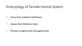
- 1. Embryology of Female Genital System • Ovary from Coelomic Epithelium • Uterus from Mullerian Ducts • Portion of Vagina from Sinovaginal bulb
- 2. APPLIED ANATOMY -IMPERFORATE HYMEN
- 8. MANAGEMENT
- 9. ISTHMUS- Narrow part between cervix & corpus of uterus. Same histological structures as corpus of uterus incorporated into cavity of uterus during pregnancy – isthmus opens up, so anatomical internal os ceases to exist, only histological os remains.
- 11. Hegar’s sign- At 6-10 wks. Seen in 2/3rd of cases Bimanual palpation Fingers of both hands approximate ,there is softening of isthmus with gestational sac in the upper cavity and empty lower cavity
- 12. Symphysio fundal height ( SFH ) in cm, after 24 weeks upto 36 weeks corresponds to number of weeks SFH can be used to clinically calculate the estimated fetal weight using Johnson’s formula Fetal weight = (SFH – n) x 155 n=12 if vertex is above ischial spine, n=11 is vertex is below ischial spine
- 14. Arrangement of muscle fibres- 1.Outer longitudinal: hoodlike layer ,arches over fundus and extends into various ligaments. 2. Middle layer: thickest and strongest, arranged in criss cross fashion. Apposition of 2 double curve muscle fibres – fig of 8 form which form the “ living ligatures”. 3. Inner circular: sphincter like fibres around fallopian tube orifices and internal os of cervix.
- 15. • Muscle fibre undergoes elongation, apposition of 2 double curves make figure of 8 that contract to occlude the blood vessels running in between them. • CLINICAL SIGNIFICANCE : occlusion of vessels during contractions helps in reducing the amount of blood loss postpartum