
Recommended
More Related Content
What's hot
What's hot (20)
Similar to Anatomy, Physiology & Classification of Varicose Veins
Similar to Anatomy, Physiology & Classification of Varicose Veins (20)
More from RavulJindal
More from RavulJindal (15)
Recently uploaded
Recently uploaded (20)
Anatomy, Physiology & Classification of Varicose Veins
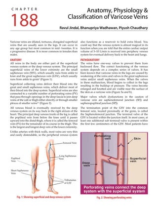
- 1. Varicose veins are dilated, tortuous, elongated superficial veins that are usually seen in the legs. It can occur in any age group but most common in mid- twenties. It is a progressive disease. It is more common in females than males. ANATOMY All veins in the body are either part of the superficial venous system or the deep venous system. The principal superficial veins of the lower extremity are the small saphenous vein (SSV), which usually runs from ankle to knee and the great saphenous vein (GSV), which usually runs from ankle to groin1 (Figure 1). Superficial collecting veins deliver their blood into the great and small saphenous veins, which deliver most of their blood into the deep system. Superficial veins are also connected to a variable number of perforating veins (PV) that pass through openings in the deep fascia to join deep veins of the calf or thigh either directly or through smaller plexus of smaller veins2,3 (Figure 2). All venous blood is eventually received by the deep venous system on its way back to the right atrium of the heart. The principal deep venous trunk of the leg is called the popliteal vein from below the knee until it passes upward into the distal thigh, where it is called the femoral vein (FV) for the remainder of its course in the thigh. This is the largest and longest deep vein of the lower extremity. Unlike arteries with thick walls, most veins are very thin and easily distendable, so the peripheral venous system also functions as a reservoir to hold extra blood. You could say that the venous system is almost magical in its function when you are told that the entire cardiac output volume of 5–10 L/min is received into periphery venous system for eventual delivery back to the heart and lungs. PATHOANATOMY The veins have one-way valves to prevent them from backward flow. The correct functioning of the venous system depends on a complex series of valves. It has been known that varicose veins in the legs are caused by weakening of the veins and valves in the great saphenous veins and/or small saphenous veins. When the valves in these malfunction, blood begins to collect in the legs resulting in the buildup of pressure. The veins become enlarged and knotted and are visible near the surface of the skin as a varicose vein (Figure 3a and b). Major valves which dysfunctions in the caution of varicose vein are saphenofemoral junction (SFJ) and saphenopopliteal junction (SPJ). The termination point of the GSV into the common femoral vein, located proximally at the groin, is called the Saphenofemoral junction. The terminal valve of the GSV is located within the junction itself. In most cases, at least one additional sub terminal valve is present within the first few centimeters of the GSV. Most patients have Anatomy, Physiology & Classification of Varicose Veins Ravul Jindal, Bhanupriya Wadhawan, Piyush Chaudhary Fig. 1: Showing superficial and deep venous system Fig. 2: Showing perforating veins Femoral Vein Long Saphenous Vein Tributaries of LSV Popliteal Vein Short Saphenous Vein C H A P T E R 188
- 2. VENOUSDISORDERS 868 a single sub terminal valve that can be readily identified approximately 1 cm distal to the junctional valve. PATHOPHYSIOLOGY The pathophysiology behind their formation is complicated and involves the concept of ambulatory venous hypertension. In healthy veins, the flow of venous blood is through the superficial system into the deep system and up the leg and toward the heart. One-way venous valves are found in both systems and the perforating veins. Incompetence in any of these valves can lead to a disruption in the unidirectional flow of blood toward the heart and result in ambulatory venous hypertension (AVH). 6 Incompetence in the superficial venous system alone usually results from failure at valves located at the SFJ and SPJ. The gravitational weight of the column of blood along the length of the vein creates hydrostatic pressure, which is worse at the more distal aspect of the length of vein. Reflux at or near the SFJ does not always come through the terminal valve of the GSV, nor does it always involve the entire trunk of the GSV. Reflux can enter the GSV below the sub terminal valve or even immediately below the junction, passing through a failed sub terminal valve to mimic true SFJ incompetence. Reflux can also pass directly into any of the other veins that join the GSV at that level, or it may pass a few centimeters along the GSV and then abandon the GSV for another branch vessel. Incompetence of the perforating veins leads to hydrodynamic pressure. The calf pump mechanism helps to empty the deep venous system, but if perforating vein valves fail, then the pressure generated in the deep venous system by the calf pump mechanism are transmitted into the superficial system via the incompetent perforating veins. Once venous hypertension is present, the venous dysfunction continues to worsen through a vicious circle. Pooled blood and venous hypertension leads to venous dilatation, which then causes greater valvular insufficiency. Over time, with more local dilatation, other adjacent valves sequentially fail, and after a series of valves has failed, the entire superficial venous system is incompetent. This can then cause subsequent perforator and deep venous valvular dysfunction. The clinical findings of varicose veins, reticular veins, and telangiectasias are due to the hypertension in the superficial venous system that spreads to collateral veins and tributary veins, causing dilated tortuous structures. Treatment modalities are geared towards correcting the superficial venous hypertension. In contrast to the superficial veins, the deep veins do not become excessively distended. They can withstand the increased pressure because of their construction and the confining fascia. THE CLASSIFICATION OF VENOUS DISEASE Venous disease of the legs can be classified according to the severity, cause, site and specific abnormality using the CEAP classification. The elements of the CEAP classification are: Fig. 3a: Showing normal veins and diseased veins Fig. 3b: Showing normal veins and diseased veins Fig. 4: Showing terminal and preterminal valve VF=femoral vein GSV=Great saphenous vein SSV=Supra-saphenic valve TV=Terminal valve PTV=Pre-terminal valve A B Deep veins Iliac vein Common femoral vein Popliteal vein Normal blood flow back to heart Deep vein Deep vein Perforating veins Superficial veins Normal perforating valve and normal blood flow Incompetent perforating valve causing abnormal blood flow Fascia layer Perforating vein Superficial vein Superficial veins Great saphenous vein Normal vein Open valve Venous insuficiency Normal vein Closed valve Varicose vein Damaged/nonfunctional valve Incompetent valve Vein wall thinned and bulging Abnormal blood flow backward down leg Damaged nonfunctional valve cannot close properly and blood flow is impaired Normal valve closes to prevent reverse of blood flow Normal valve opens to allow blood flow toward heart
- 3. CHAPTER188 869 • Clinical severity • Etiology or cause • Anatomy • Pathophysiology For the initial assessment of a patient, the clinical severity is the most important and can be made by simple observation and does not need special tests. There are seven grades of increasing clinical severity 3,4,5 : Grade Description C 0 No evidence of venous disease. C 1 Superficial spider veins (reticular veins) only C 2 Simple varicose veins only C 3 Ankle edema of venous origin (not foot edema) C 4 a. Pigmentation or Eczema b. Lipodermatosclerosis or athrophie blanche Skin pigmentation in the gaiter area (lipodermatosclerosis) C 5 A healed venous ulcer C 6 An open venous ulcer The majority of patients referred to the vascular surgical clinic have grade 2 diseases (simple varicose veins). Patients with C3-6 disease are demonstrating increase severity of chronic venous insufficiency, and all have a functional abnormality of the venous system. These patients are most at risk of chronic ulceration and require specialized tests such as venous duplex and ambulatory venous pressure measurement to diagnose and characterize the underlying venous abnormality. If we correct the venous abnormality in the disease process then the risk of complications associated with the venous disease are much lower.
- 4. VENOUSDISORDERS 870 REFERENCES 1. Souroullas P, Barnes R, Smith G, Nandhra S, Carradice D, Chetter I.The classic saphenofemoral junction and its anatomical variations. Phlebology 2016 Feb 2. Goldman MP, Fronek A. Anatomy and pathophysiology of varicose veins. J Dermatol Surg Oncol 1989; 15:138-45. 3. Rabe E, Pannier F. Clinical, aetiological, anatomical and pathological classification (CEAP): gold standard and limits. Phlebology 2012; 27 Suppl 1:114-8. 4. Gloviczki P, Comerota AJ, Dalsing MC, Eklof BG, Gillespie DL, Gloviczki ML, Lohr JM, McLafferty RB, Meissner MH, Murad MH, Padberg FT, Pappas PJ, Passman MA, Raffetto JD, Vasquez MA, Wakefield TW; The care of patients with varicose veins and associated chronic venous diseases: clinical practice guidelines of the Society for Vascular Surgery and the American Venous Forum.Society for Vascular Surgery; American Venous Forum. J Vasc Surg 2011; 53(5 Suppl):2S-48S. 5. Vasquez MA, Rabe E, McLafferty RB, Shortell CK, Marston WA, Gillespie D, Meissner MH, Rutherford RB; Revision of the venous clinical severity score: venous outcomes consensus statement: special communication of the American Venous Forum Ad Hoc Outcomes Working Group.American Venous Forum Ad Hoc Outcomes Working Group. J Vasc Surg 2010; 52:1387-96. 6. Yetkin E, Ileri M. Dilating venous disease: Pathophysiology and a systematic aspect to different vascular territories. Med Hypotheses 2016; 91:73-6.