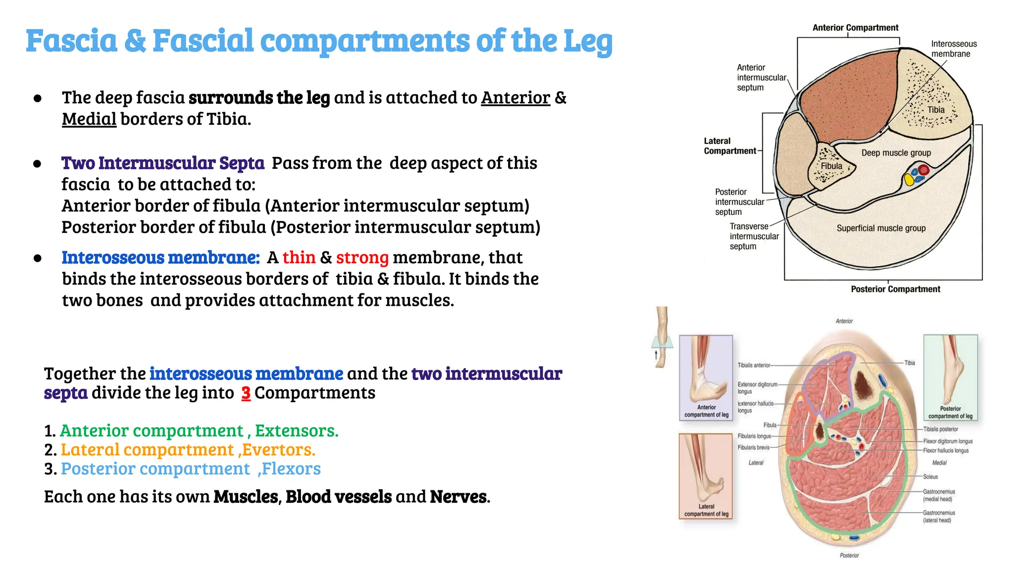
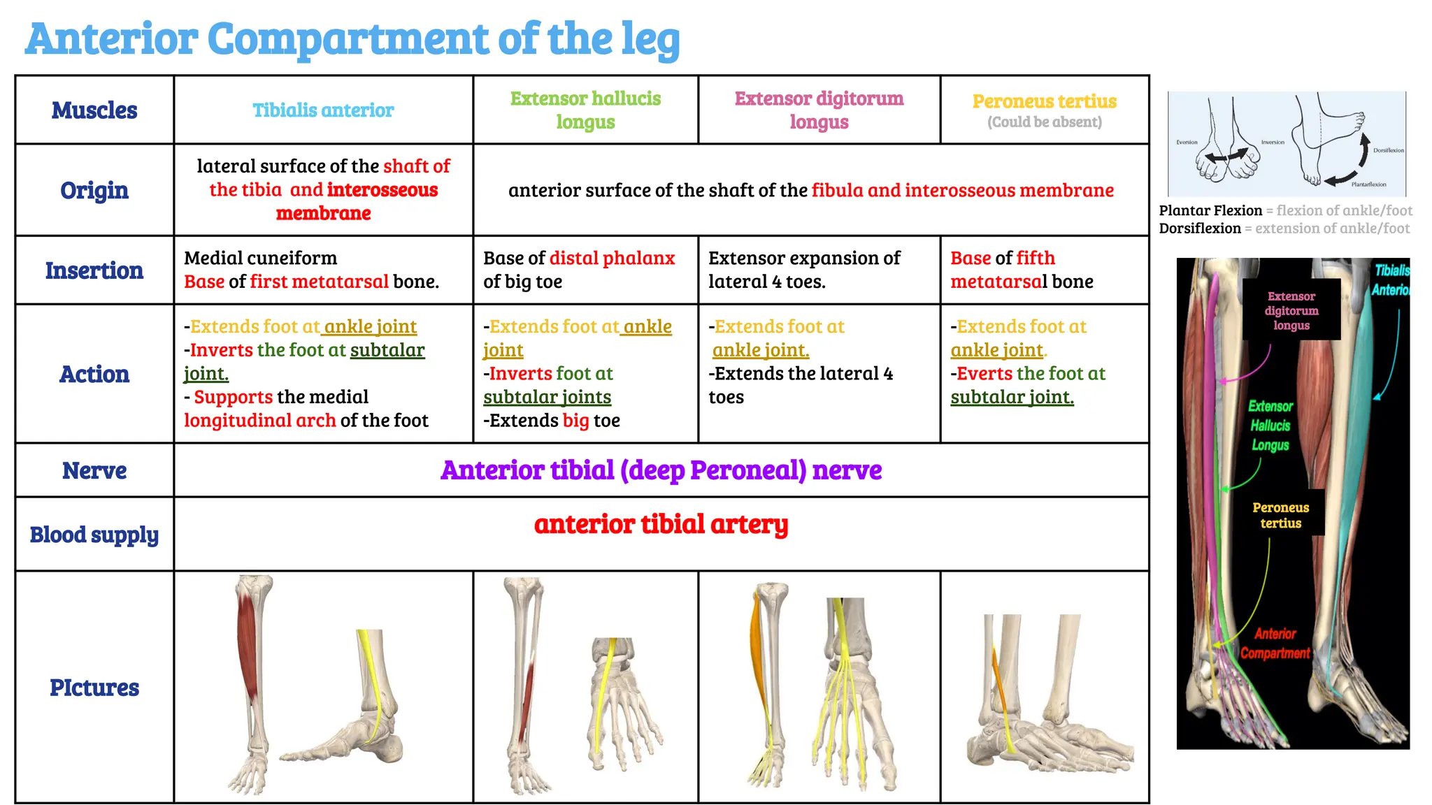
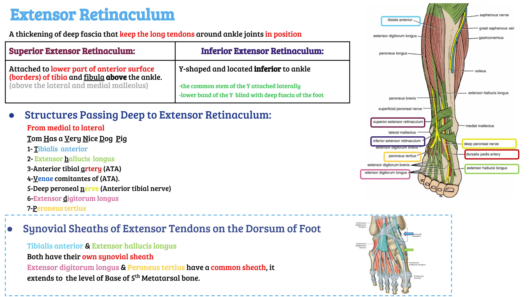
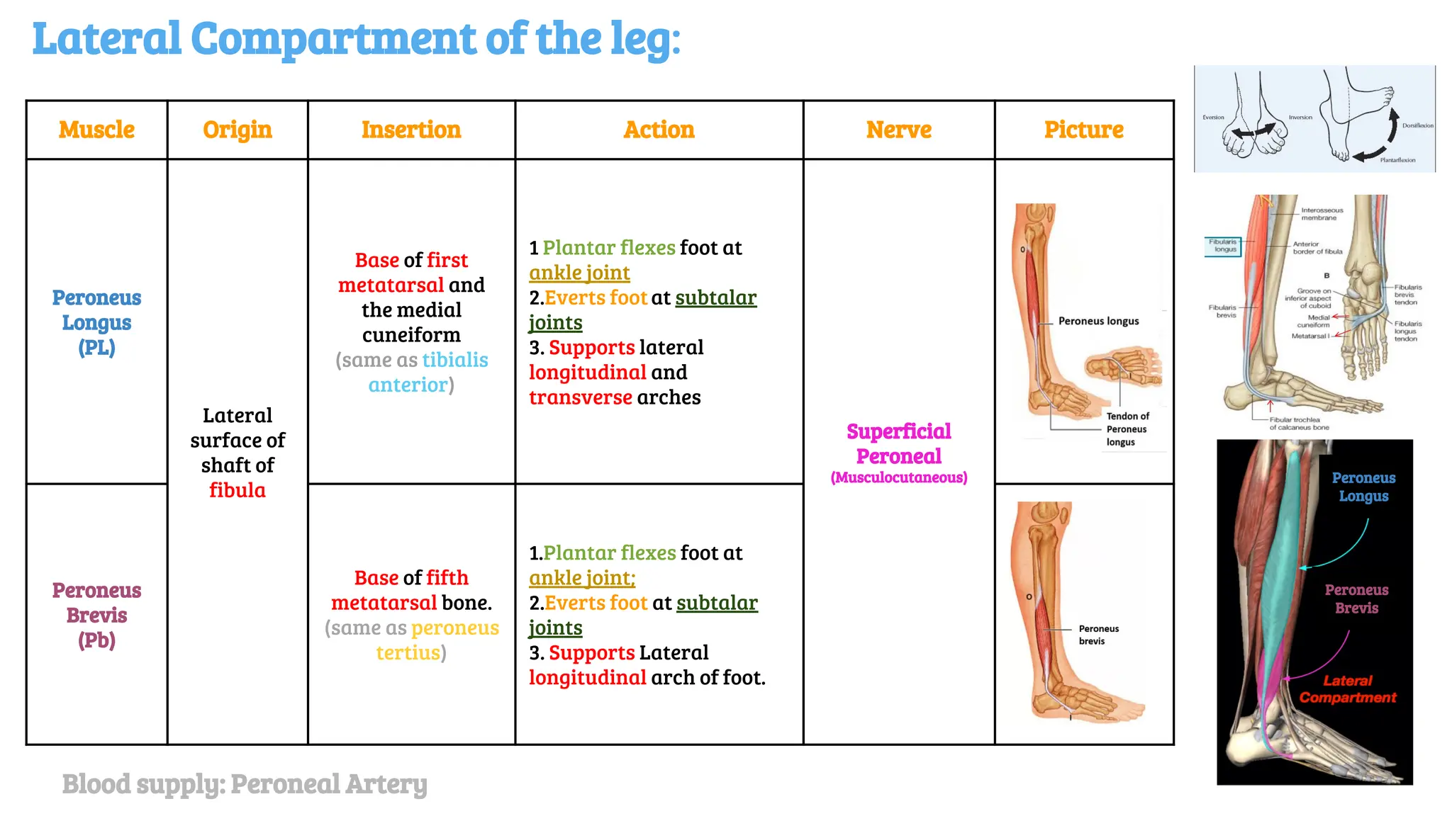
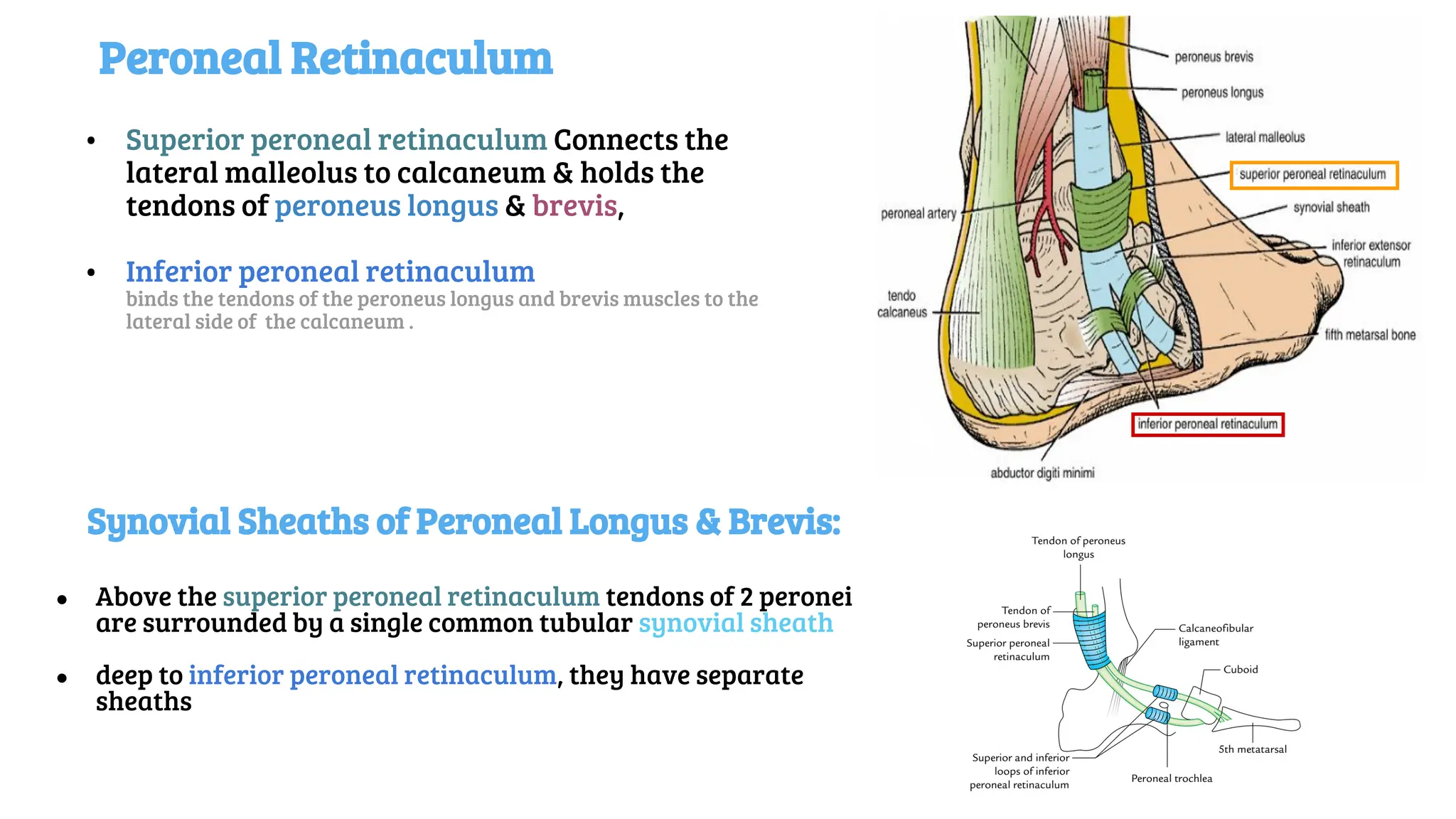
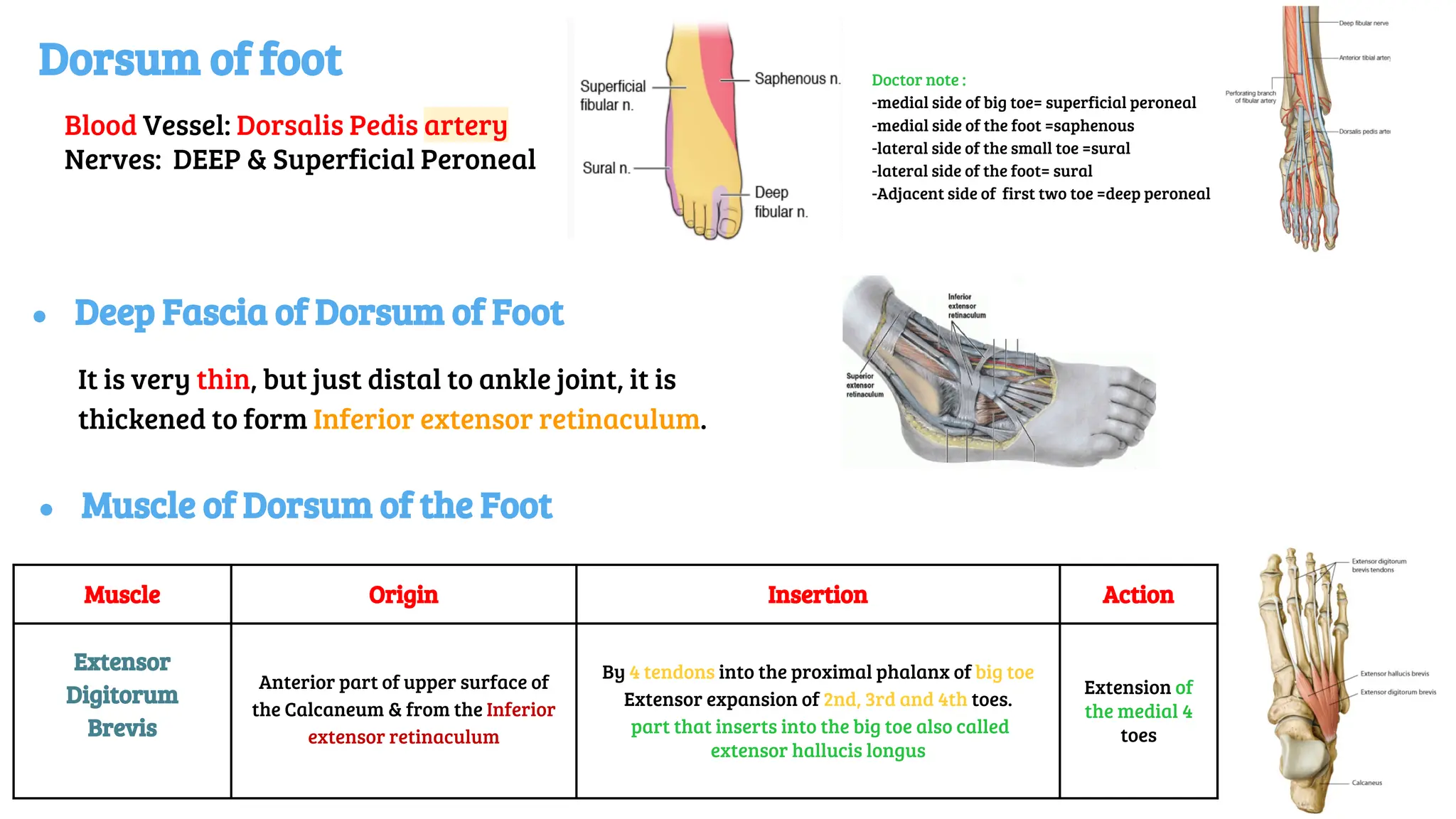
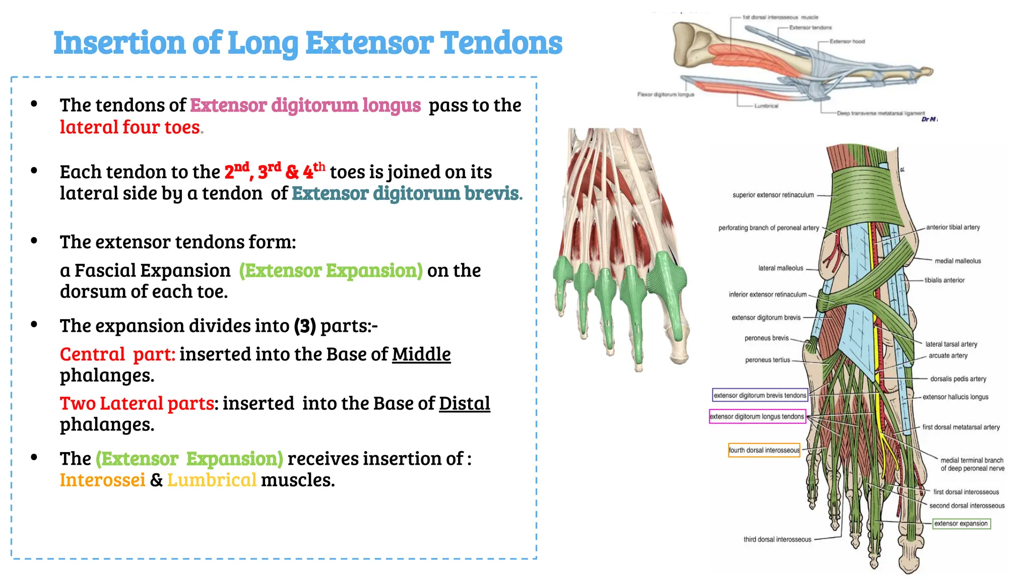
Musculoskeletal block- Anatomy-lecture 15
Editing file
Frontal, Lateral compartment
of Leg and Dorsum of Foot
Objectives
✓ Identify the deep fascia of leg
✓ Identify the fascial compartments of the leg
✓ Describe the anatomy of the anterior & lateral compartments
✓ List the contents of each compartment (muscles, vessels & nerves)
✓ Describe the anatomy and contents of the dorsum of the foot
Color guide :
Only in boys slides in Blue
Only in girls slides in Purple
important in Red
Doctor note in Green
Extra information in Grey
Fascia & Fascial compartments of the Leg
● Interosseous membrane: A thin & strong membrane, that
binds the interosseous borders of tibia & fibula. It binds the
two bones and provides attachment for muscles.
● The deep fascia surrounds the leg and is attached to Anterior &
Medial borders of Tibia.
Together the interosseous membrane and the two intermuscular
septa divide the leg into 3 Compartments
1. Anterior compartment , Extensors.
2. Lateral compartment ,Evertors.
3. Posterior compartment ,Flexors
Each one has its own Muscles, Blood vessels and Nerves.
● Two Intermuscular Septa Pass from the deep aspect of this
fascia to be attached to:
Anterior border of fibula (Anterior intermuscular septum)
Posterior border of fibula (Posterior intermuscular septum)
Muscles Tibialis anterior
Extensor hallucis
longus
Extensor digitorum
longus
Peroneus tertius
(Could be absent)
Origin
lateral surface of the shaft of
the tibia and interosseous
membrane
anterior surface of the shaft of the fibula and interosseous membrane
Insertion
Medial cuneiform
Base of first metatarsal bone.
Base of distal phalanx
of big toe
Extensor expansion of
lateral 4 toes.
Base of fifth
metatarsal bone
Action
-Extends foot at ankle joint
-Inverts the foot at subtalar
joint.
- Supports the medial
longitudinal arch of the foot
-Extends foot at ankle
joint
-Inverts foot at
subtalar joints
-Extends big toe
-Extends foot at
ankle joint.
-Extends the lateral 4
toes
-Extends foot at
ankle joint.
-Everts the foot at
subtalar joint.
Nerve Anterior tibial (deep Peroneal) nerve
Blood supply anterior tibial artery
PIctures
Anterior Compartment of the leg
Plantar Flexion = flexion of ankle/foot
Dorsiflexion = extension of ankle/foot
Peroneus
tertius
Extensor
digitorum
longus
Extensor Retinaculum
● Structures Passing Deep to Extensor Retinaculum:
From medial to lateral
Tom Has a Very Nice Dog Pig
1- Tibialis anterior
2- Extensor hallucis longus
3-Anterior tibial artery (ATA)
4-Venae comitantes of (ATA).
5-Deep peroneal nerve (Anterior tibial nerve)
6-Extensor digitorum longus
7-Peroneus tertius
A thickening of deep fascia that keep the long tendons around ankle joints in position
Superior Extensor Retinaculum: Inferior Extensor Retinaculum:
Attached to lower part of anterior surface
(borders) of tibia and fibula above the ankle.
(above the lateral and medial malleolus)
Y-shaped and located inferior to ankle
-the common stem of the Y attached laterally
-lower band of the Y blind with deep fascia of the foot
● Synovial Sheaths of Extensor Tendons on the Dorsum of Foot
Tibialis anterior & Extensor hallucis longus
Both have their own synovial sheath
Extensor digitorum longus & Peroneus tertius have a common sheath, it
extends to the level of Base of 5th
Metatarsal bone.
Lateral Compartment of the leg:
Muscle Origin Insertion Action Nerve Picture
Peroneus
Longus
(PL)
Lateral
surface of
shaft of
fibula
Base of first
metatarsal and
the medial
cuneiform
(same as tibialis
anterior)
1 Plantar flexes foot at
ankle joint
2.Everts footat subtalar
joints
3. Supports lateral
longitudinal and
transverse arches
Superficial
Peroneal
(Musculocutaneous)
Peroneus
Brevis
(Pb)
Base of fifth
metatarsal bone.
(same as peroneus
tertius)
1.Plantar flexes foot at
ankle joint;
2.Everts foot at subtalar
joints
3. Supports Lateral
longitudinal arch of foot.
Peroneus
Longus
Peroneus
Brevis
Blood supply: Peroneal Artery
Peroneal Retinaculum
• Superior peroneal retinaculum Connects the
lateral malleolus to calcaneum & holds the
tendons of peroneus longus & brevis,
• Inferior peroneal retinaculum
binds the tendons of the peroneus longus and brevis muscles to the
lateral side of the calcaneum .
Synovial Sheaths of Peroneal Longus & Brevis:
● Above the superior peroneal retinaculum tendons of 2 peronei
are surrounded by a single common tubular synovial sheath
● deep to inferior peroneal retinaculum, they have separate
sheaths
Dorsum of foot
Blood Vessel: Dorsalis Pedis artery
Nerves: DEEP & Superficial Peroneal
● Deep Fascia of Dorsum of Foot
It is very thin, but just distal to ankle joint, it is
thickened to form Inferior extensor retinaculum.
Muscle Origin Insertion Action
Extensor
Digitorum
Brevis
Anterior part of upper surface of
the Calcaneum & from the Inferior
extensor retinaculum
By 4 tendons into the proximal phalanx of big toe
Extensor expansion of 2nd, 3rd and 4th toes.
part that inserts into the big toe also called
extensor hallucis longus
Extension of
the medial 4
toes
● Muscle of Dorsum of the Foot
Doctor note :
-medial side of big toe= superficial peroneal
-medial side of the foot =saphenous
-lateral side of the small toe =sural
-lateral side of the foot= sural
-Adjacent side of first two toe =deep peroneal
Insertion of Long Extensor Tendons
• The tendons of Extensor digitorum longus pass to the
lateral four toes.
• Each tendon to the 2nd
, 3rd
& 4th
toes is joined on its
lateral side by a tendon of Extensor digitorum brevis.
• The extensor tendons form:
a Fascial Expansion (Extensor Expansion) on the
dorsum of each toe.
• The expansion divides into (3) parts:-
Central part: inserted into the Base of Middle
phalanges.
Two Lateral parts: inserted into the Base of Distal
phalanges.
• The (Extensor Expansion) receives insertion of :
Interossei & Lumbrical muscles.
MCQs
Question 1:The Peroneus tertius is inserting from anterior surface of fibula to:
A. Extensor expansion of lateral four toes
B. Base of 5th
metatarsal bone.
C. Medial cuneiform & base of 1st
metatarsal bone.
D. Base of distal phalanx of great toe.
Question 2: Which muscle can evert the foot?
A. Peroneus Tertius.
B.Tibialis Anterior.
C.Extensor Hallucis Longus.
D. Extensor Digitorum Longus.
Question 3: The peroneus longus is inserted into:
A.Base of first metatarsal
B. The lateral cuneiform
C. The medial cuneiform
D.Both a&c
Question 4:The Extensor Expansion receives insertion of
A.Interossei
B. Lumbricals
C. Tibialis anterior
D. A & B
Question 5:Which nerve supplies the anterior compartment of the leg?
A.Anterior tibial
B.Lateral fibular
C.Deep peroneal
D.superficial peroneal
Question 6: Which of the following is medial to vessels in the extensor retinaculum?
A.Extensor Hallucis Longus Tendon
B. Deep Peroneal nerve
C. Peroneus Tertius Tendon
D.Extensor Digitorum Longus Tendon
Question 7: All of the following are attached to the fibula except?
A.Extensor Digitorum Longus
B. Peroneus Brevis
C. Tibialis Anterior
D.Extensor Hallucis Longus
Question 8: How many Intermuscular septa pass from deep fascia to attach to fibula?
A. 3
B. 2
C.1
D.4
Answers: Q1.B Q2.A- Q3.D -Q4.D- Q5.C- Q6.A-Q7.C- Q8.B
Give us your feedback:
Boys team:
● Khalid AL-Dossari
● Naif Al-Dossari
● Faisal Alqifari
● Salman Alagla
● Ziyad Al-jofan
● Suhail Basuhail
● Ali Aldawood
● Khalid Nagshabandi
● Mohammed Al-huqbani
● Jehad Alorainy
● Khalid AlKhani
● Omar Alammari
Girls team :
● Ajeed Al Rashoud
● Taif Alotaibi
● Noura Al Turki
● Amirah Al-Zahrani
● Alhanouf Al-haluli
● Sara Al-Abdulkarem
● Rawan Al Zayed
● Reema Al Masoud
● Renad Al Haqbani
● Nouf Al Humaidhi
● Fay Al Buqami
● Jude Al Khalifah
● Nouf Al Hussaini
● Alwateen Al Balawi
● Rahaf Al Shabri
● Danah Al Halees
● Haifa Al Waily
● Rema Al Mutawa
● Amirah Al Dakhilallah
● Maha Al Nahdi
● Renad Al Mutawa
● Ghaida Al Braithen
● Reham Yousef
Team members
Team leaders
● Abdulrahman Shadid
● Ateen Almutairi
Good luck
Special thank for
Anatomy team 436
=This lecture done by

Lecture (15) Frontal, Lateral compartment of Leg and Dorsum.pdf

  • 1.
    Musculoskeletal block- Anatomy-lecture15 Editing file Frontal, Lateral compartment of Leg and Dorsum of Foot
  • 2.
    Objectives ✓ Identify thedeep fascia of leg ✓ Identify the fascial compartments of the leg ✓ Describe the anatomy of the anterior & lateral compartments ✓ List the contents of each compartment (muscles, vessels & nerves) ✓ Describe the anatomy and contents of the dorsum of the foot Color guide : Only in boys slides in Blue Only in girls slides in Purple important in Red Doctor note in Green Extra information in Grey
  • 3.
    Fascia & Fascialcompartments of the Leg ● Interosseous membrane: A thin & strong membrane, that binds the interosseous borders of tibia & fibula. It binds the two bones and provides attachment for muscles. ● The deep fascia surrounds the leg and is attached to Anterior & Medial borders of Tibia. Together the interosseous membrane and the two intermuscular septa divide the leg into 3 Compartments 1. Anterior compartment , Extensors. 2. Lateral compartment ,Evertors. 3. Posterior compartment ,Flexors Each one has its own Muscles, Blood vessels and Nerves. ● Two Intermuscular Septa Pass from the deep aspect of this fascia to be attached to: Anterior border of fibula (Anterior intermuscular septum) Posterior border of fibula (Posterior intermuscular septum)
  • 4.
    Muscles Tibialis anterior Extensorhallucis longus Extensor digitorum longus Peroneus tertius (Could be absent) Origin lateral surface of the shaft of the tibia and interosseous membrane anterior surface of the shaft of the fibula and interosseous membrane Insertion Medial cuneiform Base of first metatarsal bone. Base of distal phalanx of big toe Extensor expansion of lateral 4 toes. Base of fifth metatarsal bone Action -Extends foot at ankle joint -Inverts the foot at subtalar joint. - Supports the medial longitudinal arch of the foot -Extends foot at ankle joint -Inverts foot at subtalar joints -Extends big toe -Extends foot at ankle joint. -Extends the lateral 4 toes -Extends foot at ankle joint. -Everts the foot at subtalar joint. Nerve Anterior tibial (deep Peroneal) nerve Blood supply anterior tibial artery PIctures Anterior Compartment of the leg Plantar Flexion = flexion of ankle/foot Dorsiflexion = extension of ankle/foot Peroneus tertius Extensor digitorum longus
  • 5.
    Extensor Retinaculum ● StructuresPassing Deep to Extensor Retinaculum: From medial to lateral Tom Has a Very Nice Dog Pig 1- Tibialis anterior 2- Extensor hallucis longus 3-Anterior tibial artery (ATA) 4-Venae comitantes of (ATA). 5-Deep peroneal nerve (Anterior tibial nerve) 6-Extensor digitorum longus 7-Peroneus tertius A thickening of deep fascia that keep the long tendons around ankle joints in position Superior Extensor Retinaculum: Inferior Extensor Retinaculum: Attached to lower part of anterior surface (borders) of tibia and fibula above the ankle. (above the lateral and medial malleolus) Y-shaped and located inferior to ankle -the common stem of the Y attached laterally -lower band of the Y blind with deep fascia of the foot ● Synovial Sheaths of Extensor Tendons on the Dorsum of Foot Tibialis anterior & Extensor hallucis longus Both have their own synovial sheath Extensor digitorum longus & Peroneus tertius have a common sheath, it extends to the level of Base of 5th Metatarsal bone.
  • 6.
    Lateral Compartment ofthe leg: Muscle Origin Insertion Action Nerve Picture Peroneus Longus (PL) Lateral surface of shaft of fibula Base of first metatarsal and the medial cuneiform (same as tibialis anterior) 1 Plantar flexes foot at ankle joint 2.Everts footat subtalar joints 3. Supports lateral longitudinal and transverse arches Superficial Peroneal (Musculocutaneous) Peroneus Brevis (Pb) Base of fifth metatarsal bone. (same as peroneus tertius) 1.Plantar flexes foot at ankle joint; 2.Everts foot at subtalar joints 3. Supports Lateral longitudinal arch of foot. Peroneus Longus Peroneus Brevis Blood supply: Peroneal Artery
  • 7.
    Peroneal Retinaculum • Superiorperoneal retinaculum Connects the lateral malleolus to calcaneum & holds the tendons of peroneus longus & brevis, • Inferior peroneal retinaculum binds the tendons of the peroneus longus and brevis muscles to the lateral side of the calcaneum . Synovial Sheaths of Peroneal Longus & Brevis: ● Above the superior peroneal retinaculum tendons of 2 peronei are surrounded by a single common tubular synovial sheath ● deep to inferior peroneal retinaculum, they have separate sheaths
  • 8.
    Dorsum of foot BloodVessel: Dorsalis Pedis artery Nerves: DEEP & Superficial Peroneal ● Deep Fascia of Dorsum of Foot It is very thin, but just distal to ankle joint, it is thickened to form Inferior extensor retinaculum. Muscle Origin Insertion Action Extensor Digitorum Brevis Anterior part of upper surface of the Calcaneum & from the Inferior extensor retinaculum By 4 tendons into the proximal phalanx of big toe Extensor expansion of 2nd, 3rd and 4th toes. part that inserts into the big toe also called extensor hallucis longus Extension of the medial 4 toes ● Muscle of Dorsum of the Foot Doctor note : -medial side of big toe= superficial peroneal -medial side of the foot =saphenous -lateral side of the small toe =sural -lateral side of the foot= sural -Adjacent side of first two toe =deep peroneal
  • 9.
    Insertion of LongExtensor Tendons • The tendons of Extensor digitorum longus pass to the lateral four toes. • Each tendon to the 2nd , 3rd & 4th toes is joined on its lateral side by a tendon of Extensor digitorum brevis. • The extensor tendons form: a Fascial Expansion (Extensor Expansion) on the dorsum of each toe. • The expansion divides into (3) parts:- Central part: inserted into the Base of Middle phalanges. Two Lateral parts: inserted into the Base of Distal phalanges. • The (Extensor Expansion) receives insertion of : Interossei & Lumbrical muscles.
  • 10.
    MCQs Question 1:The Peroneustertius is inserting from anterior surface of fibula to: A. Extensor expansion of lateral four toes B. Base of 5th metatarsal bone. C. Medial cuneiform & base of 1st metatarsal bone. D. Base of distal phalanx of great toe. Question 2: Which muscle can evert the foot? A. Peroneus Tertius. B.Tibialis Anterior. C.Extensor Hallucis Longus. D. Extensor Digitorum Longus. Question 3: The peroneus longus is inserted into: A.Base of first metatarsal B. The lateral cuneiform C. The medial cuneiform D.Both a&c Question 4:The Extensor Expansion receives insertion of A.Interossei B. Lumbricals C. Tibialis anterior D. A & B Question 5:Which nerve supplies the anterior compartment of the leg? A.Anterior tibial B.Lateral fibular C.Deep peroneal D.superficial peroneal Question 6: Which of the following is medial to vessels in the extensor retinaculum? A.Extensor Hallucis Longus Tendon B. Deep Peroneal nerve C. Peroneus Tertius Tendon D.Extensor Digitorum Longus Tendon Question 7: All of the following are attached to the fibula except? A.Extensor Digitorum Longus B. Peroneus Brevis C. Tibialis Anterior D.Extensor Hallucis Longus Question 8: How many Intermuscular septa pass from deep fascia to attach to fibula? A. 3 B. 2 C.1 D.4 Answers: Q1.B Q2.A- Q3.D -Q4.D- Q5.C- Q6.A-Q7.C- Q8.B
  • 11.
    Give us yourfeedback: Boys team: ● Khalid AL-Dossari ● Naif Al-Dossari ● Faisal Alqifari ● Salman Alagla ● Ziyad Al-jofan ● Suhail Basuhail ● Ali Aldawood ● Khalid Nagshabandi ● Mohammed Al-huqbani ● Jehad Alorainy ● Khalid AlKhani ● Omar Alammari Girls team : ● Ajeed Al Rashoud ● Taif Alotaibi ● Noura Al Turki ● Amirah Al-Zahrani ● Alhanouf Al-haluli ● Sara Al-Abdulkarem ● Rawan Al Zayed ● Reema Al Masoud ● Renad Al Haqbani ● Nouf Al Humaidhi ● Fay Al Buqami ● Jude Al Khalifah ● Nouf Al Hussaini ● Alwateen Al Balawi ● Rahaf Al Shabri ● Danah Al Halees ● Haifa Al Waily ● Rema Al Mutawa ● Amirah Al Dakhilallah ● Maha Al Nahdi ● Renad Al Mutawa ● Ghaida Al Braithen ● Reham Yousef Team members Team leaders ● Abdulrahman Shadid ● Ateen Almutairi Good luck Special thank for Anatomy team 436 =This lecture done by