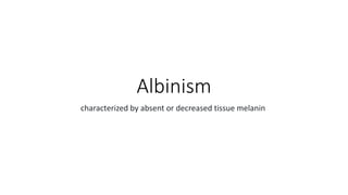
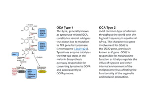
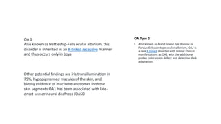
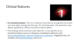
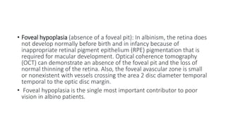
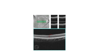
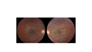
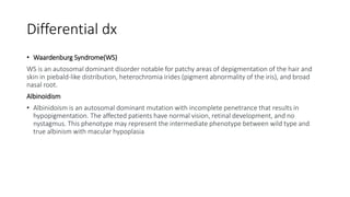
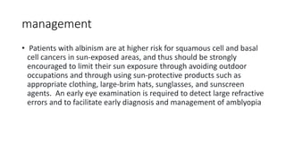
Albinism
characterized by absent or decreased tissue melanin
The disorder can be divided into two groups:
• oculocutaneous albinism (OCA)-—the most common condition
hypopigmentation disorders with varying degrees of involvement of
the eyes, hair, and skin
• the second group is the less common group called ocular albinism
(OA) with disease involvement limited to the eyes
• Oculocutaneous albinism is a group of autosomal recessive conditions
associated with mutations in several enzymes or membrane proteins
that contribute to melanin synthesis.
• classically been organized into two broad categories, tyrosinase-
positive (mild to moderate) and tyrosinas-negative (severe) albinism
OCA Type 1
This type, generally known
as tyrosinase-related OCA,
constitutes several subtypes
that occur due to mutation
in TYR gene for tyrosinase
(chromosome 11q14-q21).
Tyrosinase enzyme catalyzes
the first two steps in the
melanin biosynthesis
pathway, responsible for
converting tyrosine to DOPA
and subsequently to
DOPAquinone.
OCA Type 2
most common type of albinism
throughout the world with the
highest frequency in equatorial
Africa. The characteristic gene
involvement for OCA2 is
the OCA2 gene, previously
known as P gene. OCA2 is
responsible for melanosome
function as it helps regulate the
influx of tyrosine and other
internal environment of the
melanosome thus affecting the
functionality of the organelle
and melanin production.
OA Type 2
• Also known as Aland Island eye disease or
Forsius-Eriksson type ocular albinism, OA2 is
a rare X-linked disorder with similar clinical
manifestations as OA1 with the additional
protan color vision defect and defective dark
adaptation.
OA 1
Also known as Nettleship-Falls ocular albinism, this
disorder is inherited in an X-linked recessive manner
and thus occurs only in boys
Other potential findings are iris transillumination in
75%, hypopigmented macules of the skin, and
biopsy evidence of macromelanosomes in those
skin segments.OA1 has been associated with late-
onset sensorineural deafness (OASD
Clinical features
• Iris transillumination: The iris in albinism has little to no pigment to screen
out stray light coming into the eye. On slit lamp exam, the examiner may
detect speckled or diffuse transillumination defect.
• This finding, while common with albinism, is not specific as iris
transillumination occurs in diseases unrelated to albinism such
as pseudoexfoliation, pigment dispersion syndrome, megalocornea, iris
atrophy, and Axenfeld-Rieger spectrum.
• Foveal hypoplasia (absence of a foveal pit): In albinism, the retina does
not develop normally before birth and in infancy because of
inappropriate retinal pigment epithelium (RPE) pigmentation that is
required for macular development. Optical coherence tomography
(OCT) can demonstrate an absence of the foveal pit and the loss of
normal thinning of the retina. Also, the foveal avascular zone is small
or nonexistent with vessels crossing the area 2 disc diameter temporal
temporal to the optic disc margin.
• Foveal hypoplasia is the single most important contributor to poor
vision in albino patients.
• Abnormal decussation of the visual pathways: Normally about half of
optic nerve fibers from each eye decussates at the optic chiasm to the
contralateral side, contributing to stereopsis. Albinism is associated
with fiber over-decussation, resulting in crossing of up to 90% of fibers
fibers to the contralateral side and thus strabismus and loss of
stereopsis.
• Photophobia: Sensitivity to bright light and glare can occur due to
scattering of light within the eye. Patients may prefer to wear
sunglasses to reduce their sensitivity to light.
• Refractive Errors: Both myopia and hyperopia can occur, and
astigmatism is very common.
Differential dx
• Waardenburg Syndrome(WS)
WS is an autosomal dominant disorder notable for patchy areas of depigmentation of the hair and
skin in piebald-like distribution, heterochromia irides (pigment abnormality of the iris), and broad
nasal root.
Albinoidism
• Albinidoism is an autosomal dominant mutation with incomplete penetrance that results in
hypopigmentation. The affected patients have normal vision, retinal development, and no
nystagmus. This phenotype may represent the intermediate phenotype between wild type and
true albinism with macular hypoplasia
management
• Patients with albinism are at higher risk for squamous cell and basal
cell cancers in sun-exposed areas, and thus should be strongly
encouraged to limit their sun exposure through avoiding outdoor
occupations and through using sun-protective products such as
appropriate clothing, large-brim hats, sunglasses, and sunscreen
agents. An early eye examination is required to detect large refractive
errors and to facilitate early diagnosis and management of amblyopia

Albinism.pptx

  • 1.
    Albinism characterized by absentor decreased tissue melanin
  • 2.
    The disorder canbe divided into two groups: • oculocutaneous albinism (OCA)-—the most common condition hypopigmentation disorders with varying degrees of involvement of the eyes, hair, and skin • the second group is the less common group called ocular albinism (OA) with disease involvement limited to the eyes • Oculocutaneous albinism is a group of autosomal recessive conditions associated with mutations in several enzymes or membrane proteins that contribute to melanin synthesis. • classically been organized into two broad categories, tyrosinase- positive (mild to moderate) and tyrosinas-negative (severe) albinism
  • 3.
    OCA Type 1 Thistype, generally known as tyrosinase-related OCA, constitutes several subtypes that occur due to mutation in TYR gene for tyrosinase (chromosome 11q14-q21). Tyrosinase enzyme catalyzes the first two steps in the melanin biosynthesis pathway, responsible for converting tyrosine to DOPA and subsequently to DOPAquinone. OCA Type 2 most common type of albinism throughout the world with the highest frequency in equatorial Africa. The characteristic gene involvement for OCA2 is the OCA2 gene, previously known as P gene. OCA2 is responsible for melanosome function as it helps regulate the influx of tyrosine and other internal environment of the melanosome thus affecting the functionality of the organelle and melanin production.
  • 4.
    OA Type 2 •Also known as Aland Island eye disease or Forsius-Eriksson type ocular albinism, OA2 is a rare X-linked disorder with similar clinical manifestations as OA1 with the additional protan color vision defect and defective dark adaptation. OA 1 Also known as Nettleship-Falls ocular albinism, this disorder is inherited in an X-linked recessive manner and thus occurs only in boys Other potential findings are iris transillumination in 75%, hypopigmented macules of the skin, and biopsy evidence of macromelanosomes in those skin segments.OA1 has been associated with late- onset sensorineural deafness (OASD
  • 5.
    Clinical features • Iristransillumination: The iris in albinism has little to no pigment to screen out stray light coming into the eye. On slit lamp exam, the examiner may detect speckled or diffuse transillumination defect. • This finding, while common with albinism, is not specific as iris transillumination occurs in diseases unrelated to albinism such as pseudoexfoliation, pigment dispersion syndrome, megalocornea, iris atrophy, and Axenfeld-Rieger spectrum.
  • 6.
    • Foveal hypoplasia(absence of a foveal pit): In albinism, the retina does not develop normally before birth and in infancy because of inappropriate retinal pigment epithelium (RPE) pigmentation that is required for macular development. Optical coherence tomography (OCT) can demonstrate an absence of the foveal pit and the loss of normal thinning of the retina. Also, the foveal avascular zone is small or nonexistent with vessels crossing the area 2 disc diameter temporal temporal to the optic disc margin. • Foveal hypoplasia is the single most important contributor to poor vision in albino patients.
  • 8.
    • Abnormal decussationof the visual pathways: Normally about half of optic nerve fibers from each eye decussates at the optic chiasm to the contralateral side, contributing to stereopsis. Albinism is associated with fiber over-decussation, resulting in crossing of up to 90% of fibers fibers to the contralateral side and thus strabismus and loss of stereopsis.
  • 9.
    • Photophobia: Sensitivityto bright light and glare can occur due to scattering of light within the eye. Patients may prefer to wear sunglasses to reduce their sensitivity to light. • Refractive Errors: Both myopia and hyperopia can occur, and astigmatism is very common.
  • 11.
    Differential dx • WaardenburgSyndrome(WS) WS is an autosomal dominant disorder notable for patchy areas of depigmentation of the hair and skin in piebald-like distribution, heterochromia irides (pigment abnormality of the iris), and broad nasal root. Albinoidism • Albinidoism is an autosomal dominant mutation with incomplete penetrance that results in hypopigmentation. The affected patients have normal vision, retinal development, and no nystagmus. This phenotype may represent the intermediate phenotype between wild type and true albinism with macular hypoplasia
  • 12.
    management • Patients withalbinism are at higher risk for squamous cell and basal cell cancers in sun-exposed areas, and thus should be strongly encouraged to limit their sun exposure through avoiding outdoor occupations and through using sun-protective products such as appropriate clothing, large-brim hats, sunglasses, and sunscreen agents. An early eye examination is required to detect large refractive errors and to facilitate early diagnosis and management of amblyopia