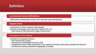
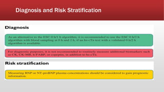
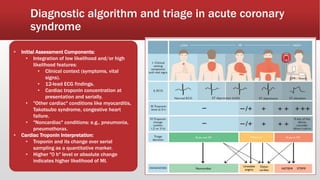
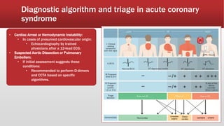
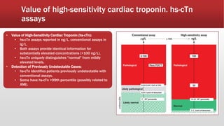
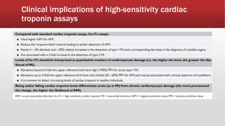
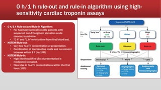
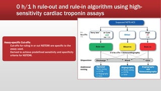
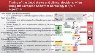
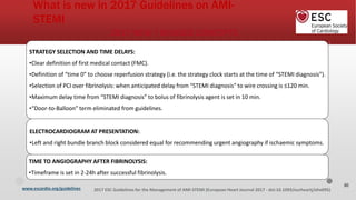
2. Definition
• AMI involves cardiomyocyte necrosis in line with acute myocardial ischemia.
Acute Myocardial Infarction (AMI) Definition:
• Combination of criteria needed for AMI diagnosis.
• Elevated or decreased cardiac biomarker, ideally hs-cTn T or I.
• Value above the 99th percentile of upper reference limit.
Diagnostic Criteria:
• Symptoms of myocardial ischemia.
• New ischemic ECG changes.
• Development of pathological Q waves on ECG.
• Imaging shows loss of viable myocardium or new regional wall motion abnormality consistent with ischemia.
• Intracoronary thrombus detected via angiography or autopsy.
Plus at least one of the following:
9. Diagnostic algorithm and triage in acute coronary
syndrome
• Initial Assessment Components:
• Integration of low likelihood and/or high
likelihood features:
• Clinical context (symptoms, vital
signs).
• 12-lead ECG findings.
• Cardiac troponin concentration at
presentation and serially.
• "Other cardiac" conditions like myocarditis,
Takotsubo syndrome, congestive heart
failure.
• "Noncardiac" conditions: e.g., pneumonia,
pneumothorax.
• Cardiac Troponin Interpretation:
• Troponin and its change over serial
sampling as a quantitative marker.
• Higher "0 h" level or absolute change
indicates higher likelihood of MI.
10. Diagnostic algorithm and triage in acute coronary
syndrome
• Cardiac Arrest or Hemodynamic Instability:
• In cases of presumed cardiovascular origin:
• Echocardiography by trained
physicians after a 12-lead ECG.
• Suspected Aortic Dissection or Pulmonary
Embolism:
• If initial assessment suggests these
conditions:
• Recommended to perform D-dimers
and CCTA based on specific
algorithms.
11. Value of high-sensitivity cardiac troponin. hs-cTn
assays
• Value of High-Sensitivity Cardiac Troponin (hs-cTn):
• hs-cTn assays reported in ng/L, conventional assays in
lg/L.
• Both assays provide identical information for
substantially elevated concentrations (>100 ng/L).
• hs-cTn uniquely distinguishes "normal" from mildly
elevated levels.
• Detection of Previously Undetectable Cases:
• hs-cTn identifies patients previously undetectable with
conventional assays.
• Some have hs-cTn >99th percentile (possibly related to
AMI).
13. 0 h/1 h rule-out and rule-in algorithm using high-
sensitivity cardiac troponin assays
• 0 h/1 h Rule-out and Rule-in Algorithm:
• For haemodynamically stable patients with
suspected non-ST-segment elevation acute
coronary syndrome.
• "0 h" and "1 h" refer to time from first blood test.
• NSTEMI Rule-out:
• Very low hs-cTn concentration at presentation.
• Combination of low baseline levels and no relevant
increase within 1 h (no 1hD).
• NSTEMI Rule-in:
• High likelihood if hs-cTn at presentation is
moderately elevated.
• Clear rise in hs-cTn concentrations within the first
hour (1hD).
14. 0 h/1 h rule-out and rule-in algorithm using high-
sensitivity cardiac troponin assays
Assay-specific Cut-offs:
Cut-offs for ruling in or out NSTEMI are specific to the
assay used.
Derived to achieve predefined sensitivity and specificity
criteria for NSTEMI.
15. Differential diagnoses of acute coronary
syndromes in the setting of acute chest pain
Bold = common and/or important differential diagnoses.
Dilated, hypertrophic and restrictive cardiomyopathies may cause angina or chest discomfort.
16. Timing of the blood draws and clinical decisions when
using the European Society of Cardiology 0 h/1 h
algorithm
CPO = Chest Pain Onset
•CPR = Cardiopulmonary Resuscitation
•ECG = Electrocardiogram/Electrocardiography
•hs-cTn = High-Sensitivity Cardiac Troponin
•MACE = Major Adverse Cardiovascular Events
•MI = Myocardial Infarction
•Time Points and Blood Collection:
• "0 h" and "1 h" are blood collection time points.
•Turn-around Time:
• Time from blood draw to reporting results (usually 1 h).
• Includes transportation, scanning, centrifugation,
analysis, and reporting.
•Consistency Across Assays:
• Turn-around time same for hs-cTn and conventional
assays on automated platforms.
•Clinical Decision Time:
• Add local turn-around time to blood draw for earliest
decision point.
• "0 h" blood: decision at 1 h with 1 h turn-around.
• "1 h" blood: results at 2 h (1 h + 1 h) with 1 h turn-
around.
European Heart Journal (2021) 42, 12891367
17. Timing of the blood draws and clinical decisions when
using the European Society of Cardiology 0 h/1 h
algorithm
•Abbreviations:CPO = Chest Pain Onset
•CPR = Cardiopulmonary Resuscitation
•ECG = Electrocardiogram/Electrocardiography
•hs-cTn = High-Sensitivity Cardiac Troponin
•MACE = Major Adverse Cardiovascular Events
•MI = Myocardial Infarction
•Time Points and Blood Collection:
• "0 h" and "1 h" are blood collection time points.
•Turn-around Time:
• Time from blood draw to reporting results (usually
1 h).
• Includes transportation, scanning, centrifugation,
analysis, and reporting.
•Consistency Across Assays:
• Turn-around time same for hs-cTn and
conventional assays on automated platforms.
•Clinical Decision Time:
• Add local turn-around time to blood draw for
earliest decision point.
• "0 h" blood: decision at 1 h with 1 h turn-around.
• "1 h" blood: results at 2 h (1 h + 1 h) with 1 h turn-
around.
18. Timing of the blood draws and clinical decisions when
using the European Society of Cardiology 0 h/1 h
algorithm
•Abbreviations:CPO = Chest Pain Onset
•CPR = Cardiopulmonary Resuscitation
•ECG = Electrocardiogram/Electrocardiography
•hs-cTn = High-Sensitivity Cardiac Troponin
•MACE = Major Adverse Cardiovascular Events
•MI = Myocardial Infarction
•Time Points and Blood Collection:
• "0 h" and "1 h" represent the time points for blood collection.
•Turn-around Time:
• Turn-around time is the duration from blood draw to reporting results to
the clinician.
• Usually around 1 h using an automated platform in the central
laboratory.
• Includes processes like blood tube transport, probe scanning,
centrifugation, analysis, and result reporting to electronic records.
•Consistency Across Assays:
• Turn-around time remains the same for hs-cTn and conventional assays
on automated platforms.
•Clinical Decision Time:
• Adding local turn-around time to blood draw time determines the earliest
clinical decision point.
• For example, at the "0 h" time point, decision time is at 1 h if local turn-
around time is 1 h.
• For blood drawn at "1 h," results arrive at 2 h (1 h + 1 h) with a 1-hour
local turn-around time.
20. STEMI Chain of Survival
Early
Symptom
Recognition &
Call for Help
EMS Evaluation
& Treatment
Emergency
Department
Evaluation &
Treatment
Reperfusion
Therapy
Total Ischaemic Time
Ibanez B, et al. Eur Heart J. 2018 Jan 7;39(2):119-177.
21. NEW
The time point when the patient is either initially
assessed by a physician, paramedic, nurse or other
trained EMS personnel who can obtain and
interpret the ECG, and deliver initial interventions
(e.g. defibrillation). FMC can be either in the
prehospital setting or upon patient arrival at the
hospital (e.g. emergency department).
Modes of patient presentation
Ibanez B, et al. Eur Heart J. 2018 Jan 7;39(2):119-177.
FMC: first Medical Contact
EMS: Emergency Medical System
PCI: Percutaneous Coronary
Intervention
23. In some cases the ECG diagnosis may be
difficult:
bundle branch block
Stable STEMI Patients
Ibanez B, et al. Eur Heart J. 2018 Jan 7;39(2):119-177.
,
FMC: first Medical Contact
EMS: Emergency Medical System
PCI: Percutaneous Coronary
Intervention
27. Modes of patient presentation
Left and right bundle branch block are considered equal for
recommending urgent angiography if ischaemic symptoms.
bundle branch block,
ventricular pacing,
hyper-acute T waves,
isolated depression in anterior
leads,
universal ST depression with
elevation in aVR
In these cases, and in the
presence of symptoms,
A primary PCI strategy (urgent
angiography and PCI if
indicated) should be followed.
28. Summary of indications for imaging and stress testing in
ST-elevation myocardial infarction patients
29. www.escardio.org/guidelines 2017 ESC Guidelines for the Management of AMI-STEMI (European Heart Journal 2017 - doi:10.1093/eurheartj/ehx095)
Relief of hypoxaemia and symptoms
Recommendations Class Level
Hypoxia
Oxygen is indicated in patients with hypoxaemia (SaO2 <90% or PaO2
<60 mmHg). I C
Routine oxygen is not recommended in patients with SaO2 ≥90%. III B
Symptoms
Titrated i.v. opioids should be considered to relieve pain. IIa C
A mild tranquillizer (usually a benzodiazepine) should be considered
in very anxious patients. IIa C
NEW
I IIa
30. Stable STEMI Patients
Modes of patient presentation, components of ischaemia time and flowchart for reperfusion strategy selection
31. FMC:EMS
FMC: Non-PCI Center
FMC: PCI Center
<10’
<10’
<10’ Time
to PCI
≤ 120’
> 120’
Strategy (Wire
crossing)
Primary PCI <90’ Reperfusion
Fibrinolysis <10’ Reperfusion
strategy (Lytic bolus)
Reperfusion
(Wire
crossing)
Primary PCI <60’
Strategy
Transfer to
PCI centre
FMC: first Medical Contact
EMS: Emergency Medical System
PCI: Percutaneous Coronary
Intervention
Patient Delay SystemDelay
Total ischaemic Time
Stable STEMI
Patients
32. Stable STEMI Patients
Strategy Clock
Time to PCI?
0
≤ 120’ > 120’
Alert & transfer to Primary PCI Fibrinolysis
PCI Centre Strategy Strategy
Reperfusion
10’ (Lytic bolus)
Transfer to
6
0
-
9
0
PCI centre ’
≥
Reperfusion
90’ (Wire
120
’
crossing)
Meet reperfusion
Rescue PCI No
criteria Yes
2hrs.
Routine PCI
24hrs. Strategy
NEW
STEMI diagnosis (defined as the time at which the ECG of a patient
with ischaemic symptoms is interpreted as presenting ST-segment
elevation or equivalent) is the time zero in the reperfusion strategy
clock.
36. NEW
NEW
NEW!
Strategy should be guided as in other STEMI patients (if
time from STEMI diagnosis to wire crossing is >120min
immediate fibrinolysis & transfer to PCI center.
Urgent angiography upon arrival regardless time from
lytics.
38. www.escardio.org/guidelines 2017 ESC Guidelines for the Management of AMI-STEMI (European Heart Journal 2017 - doi:10.1093/eurheartj/ehx095)
Acute Heart Failure and Cardiogenic
shock
Opiates to relief dyspnea and anxiety IIbB
IC
Inotropic/vasopresor agents IIbC
IIaC
Ultrafiltration IIbB
IIaB
Mechanical support IIbC
IIbC
IABP
IIbB
routine
Mechanical complications
IIIB
IIaC
39. www.escardio.org/guidelines 2017 ESC Guidelines for the Management of AMI-STEMI (European Heart Journal 2017 - doi:10.1093/eurheartj/ehx095)
Cardiac
arrest
Recommendations Class Leve
l
A primary PCI strategy is recommended in patients with
resuscitated cardiac arrest and an ECG consistent with STEMI. I B
Targeted temperature management is indicated early after
resuscitation of cardiac arrest patients who remain unresponsive. I B
It is indicated that healthcare systems implement strategies to
facilitate transfer of all patients in whom a myocardial infarction is
suspected directly to the hospital offering 24/7 PCI-mediated
reperfusion therapy via one specialized EMS.
I C
22
NEW
40. Cardiac Arrest Patients
Recommendations Classa Levelb
A primary PCI strategy is recommended in patients with I B
resuscitated cardiac arrest and an ECG consistent with STEMI.
Targeted temperature managementd is indicated early after I B
resuscitation of cardiac arrest patients who remain unresponsive.
It is indicated that healthcareIn casessystems implementwithoutstrategiesST-segmentto
I elevationC on post-
facilitate that all patientsresucitationinwhom a myocardialECGinfarctionbutis with a
h
i
g
hsuspicion of suspected are transferred directlyongoingtothe
hospitalmyocardialoffering 24/7 ischaemia, urgent reperfusion
therapy (preferablyangiographyprimaryPCI) viashouldonespecializedbe done < 2
hours after a
EMS. quick evaluation to exclude non-coronary causes.
It is indicated that all medical and paramedical personnel caring I C
for suspected myocardial infarction have access to defibrillation
equipment and are trained in basic cardiac life support.
Urgent angiography (and PCI if indicated) should be considered IIa C
in patients with resuscitated cardiac arrest without diagnostic
ST-segment elevation but with a high suspicion of ongoing
myocardial ischemia.
Prehospital cooling using a rapid infusion of large volumes of cold III B
NEW
NEW
41. www.escardio.org/guidelines 2017 ESC Guidelines for the Management of AMI-STEMI (European Heart Journal 2017 - doi:10.1093/eurheartj/ehx095)
Management of atrial fibrillation
Recommendations Class Level
Acute rate control of AF
Intravenous beta-blockers are indicated for rate control if necessary
and there are no clinical signs of acute heart failure or hypotension. I C
Intravenous amiodarone is indicated for rate control if necessary in
the presence of concomitant acute heart failure and no hypotension. I C
Intravenous digitalis should be considered for rate control if necessary
in the presence of concomitant acute heart failure and hypotension. Ila B
Cardioversion
Immediate electrical cardioversion is indicated when adequate rate
control cannot be achieved promptly with pharmacological agents
in patients with AF and ongoing ischaemia, severe haemodynamic
compromise or heart failure.
I C
A -> C
B -> C
I -> IIa
42. www.escardio.org/guidelines 2017 ESC Guidelines for the Management of AMI-STEMI (European Heart Journal 2017 - doi:10.1093/eurheartj/ehx095)
Management of atrial fibrillation
Recommendations Class Level
Intravenous amiodarone is indicated to promote electrical
cardioversion and/or decrease risk for early recurrence of AF after
electrical cardioversion in unstable patients with recent onset AF.
I C
In patients with documented de novo AF during the acute phase of
STEMI, long-term oral anticoagulation should be considered depending
on CHA2DS2-VASc score and taking concomitant antithrombotic
therapy into account.
IIa C
Digoxin is ineffective in converting recent onset AF to sinus rhythm
and is not indicated for rhythm control. III A
Calcium channel blockers and beta-blockers including sotalol are
ineffective in converting recent onset AF to sinus rhythm. III B
Prophylactic treatment with antiarrhythmic drugs to prevent AF is not
indicated. III B
A -> C
NEW
43. www.escardio.org/guidelines 2017 ESC Guidelines for the Management of AMI-STEMI (European Heart Journal 2017 - doi:10.1093/eurheartj/ehx095)
Recommendations Class Level
Intravenous beta-blocker treatment is indicated for patients with
polymorphic VT and/or VF unless contra-indicated. I B
Prompt and complete revascularization is recommended to treat
myocardial ischaemia that may be present in patients with recurrent
VT and/or VF.
I C
Intravenous amiodarone is recommended for treatment of recurrent
polymorphic VT. I C
Correction of electrolyte imbalances (especially hypokalaemia and
hypomagnesemia) is recommended in patients with VT and/or VF. I C
Management of ventricular arrhythmias and
conduction disturbances in the acute phase
44. www.escardio.org/guidelines 2017 ESC Guidelines for the Management of AMI-STEMI (European Heart Journal 2017 - doi:10.1093/eurheartj/ehx095)
Recommendations Class Level
In cases of sinus bradycardia with haemodynamic intolerance or high
degree AV block without stable escape rhythm:
• i.v. positive chronotropic medication (epinephrine, vasopressin
and/or atropine) is indicated, I C
• temporary pacing is indicated in cases of failure to respond to
positive chronotropic medication, I C
• urgent angiography with a view to revascularization is indicated if
the patient has not received previous reperfusion therapy. I C
Management of ventricular arrhythmias and
conduction disturbances in the acute phase
45. www.escardio.org/guidelines 2017 ESC Guidelines for the Management of AMI-STEMI (European Heart Journal 2017 - doi:10.1093/eurheartj/ehx095)
Recommendations Class Level
Intravenous amiodarone should be considered for recurrent VT with
haemodynamic intolerance despite repetitive electrical cardioversion. IIa C
Transvenous catheter pace termination and/or overdrive pacing should
be considered if VT cannot be controlled by repetitive electrical
cardioversion.
IIa C
Radiofrequency catheter ablation at a specialized ablation centre
followed by ICD implantation should be considered in patients with
recurrent VT, VF, or electrical storm despite complete revascularization
and optimal medical therapy.
IIa C
Management of ventricular arrhythmias and
conduction disturbances in the acute phase
NEW
46. www.escardio.org/guidelines 2017 ESC Guidelines for the Management of AMI-STEMI (European Heart Journal 2017 - doi:10.1093/eurheartj/ehx095)
Recommendations Class Level
Recurrent VT with haemodynamic repercussion despite repetitive
electrical cardioversion may be treated with lidocaine if beta-blockers,
amiodarone, and overdrive stimulation are not effective/applicable.
IIb C
Prophylactic treatment with antiarrhythmic drugs is not indicated and
may be harmful. III B
Asymptomatic and haemodynamically irrelevant ventricular
arrhythmias should not be treated with antiarrhythmic drugs. III C
Management of ventricular arrhythmias and
conduction disturbances in the acute phase
NEW
NEW
IIa -> IIb
47. www.escardio.org/guidelines 2017 ESC Guidelines for the Management of AMI-STEMI (European Heart Journal 2017 - doi:10.1093/eurheartj/ehx095)
2017 NEW / REVISED CONCEPTS
STRATEGY SELECTION AND TIME DELAYS:
•Clear definition of first medical contact (FMC).
•Definition of “time 0” to choose reperfusion strategy (i.e. the strategy clock starts at the time of “STEMI diagnosis”).
•Selection of PCI over fibrinolysis: when anticipated delay from “STEMI diagnosis” to wire crossing is ≤120 min.
•Maximum delay time from “STEMI diagnosis” to bolus of fibrinolysis agent is set in 10 min.
•“Door-to-Balloon” term eliminated from guidelines.
ELECTROCARDIOGRAM AT PRESENTATION:
•Left and right bundle branch block considered equal for recommending urgent angiography if ischaemic symptoms.
TIME TO ANGIOGRAPHY AFTER FIBRINOLYSIS:
•Timeframe is set in 2-24h after successful fibrinolysis.
What is new in 2017 Guidelines on AMI-
STEMI
30
48. Title and Content Layout with List
▪ Add your first bullet point here
▪ Add your second bullet point here
▪ Add your third bullet point here
49. Title and Content Layout with Chart
4.3
2.5
3.5
4.5
2.4
4.4
1.8
2.8
2
2
3
5
CATEGORY 1 CATEGORY 2 CATEGORY 3 CATEGORY 4
Series 1 Series 2 Series 3
50. Two Content Layout with Table
▪ First bullet point here
▪ Second bullet point here
▪ Third bullet point here
Class Group A Group B
Class 1 82 95
Class 2 76 88
Class 3 84 90
51. Two Content Layout with SmartArt
▪ First bullet point here
▪ Second bullet point here
▪ Third bullet point here
Step 3
Task Description Task Description
Step 2
Task Description Task Description
Step 1
Task Description Task Description