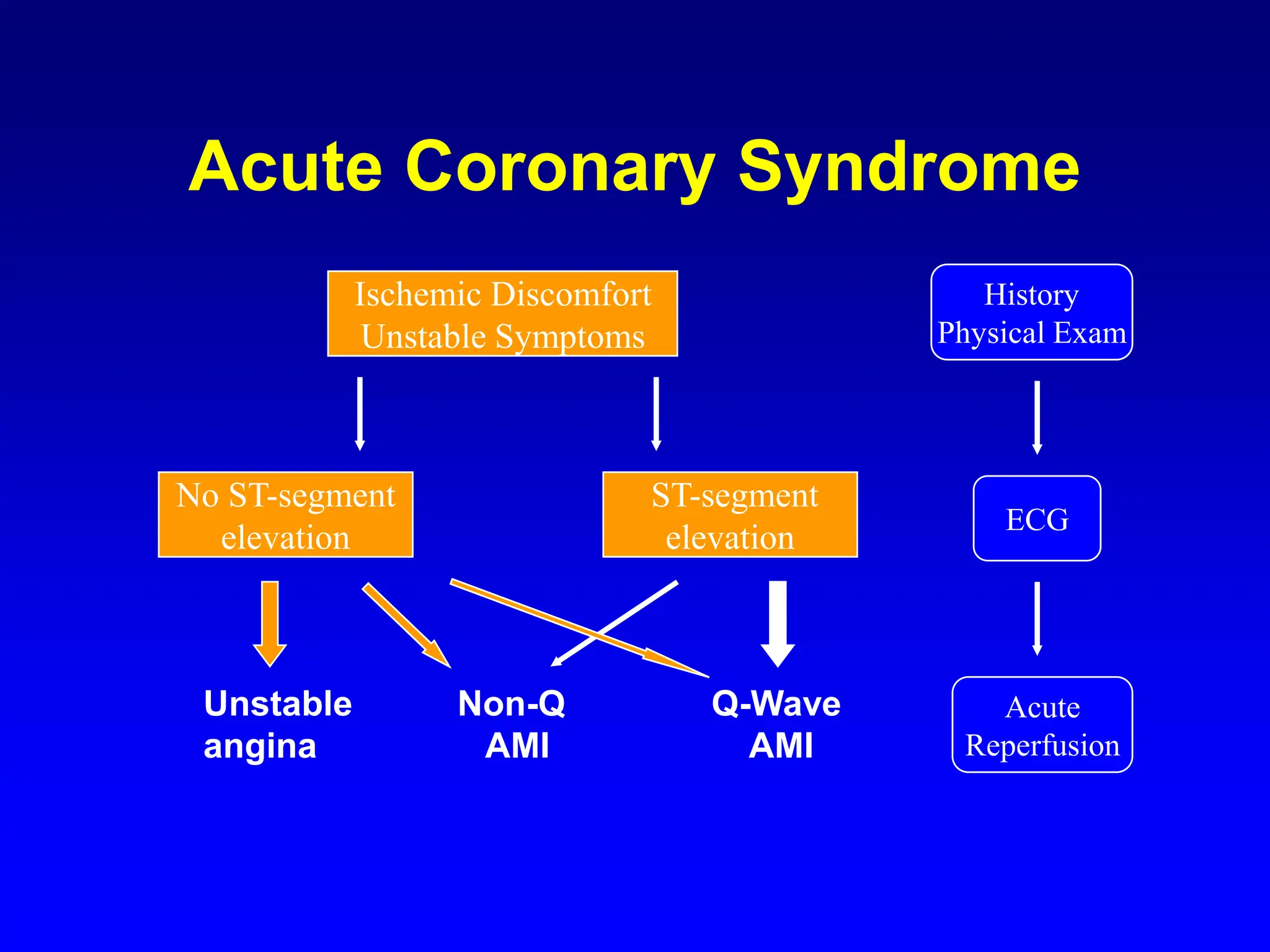
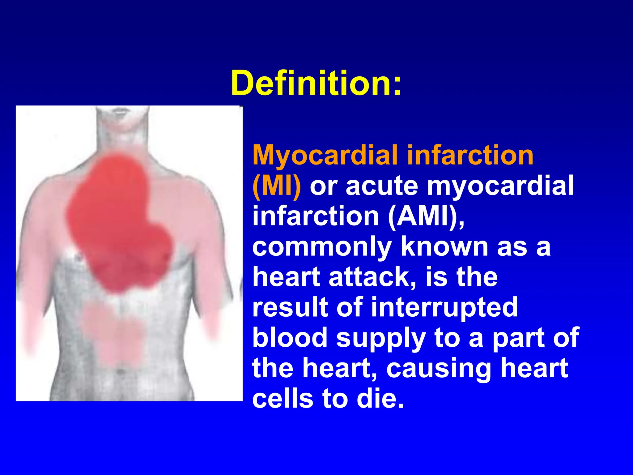
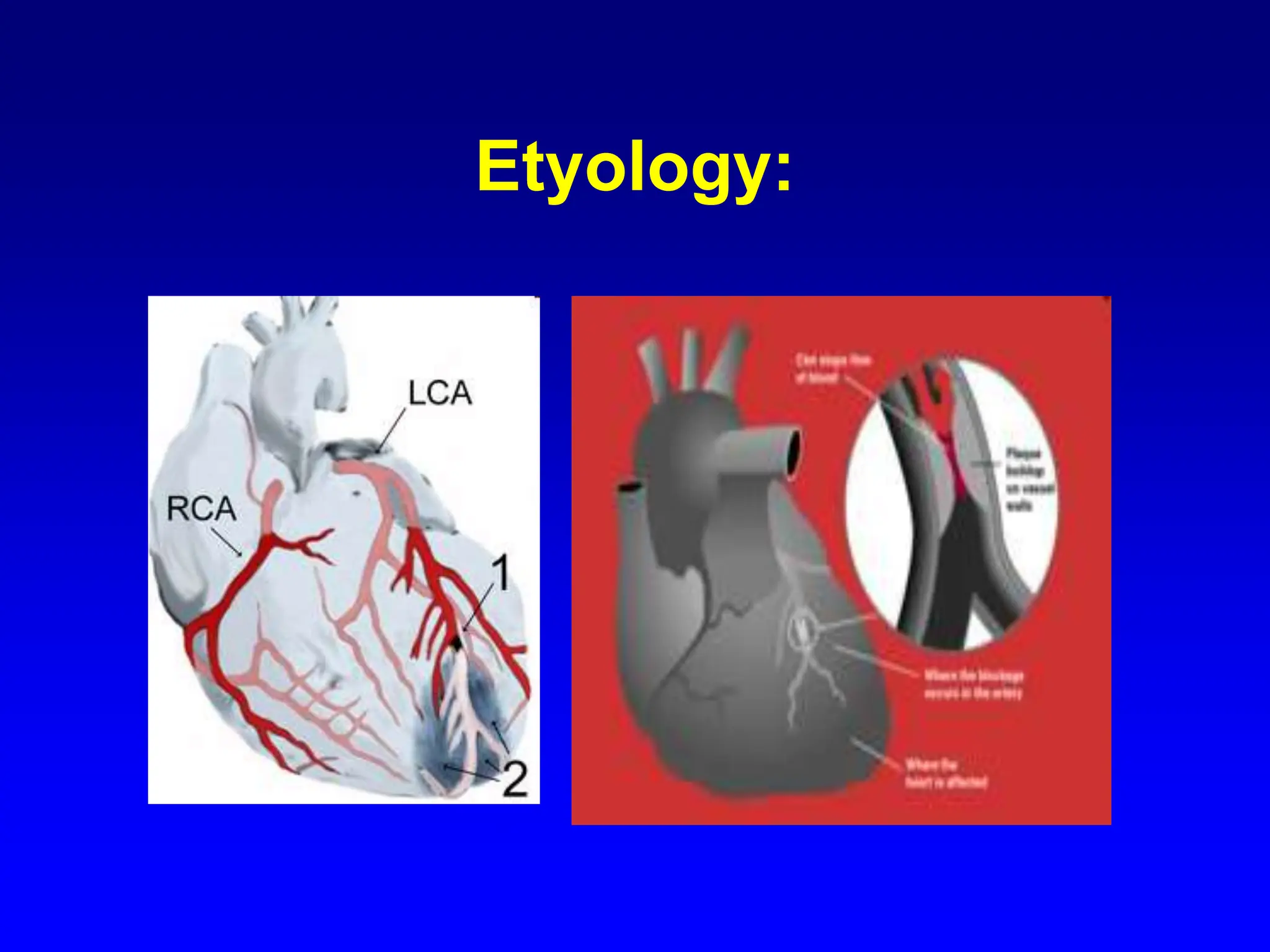
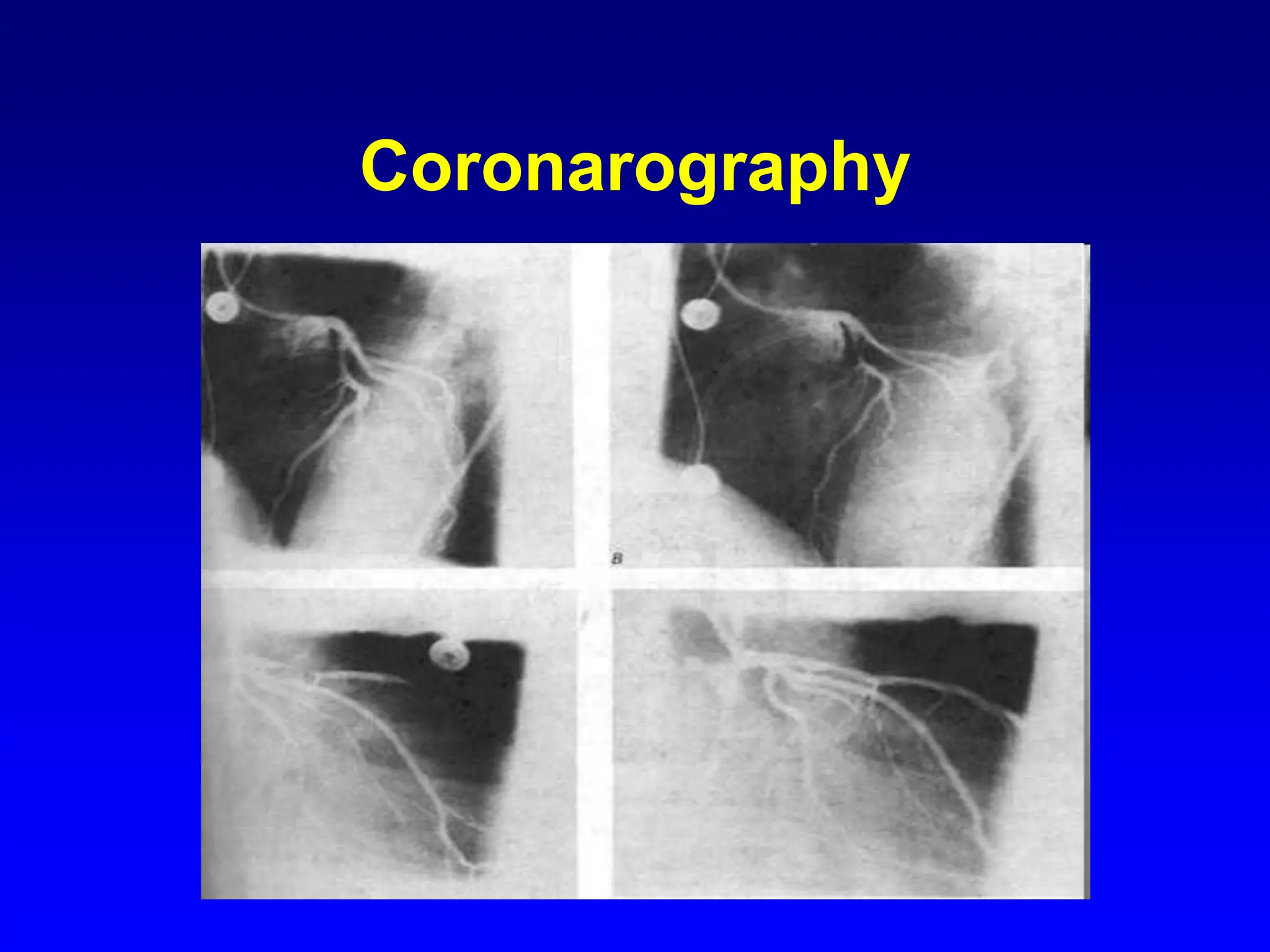
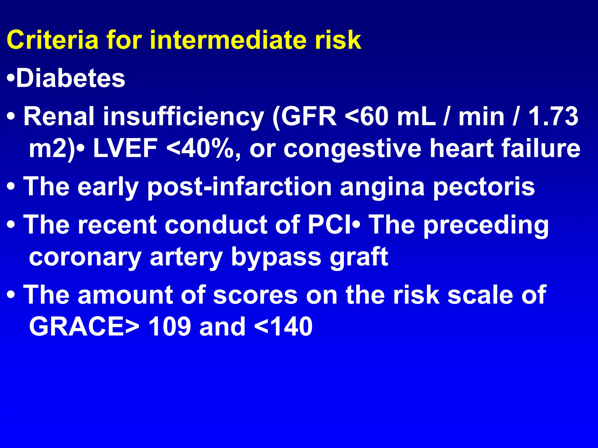
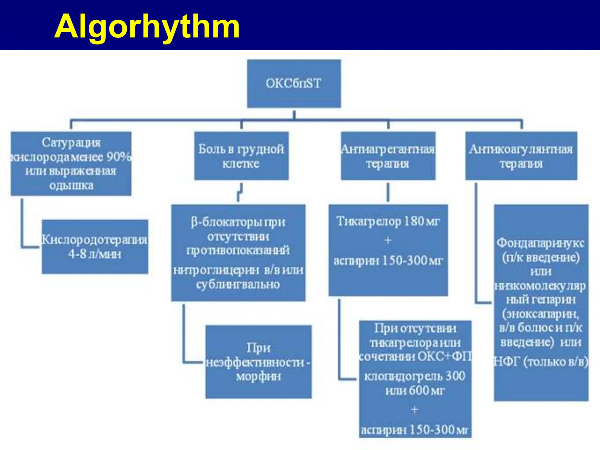
Acute coronary syndrome.
Diagnosis, treatment and
prophylaxis of myocardial
infarction.
Dr Manoj Godara
MBBS
Worldwide Statistics
Each year:
• > 4 million patients are admitted with
unstable angina and acute MI
• > 900,000 patients undergo PTCA
with or without stent
Acute Coronary Syndrome
Ischemic Discomfort
Unstable Symptoms
No ST-segment
elevation
ST-segment
elevation
Unstable Non-Q Q-Wave
angina AMI AMI
ECG
Acute
Reperfusion
History
Physical Exam
Acute Coronary Syndrome
• The spectrum of clinical conditions
ranging from:
– unstable angina
– non-Q wave MI
– Q-wave MI
• characterized by the common
pathophysiology of a disrupted
atheroslerotic plaque
Definition:
• Myocardial infarction
(MI) or acute myocardial
infarction (AMI),
commonly known as a
heart attack, is the
result of interrupted
blood supply to a part of
the heart, causing heart
cells to die.
Etyology:
Risk factors for atherosclerosis
are generally risk factors for
myocardial infarction
• Diabetes (with or without insulin
resistance) – the single most important
risk factor for ischaemic heart disease
(IHD)
• Tobacco smoking
• Air pollution
• Hypercholesterolemia (more accurately
hyperlipoproteinemia, especially high low
density lipoprotein and low high density
lipoprotein)
Risk factors for atherosclerosis
• Low HDL
• High Triglycerides
• High blood pressure
• Family history of ischaemic heart
disease (IHD)
• Obesity
Risk factors for atherosclerosis
• Age
• Hyperhomocysteinemia (high
homocysteine, a toxic blood amino acid
that is elevated when intakes of vitamins
B2, B6, B12 and folic acid are insufficient)
• Stress — Occupations with high stress
index are known to have susceptibility for
atherosclerosis
• Alcohol — Studies show that prolonged
exposure to high quantities of alcohol can
increase the risk of heart attack
• Males are more at risk than females.
The characteristics of an acute MI
depend on the following factors:
1) location, severity, rate of coronary atherosclerotic
obstruction
2) size of area perfused by occluded coronary
blood vessels
3) duration of occlusion
4) myocardial needs of affected area
5) degree of development of collateral blood vessels
6) site and severity of vasospasm (if present)
7) changes in BP, HR, heart rhythm
Symptoms:
• Patients with acute MI often complain of severe
retrosternal pain lasting more than 30 minutes
and not relieved by sublingual nitroglycerin;
• Nausea, diaphoresis, and vomiting are also
common.
• It is important to note that clinical presentation
and intensity of symptoms vary widely among
individuals. This is especially true in women, the
elderly, and diabetics who may present with
atypical or only mild symptoms that go
unrecognized. Sometimes there is no pain
present, and patients may present with a
complication of acute MI (see below) such as
pulmonary edema.
Signs:
• Increased HR and BP (but
bradycardia is also not uncommon
as a presentation). S3 or S4 sounds.
New murmurs due to papillary
muscle ischemia and/or rupture.
Investigations of Acute MI
ECG: An ECG should be obtained as soon
as possible. Look for Q waves in the
tracings although they need not be
present in acute MI. Also look for ST
segment elevation or depression and T
wave inversion. It is important to keep in
mind that 10% of patients presenting with
acute MI will have normal ECG tracings
therefore it would be prudent to repeat the
ECG later.
Investigations of Acute MI
• Cardiac Enzyme Studies: CK-MB is
the most sensitive and specific
marker of MI. The blood levels of CK-
MB rise by 6-8 hrs and peak at 12-48
hrs later. LDH levels are useful if the
patient presents days after a
suspected MI.
Investigations of Acute MI
ECG:
ECG:
ECG:
ECG:
Coronarography
Treatment of Acute MI
• supplemental oxygen
• - continuous ECG monitoring
• - aspirin 160-325 mg PO
• - morphine 2-4 mg IV q 5-15 min. if needed
• - thrombolytic therapy if patient presents
within 6 hrs of MI onset: tPA,
streptokinase
• - direct PTCA
• - bed rest for 24-36 hrs and admission to
CCU
Acute coronary syndrome without
ST elevation
• The main aim of the outpatient phase
a diagnosis and immediate
transportation to the profile hospital.
The basic diagnostic
measurement.
Laboratory tests:
• Highly sensitive troponin I at admission and
again after 1-3 hours, or 0-1 hours after the
presence of the corresponding analyzers, (all
in quantitative methods measurement).
• CBC (hemoglobin, hematocrit, platelets, etc.).
• CUT
• serum creatinine, glomerular filtration rate
• The lipid profile (cholesterol, LDL, HDL,
triglycerides)
• Blood Glucose
• Potassium
Risk stratification of patients with based
on the presence of clinical risk factors
and quantitative risk assessment on the
scale of GRACE
The criteria of very high risk
• Hemodynamic instability or cardiogenic shock
• Recurrent or continuing chest pain, resistant to
medical treatment
• life-threatening arrhythmias or cardiac arrest
• The mechanical complications of MI
• Acute heart failure with refractory angina or ST-
segment deviation
• Repeated dynamic changes in ST segment or T
wave
Risk stratification of patients with based
on the presence of clinical risk factors
and quantitative risk assessment on the
scale of GRACE
The criteria for high risk
• Elevation or lowering of cardiac
troponins associated with MI•
Dynamic changes of the ST segment
or T wave (symptomatic or
asymptomatic)
• The amount of points on the scale of
GRACE> 140
Criteria for intermediate risk
•Diabetes
• Renal insufficiency (GFR <60 mL / min / 1.73
m2)• LVEF <40%, or congestive heart failure
• The early post-infarction angina pectoris
• The recent conduct of PCI• The preceding
coronary artery bypass graft
• The amount of scores on the risk scale of
GRACE> 109 and <140
Criteria of low-risk
• Any characteristics not mentioned
above
• Electronic calculator of GRACE scale:
http://www.outcomes.org/grace
Complications of Acute MI
1. cardiogenic shock when 40+% of LV
infarcted
2. cardiac perforation - external or
interventricular
3. embolism
4. CHF
5. papillary muscle rupture
6. rhythm disturbances
7. pericarditis - autoimmune (Dressler’s
syndrome)
The list of Essential Medicines:
• 1. Oxygen inhalation (medicinal gas)
• 2. Metoprolol tartrate (1% vials 5,0ml; 50 mg
tablet)
• 3. Nitroglycerin * (0.1% solution for injection in
vials of 10 ml; 0.0005 g tablet or spray).
• 4. Morphine (injection ampoule in 1 ml of 1.0%)
• 5. Aspirin (tablet, 500 mg)
• 6. Ticagrelor (tablet 90 mg)
• 7. clopidogrel (tablet, 75 mg)
• 8. Fondaparinux (0.5 ml syringe 2.5 mg)
• 9. enoxaparin sodium (0.2 and 0.4 syringe ml)
• 10. Physiological saline (0.9% 200 ml bottle)
Algorhythm
Thank you!

Acute coronary syndrome. Diagnosis, treatment and prophylaxis of myocardial infarction

  • 1.
    Acute coronary syndrome. Diagnosis,treatment and prophylaxis of myocardial infarction. Dr Manoj Godara MBBS
  • 2.
    Worldwide Statistics Each year: •> 4 million patients are admitted with unstable angina and acute MI • > 900,000 patients undergo PTCA with or without stent
  • 3.
    Acute Coronary Syndrome IschemicDiscomfort Unstable Symptoms No ST-segment elevation ST-segment elevation Unstable Non-Q Q-Wave angina AMI AMI ECG Acute Reperfusion History Physical Exam
  • 4.
    Acute Coronary Syndrome •The spectrum of clinical conditions ranging from: – unstable angina – non-Q wave MI – Q-wave MI • characterized by the common pathophysiology of a disrupted atheroslerotic plaque
  • 9.
    Definition: • Myocardial infarction (MI)or acute myocardial infarction (AMI), commonly known as a heart attack, is the result of interrupted blood supply to a part of the heart, causing heart cells to die.
  • 10.
  • 11.
    Risk factors foratherosclerosis are generally risk factors for myocardial infarction • Diabetes (with or without insulin resistance) – the single most important risk factor for ischaemic heart disease (IHD) • Tobacco smoking • Air pollution • Hypercholesterolemia (more accurately hyperlipoproteinemia, especially high low density lipoprotein and low high density lipoprotein)
  • 12.
    Risk factors foratherosclerosis • Low HDL • High Triglycerides • High blood pressure • Family history of ischaemic heart disease (IHD) • Obesity
  • 13.
    Risk factors foratherosclerosis • Age • Hyperhomocysteinemia (high homocysteine, a toxic blood amino acid that is elevated when intakes of vitamins B2, B6, B12 and folic acid are insufficient) • Stress — Occupations with high stress index are known to have susceptibility for atherosclerosis • Alcohol — Studies show that prolonged exposure to high quantities of alcohol can increase the risk of heart attack • Males are more at risk than females.
  • 14.
    The characteristics ofan acute MI depend on the following factors: 1) location, severity, rate of coronary atherosclerotic obstruction 2) size of area perfused by occluded coronary blood vessels 3) duration of occlusion 4) myocardial needs of affected area 5) degree of development of collateral blood vessels 6) site and severity of vasospasm (if present) 7) changes in BP, HR, heart rhythm
  • 15.
    Symptoms: • Patients withacute MI often complain of severe retrosternal pain lasting more than 30 minutes and not relieved by sublingual nitroglycerin; • Nausea, diaphoresis, and vomiting are also common. • It is important to note that clinical presentation and intensity of symptoms vary widely among individuals. This is especially true in women, the elderly, and diabetics who may present with atypical or only mild symptoms that go unrecognized. Sometimes there is no pain present, and patients may present with a complication of acute MI (see below) such as pulmonary edema.
  • 16.
    Signs: • Increased HRand BP (but bradycardia is also not uncommon as a presentation). S3 or S4 sounds. New murmurs due to papillary muscle ischemia and/or rupture.
  • 17.
    Investigations of AcuteMI ECG: An ECG should be obtained as soon as possible. Look for Q waves in the tracings although they need not be present in acute MI. Also look for ST segment elevation or depression and T wave inversion. It is important to keep in mind that 10% of patients presenting with acute MI will have normal ECG tracings therefore it would be prudent to repeat the ECG later.
  • 18.
    Investigations of AcuteMI • Cardiac Enzyme Studies: CK-MB is the most sensitive and specific marker of MI. The blood levels of CK- MB rise by 6-8 hrs and peak at 12-48 hrs later. LDH levels are useful if the patient presents days after a suspected MI.
  • 19.
  • 20.
  • 21.
  • 22.
  • 23.
  • 24.
  • 25.
  • 26.
    Treatment of AcuteMI • supplemental oxygen • - continuous ECG monitoring • - aspirin 160-325 mg PO • - morphine 2-4 mg IV q 5-15 min. if needed • - thrombolytic therapy if patient presents within 6 hrs of MI onset: tPA, streptokinase • - direct PTCA • - bed rest for 24-36 hrs and admission to CCU
  • 27.
    Acute coronary syndromewithout ST elevation • The main aim of the outpatient phase a diagnosis and immediate transportation to the profile hospital.
  • 29.
    The basic diagnostic measurement. Laboratorytests: • Highly sensitive troponin I at admission and again after 1-3 hours, or 0-1 hours after the presence of the corresponding analyzers, (all in quantitative methods measurement). • CBC (hemoglobin, hematocrit, platelets, etc.). • CUT • serum creatinine, glomerular filtration rate • The lipid profile (cholesterol, LDL, HDL, triglycerides) • Blood Glucose • Potassium
  • 30.
    Risk stratification ofpatients with based on the presence of clinical risk factors and quantitative risk assessment on the scale of GRACE The criteria of very high risk • Hemodynamic instability or cardiogenic shock • Recurrent or continuing chest pain, resistant to medical treatment • life-threatening arrhythmias or cardiac arrest • The mechanical complications of MI • Acute heart failure with refractory angina or ST- segment deviation • Repeated dynamic changes in ST segment or T wave
  • 31.
    Risk stratification ofpatients with based on the presence of clinical risk factors and quantitative risk assessment on the scale of GRACE The criteria for high risk • Elevation or lowering of cardiac troponins associated with MI• Dynamic changes of the ST segment or T wave (symptomatic or asymptomatic) • The amount of points on the scale of GRACE> 140
  • 32.
    Criteria for intermediaterisk •Diabetes • Renal insufficiency (GFR <60 mL / min / 1.73 m2)• LVEF <40%, or congestive heart failure • The early post-infarction angina pectoris • The recent conduct of PCI• The preceding coronary artery bypass graft • The amount of scores on the risk scale of GRACE> 109 and <140
  • 33.
    Criteria of low-risk •Any characteristics not mentioned above • Electronic calculator of GRACE scale: http://www.outcomes.org/grace
  • 34.
    Complications of AcuteMI 1. cardiogenic shock when 40+% of LV infarcted 2. cardiac perforation - external or interventricular 3. embolism 4. CHF 5. papillary muscle rupture 6. rhythm disturbances 7. pericarditis - autoimmune (Dressler’s syndrome)
  • 37.
    The list ofEssential Medicines: • 1. Oxygen inhalation (medicinal gas) • 2. Metoprolol tartrate (1% vials 5,0ml; 50 mg tablet) • 3. Nitroglycerin * (0.1% solution for injection in vials of 10 ml; 0.0005 g tablet or spray). • 4. Morphine (injection ampoule in 1 ml of 1.0%) • 5. Aspirin (tablet, 500 mg) • 6. Ticagrelor (tablet 90 mg) • 7. clopidogrel (tablet, 75 mg) • 8. Fondaparinux (0.5 ml syringe 2.5 mg) • 9. enoxaparin sodium (0.2 and 0.4 syringe ml) • 10. Physiological saline (0.9% 200 ml bottle)
  • 38.
  • 39.