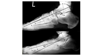
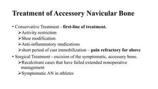
Accessory Navicular Bone
10’ talk
By Jifar J ( OSR Ⅰ)
Zewditu Tadesse 35/F
?Lt Accessory Navicular Bone
• Presented with Lt foot mass of 01 year.
• She complains the mass as associated with pain when walking.
• Otherwise, no Hx of trauma to the site/known medical chronic illness
• P/E:
• GA: well, looking
• V/S: stable
• HEENT: PC, NIS
• MSK: there is a bony prominence over Lt medial foot area with
tenderness to palpation. Bilaterally flat foot.
X - Ray
Introduction
• Accessory navicular is a normal variant seen in up to 12% of
population. ( Symptomatic < 1%)
• Accessory Navicular is a common idiopathic condition of the foot that
presents with a plantar medial enlargement of the navicular bone.
Relevant Anatomy
• An extensive network of plantar and dorsal ligaments attaches to the
navicular and rigidly stabilizes the midfoot.
plantar calcaneonavicular (spring) ligament
bifurcate ligament
dorsal talonavicular ligament
• The only tendon that inserts into the navicular is the anterior portion of
tibialis posterior tendon medially.
Cont’d
• Blood supply is provided through small branches from the dorsalis
pedis and tibialis posterior arteries from the medial pole and the dorsal
and plantar surfaces.
dorsalis pedis artery (dorsal aspect)
medial plantar artery (plantar aspect)
anastomosis between dorsalis pedis and medial plantar arteries
(medial surface of tuberosity)
Classification of Accessory Navicular Bone
Pathophysiology
• Pain generator theories
micro-fracture through the cartilaginous synchondrosis.
acute and chronic inflammation - tendinosis
cellular proliferation indicative of attempted repair.
• Chronic chondro-osseous tensile failure can occur in this condition
and is responsible for the clinical findings.
Cont’d
• The presence of type I or II accessory navicular is also a cause of
Posterior Tibial tendinopathy.
• Autosomal dominant inheritance has been reported.
• Flat feet and posterior tibial tendon insufficiency have been reported
as the association.
Clinical Presentation
Symptoms
• Majority of patients are asymptomatic.
more commonly symptomatic in females
• Medial arch pain – often worse with overuse.
• Condition may also be bilateral.
P/E
• Firm, warmth/redness and tender at the medial and plantar aspect of
the navicular bone.
Reported bilateral incidence is 50-90%
Differential Diagnosis
• Flexible pes planovalgus
• Posterior tibial tendon insufficiency ( PTTI )
• Tarsal coalition – talocalcaneal
• Stress fracture
• Bone tumor
• Kohler’s disease
Imaging
• X – ray
AP
Lateral
External oblique - best
• Ultrasonography
could detect the osseous contours of the accessory navicular and
medial navicular.
assessment of the integrity of the posterior tibialis tendon.
• MRI
Determine the size and type of AN
Assess posterior tibial tendon
Bone marrow edema
Treatment of Accessory Navicular Bone
• Conservative Treatment - first-line of treatment.
Activity restriction
Shoe modification
Anti-inflammatory medications
short period of cast immobilization – pain refractory for above
• Surgical Treatment - excision of the symptomatic, accessory bone.
Recalcitrant cases that have failed extended nonoperative
management
Symptomatic AN in athletes
Cont’d
• Conservative treatment should be maintained for at least 4- 6 months
before considering any surgical intervention.
• Most children and adolescents who have a symptomatic accessory
tarsal navicular bone become asymptomatic when they reach skeletal
maturity
Complications
• The most common complication is persistent medial prominence and
pain when the body of the navicular is not trimmed sufficiently.
Reference
• Orthobullets
• https://boneandspine.com/accessory-navicular-bone/#clinical-
presentation

Accessory Navicular Bone.pptx

  • 1.
    Accessory Navicular Bone 10’talk By Jifar J ( OSR Ⅰ)
  • 2.
    Zewditu Tadesse 35/F ?LtAccessory Navicular Bone • Presented with Lt foot mass of 01 year. • She complains the mass as associated with pain when walking. • Otherwise, no Hx of trauma to the site/known medical chronic illness • P/E: • GA: well, looking • V/S: stable • HEENT: PC, NIS • MSK: there is a bony prominence over Lt medial foot area with tenderness to palpation. Bilaterally flat foot.
  • 3.
  • 5.
    Introduction • Accessory navicularis a normal variant seen in up to 12% of population. ( Symptomatic < 1%) • Accessory Navicular is a common idiopathic condition of the foot that presents with a plantar medial enlargement of the navicular bone.
  • 6.
    Relevant Anatomy • Anextensive network of plantar and dorsal ligaments attaches to the navicular and rigidly stabilizes the midfoot. plantar calcaneonavicular (spring) ligament bifurcate ligament dorsal talonavicular ligament • The only tendon that inserts into the navicular is the anterior portion of tibialis posterior tendon medially.
  • 9.
    Cont’d • Blood supplyis provided through small branches from the dorsalis pedis and tibialis posterior arteries from the medial pole and the dorsal and plantar surfaces. dorsalis pedis artery (dorsal aspect) medial plantar artery (plantar aspect) anastomosis between dorsalis pedis and medial plantar arteries (medial surface of tuberosity)
  • 10.
  • 11.
    Pathophysiology • Pain generatortheories micro-fracture through the cartilaginous synchondrosis. acute and chronic inflammation - tendinosis cellular proliferation indicative of attempted repair. • Chronic chondro-osseous tensile failure can occur in this condition and is responsible for the clinical findings.
  • 12.
    Cont’d • The presenceof type I or II accessory navicular is also a cause of Posterior Tibial tendinopathy. • Autosomal dominant inheritance has been reported. • Flat feet and posterior tibial tendon insufficiency have been reported as the association.
  • 13.
    Clinical Presentation Symptoms • Majorityof patients are asymptomatic. more commonly symptomatic in females • Medial arch pain – often worse with overuse. • Condition may also be bilateral. P/E • Firm, warmth/redness and tender at the medial and plantar aspect of the navicular bone. Reported bilateral incidence is 50-90%
  • 14.
    Differential Diagnosis • Flexiblepes planovalgus • Posterior tibial tendon insufficiency ( PTTI ) • Tarsal coalition – talocalcaneal • Stress fracture • Bone tumor • Kohler’s disease
  • 15.
    Imaging • X –ray AP Lateral External oblique - best • Ultrasonography could detect the osseous contours of the accessory navicular and medial navicular. assessment of the integrity of the posterior tibialis tendon. • MRI Determine the size and type of AN Assess posterior tibial tendon Bone marrow edema
  • 18.
    Treatment of AccessoryNavicular Bone • Conservative Treatment - first-line of treatment. Activity restriction Shoe modification Anti-inflammatory medications short period of cast immobilization – pain refractory for above • Surgical Treatment - excision of the symptomatic, accessory bone. Recalcitrant cases that have failed extended nonoperative management Symptomatic AN in athletes
  • 20.
    Cont’d • Conservative treatmentshould be maintained for at least 4- 6 months before considering any surgical intervention. • Most children and adolescents who have a symptomatic accessory tarsal navicular bone become asymptomatic when they reach skeletal maturity
  • 24.
    Complications • The mostcommon complication is persistent medial prominence and pain when the body of the navicular is not trimmed sufficiently.
  • 25.

Editor's Notes

  • #11 Type III, "the cornuate navicular," represents the possible end stage of Type II.
  • #17 The angle between the 2 lines is referred to as Meary's angle and a plantar apex angle of greater than 4 degrees indicates collapse of the longitudinal arch.
  • #19  If shoe wear and activity modifications do not help, then a brief period of cast immobilization may be required. Condition often becomes symptomatic in young athletes from medial pressure of the accessory ossicle against athletic shoes, causing complaints of pain and tenderness along the medial midfoot region.
  • #20 Arch supports can be helpful, but the University of California, Berkely (UCBL) orthosis has shown to be particularly helpful given that it inverts the heel with walking and may directly decrease pressure on the accessory bone, thereby decreasing symptoms with activity.