By the endof
this session, you
should be able
to:
Understand the
organisation within
organisms
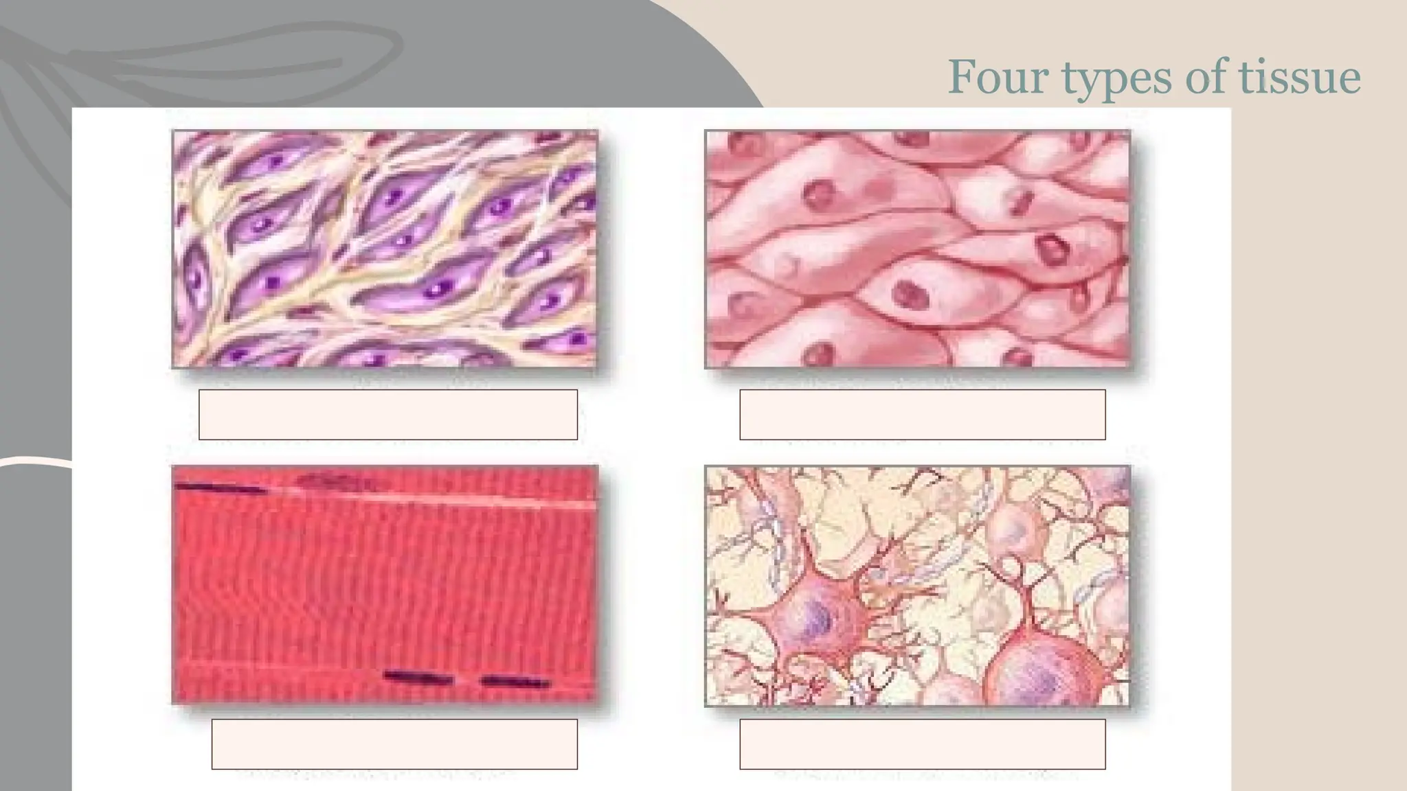
Recall the four main
classifications of tissue
types
Classify each tissue type
into sub-groups
Identify examples of
tissues within each group
Describe the structure and
function of a range of
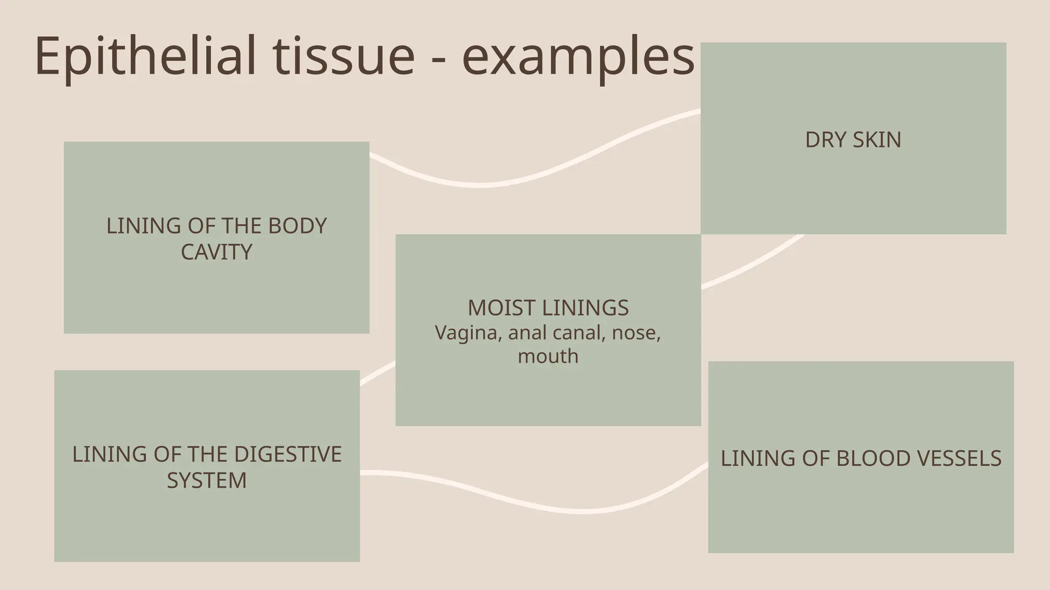
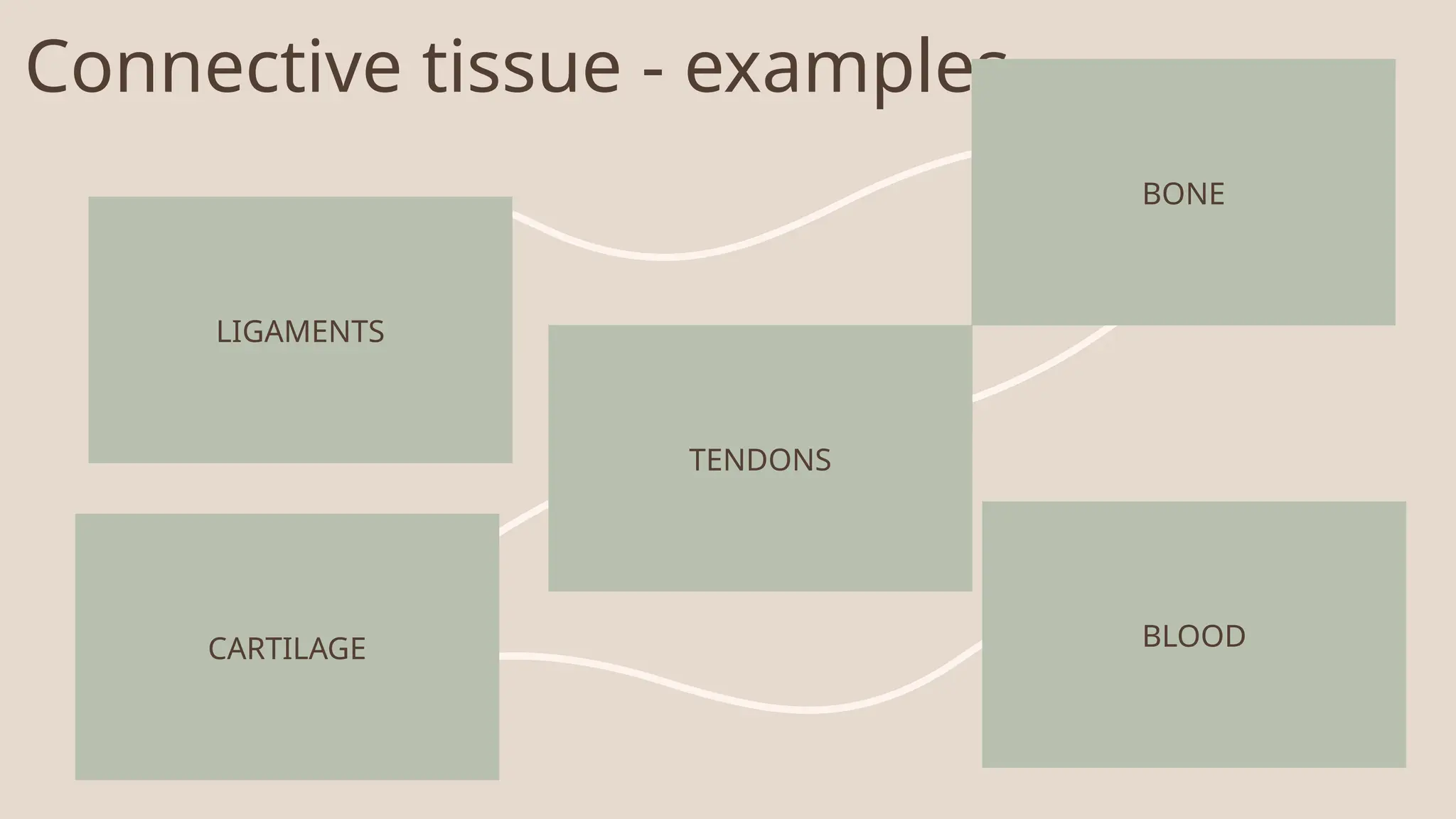
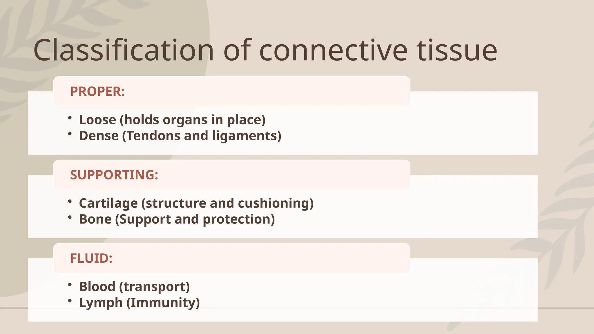
Epithelial tissue -examples
LINING OF THE BODY
CAVITY
DRY SKIN
MOIST LININGS
Vagina, anal canal, nose,
mouth
LINING OF THE DIGESTIVE
SYSTEM
LINING OF BLOOD VESSELS
8.
Functions of epithelialtissue
Protection: epithelial tissue covers all surfaces of the body, for example, the
skin and the lining of body cavities and hollow organs.
Transport: epithelial tissue is located where there is the rapid transport of
substances e.g. in the alveoli of the respiratory tract.
Lining: epithelial tissue lines the internal cavities and tubes of the body, for
example, the respiratory and digestive tracts. The functions of protection and
lining are closely interlinked.
Secretion: epithelial tissue produces a variety of substances with specific
homeostatic functions, for example, sweat and tears.
9.
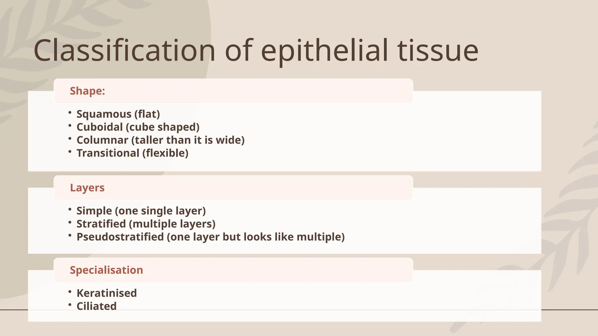
Classification of epithelialtissue
• Squamous (flat)
• Cuboidal (cube shaped)
• Columnar (taller than it is wide)
• Transitional (flexible)
Shape:
• Simple (one single layer)
• Stratified (multiple layers)
• Pseudostratified (one layer but looks like multiple)
Layers:
• Keratinised
• Ciliated
Specialisation:
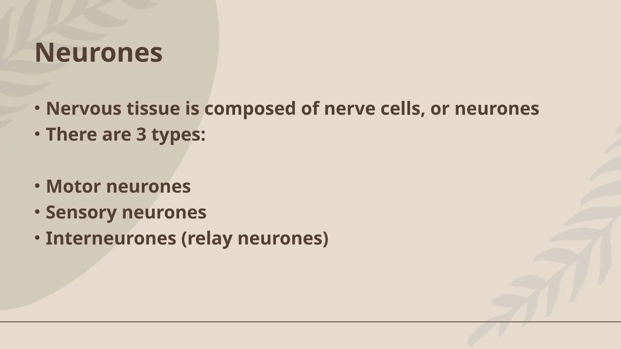
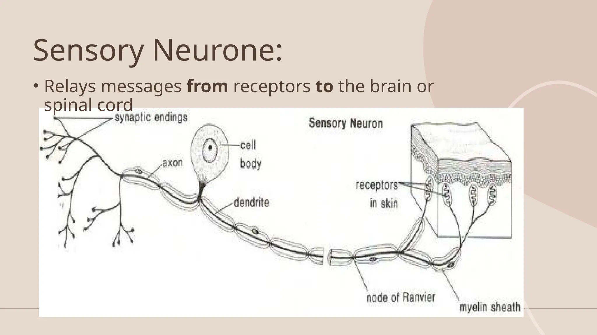
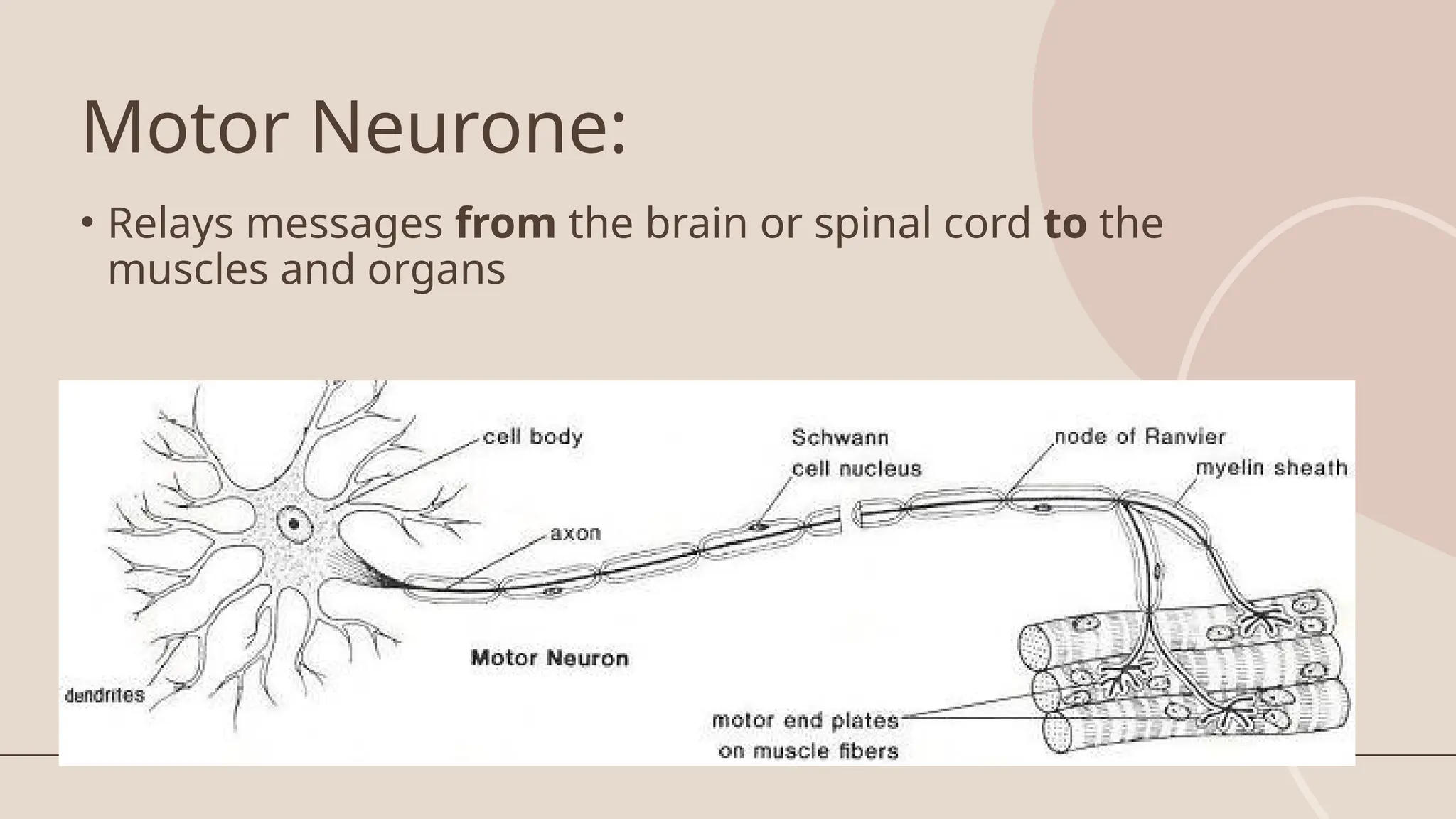
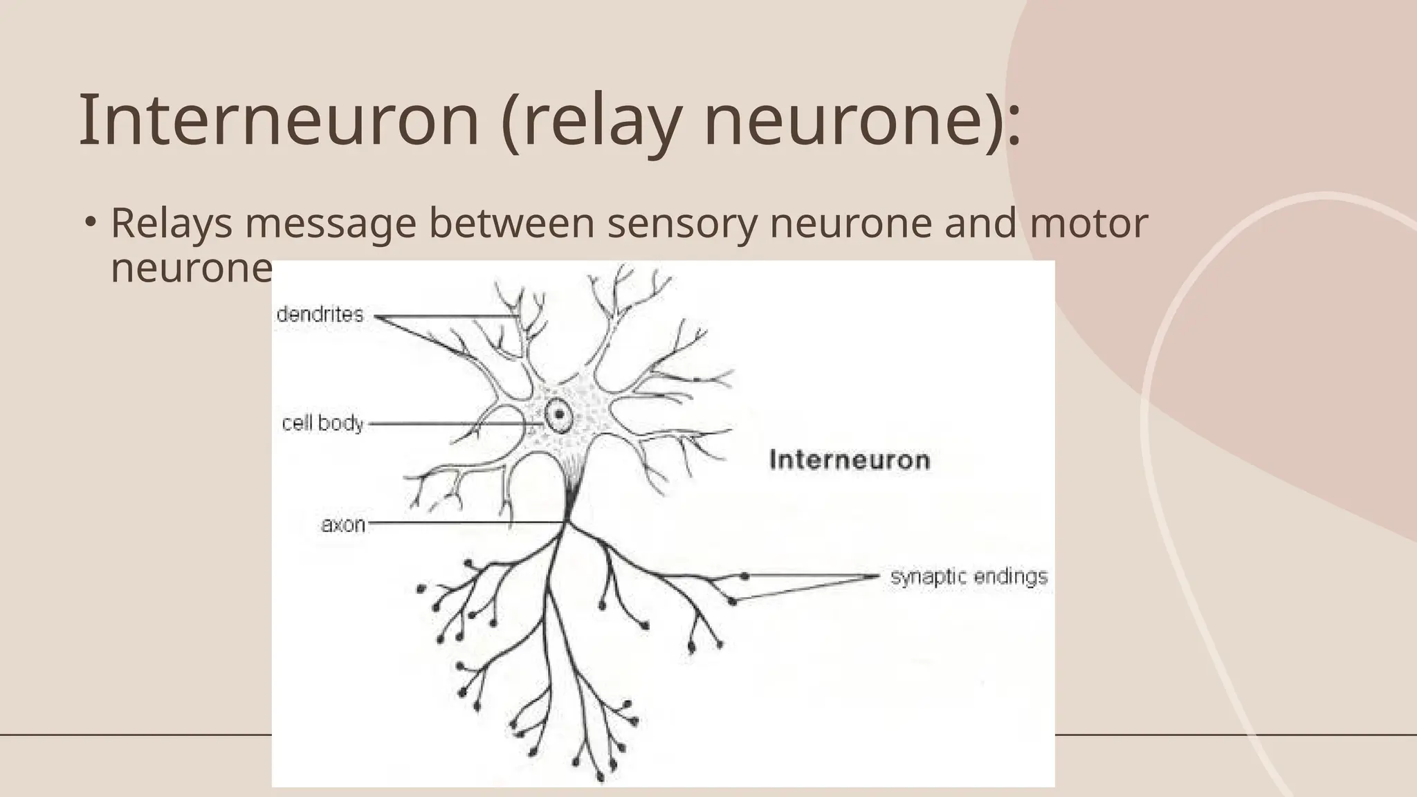
Neurones
• Nervous tissueis composed of nerve cells, or neurones
• There are 3 types:
• Motor neurones
• Sensory neurones
• Interneurones (relay neurones)
29.
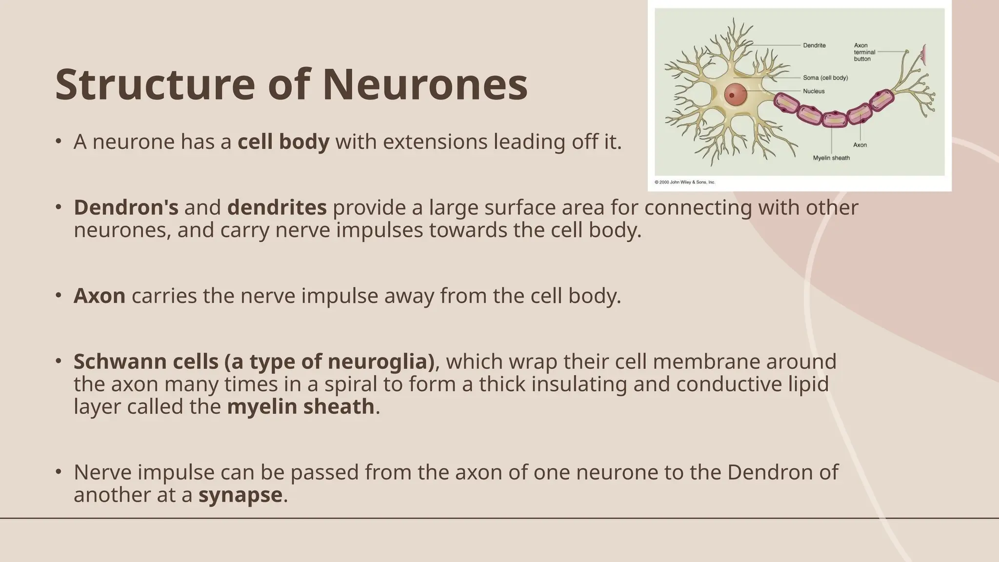
• A neuronehas a cell body with extensions leading off it.
• Dendron's and dendrites provide a large surface area for connecting with other
neurones, and carry nerve impulses towards the cell body.
• Axon carries the nerve impulse away from the cell body.
• Schwann cells (a type of neuroglia), which wrap their cell membrane around
the axon many times in a spiral to form a thick insulating and conductive lipid
layer called the myelin sheath.
• Nerve impulse can be passed from the axon of one neurone to the Dendron of
another at a synapse.
Structure of Neurones
Structure and functionof Neurones
SENSORY NEURON INTERNEURON MOTOR NEURON
Length of
Fibers
Long dendrites and short
axon
Short dendrites and short or
long axon
Short dendrites and long axons
Location
Cell body and dendrite are
outside of the spinal cord
Entirely within the spinal cord
or CNS
Dendrites and the cell body are
located in the spinal cord; the
axon is outside of the spinal
cord
Function
Conduct impulse to the spinal
cord
Interconnect the sensory
neuron with appropriate motor
neuron
Conduct impulse to an effector
(muscle or gland)
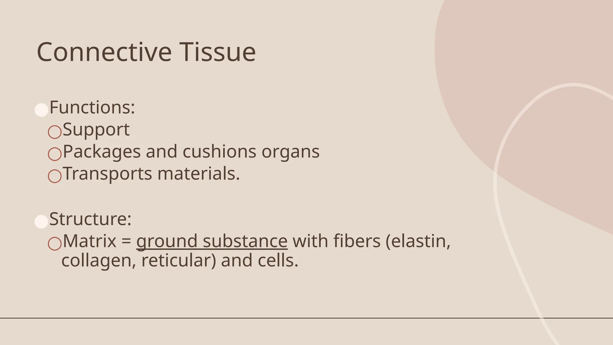
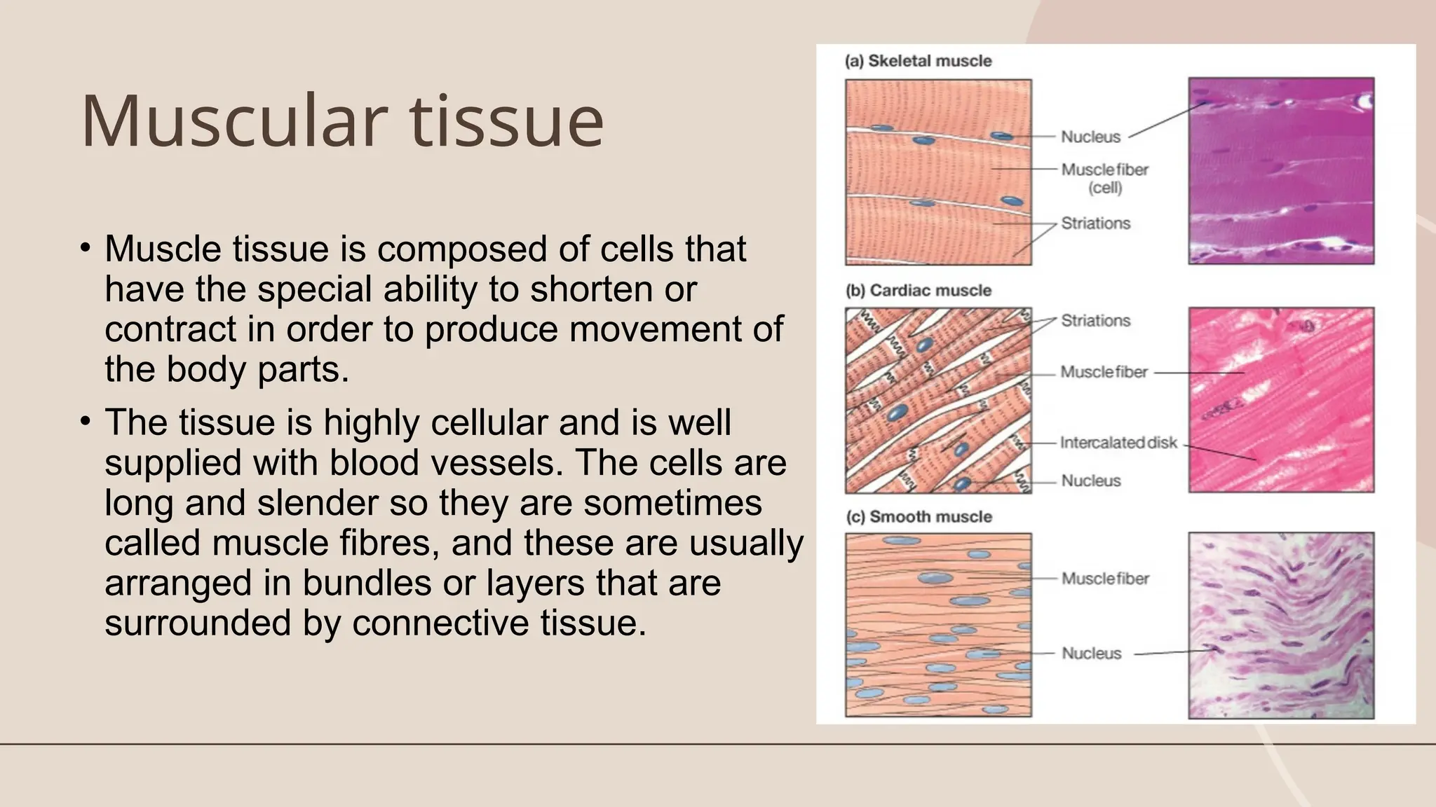
Muscular tissue
• Muscletissue is composed of cells that
have the special ability to shorten or
contract in order to produce movement of
the body parts.
• The tissue is highly cellular and is well
supplied with blood vessels. The cells are
long and slender so they are sometimes
called muscle fibres, and these are usually
arranged in bundles or layers that are
surrounded by connective tissue.
36.
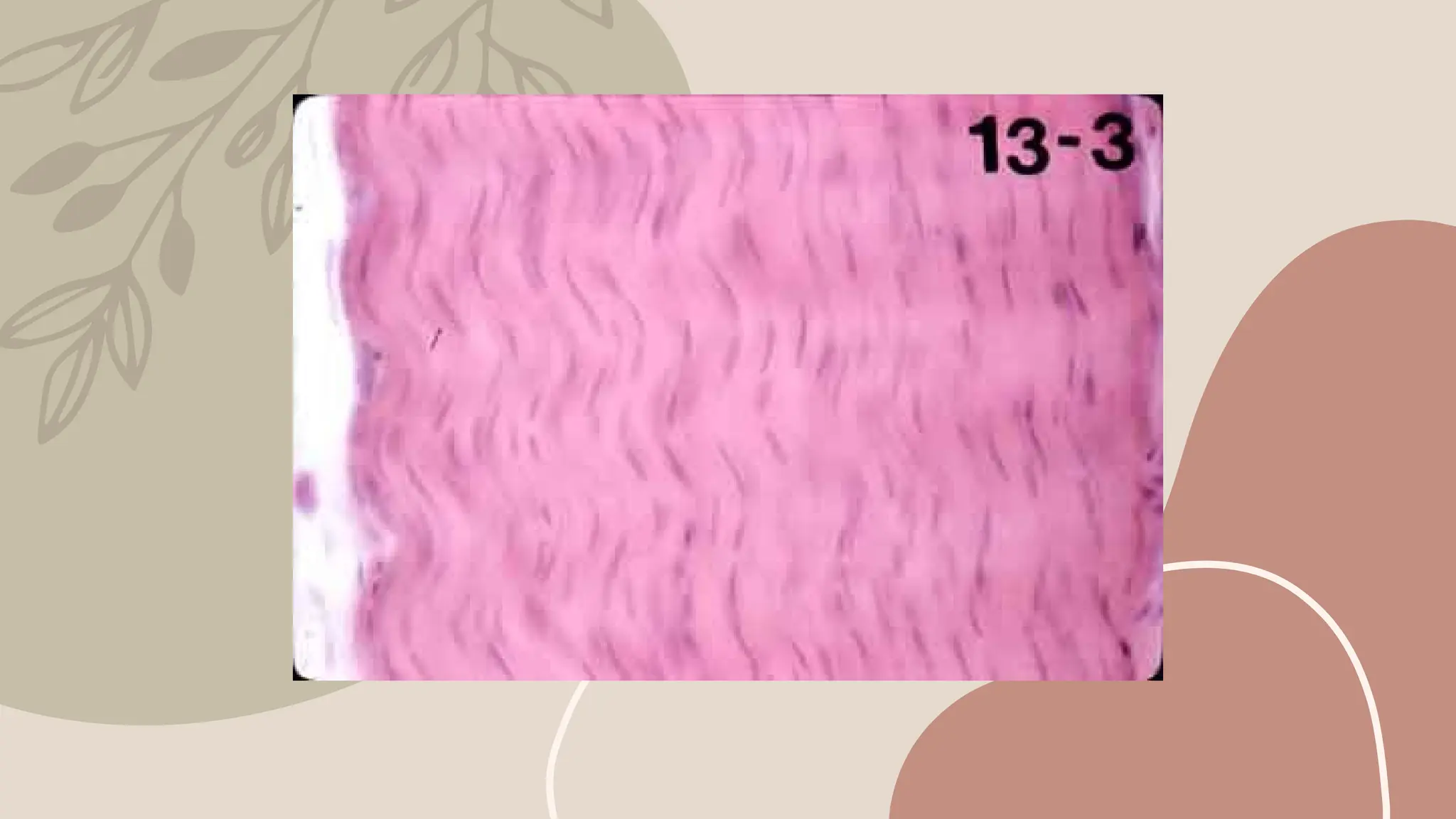
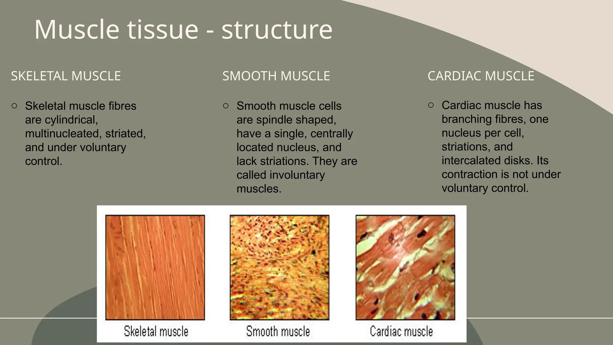
SKELETAL MUSCLE
o Skeletalmuscle fibres
are cylindrical,
multinucleated, striated,
and under voluntary
control.
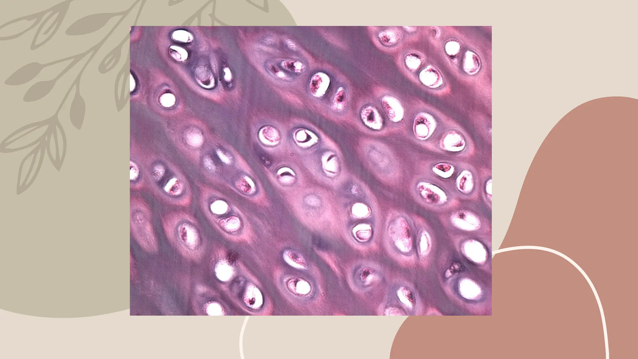
SMOOTH MUSCLE
o Smooth muscle cells
are spindle shaped,
have a single, centrally
located nucleus, and
lack striations. They are
called involuntary
muscles.
Muscle tissue - structure
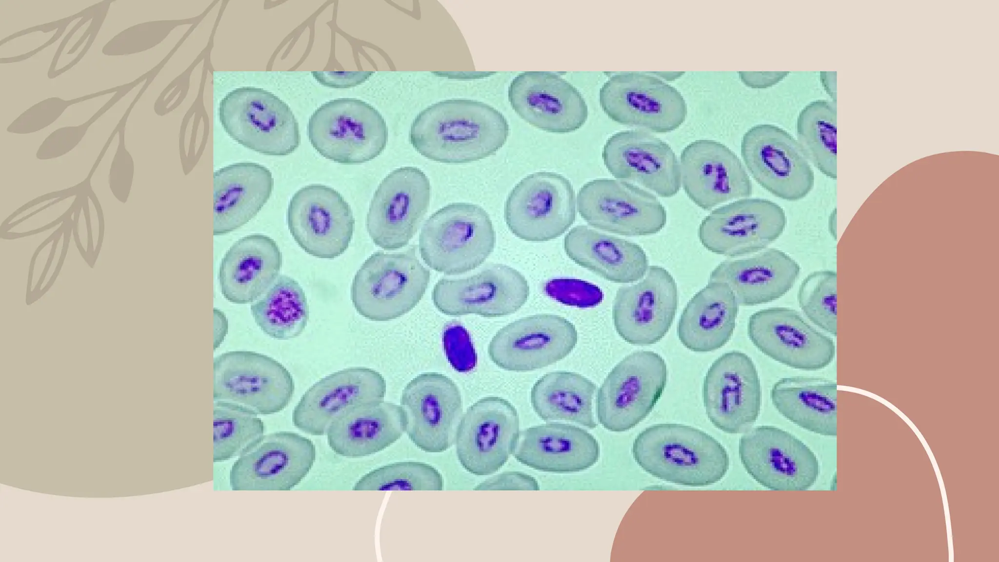
CARDIAC MUSCLE
o Cardiac muscle has
branching fibres, one
nucleus per cell,
striations, and
intercalated disks. Its
contraction is not under
voluntary control.
37.
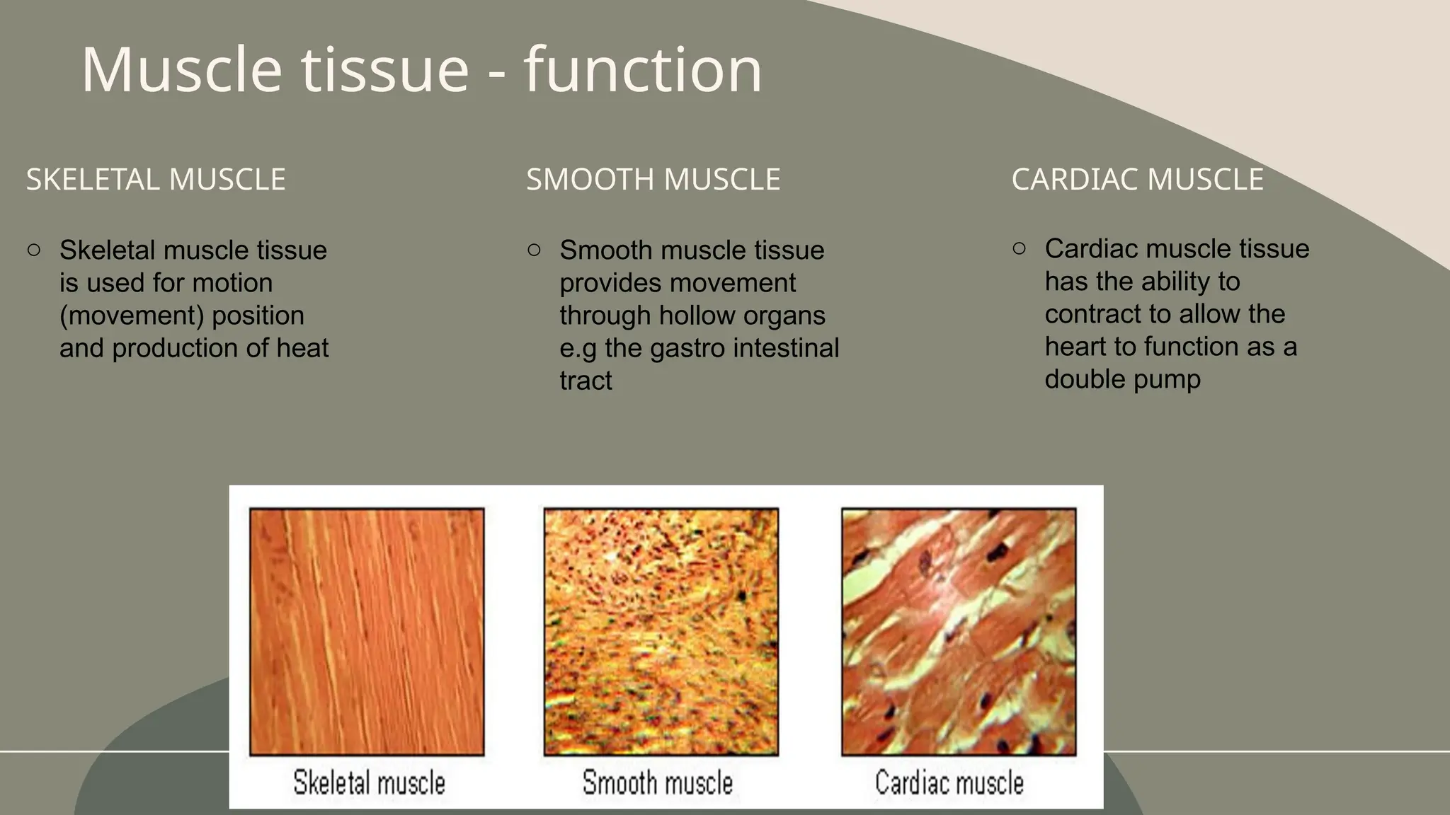
SKELETAL MUSCLE
o Skeletalmuscle tissue
is used for motion
(movement) position
and production of heat
SMOOTH MUSCLE
o Smooth muscle tissue
provides movement
through hollow organs
e.g the gastro intestinal
tract
Muscle tissue - function
CARDIAC MUSCLE
o Cardiac muscle tissue
has the ability to
contract to allow the
heart to function as a
double pump
38.
By the endof
this session, you
should be able
to:
Understand the
organisation within
organisms
Recall the four main
classifications of tissue
types
Classify each tissue type
into sub-groups
Identify examples of
tissues within each group
Describe the structure and
function of a range of
Editor's Notes
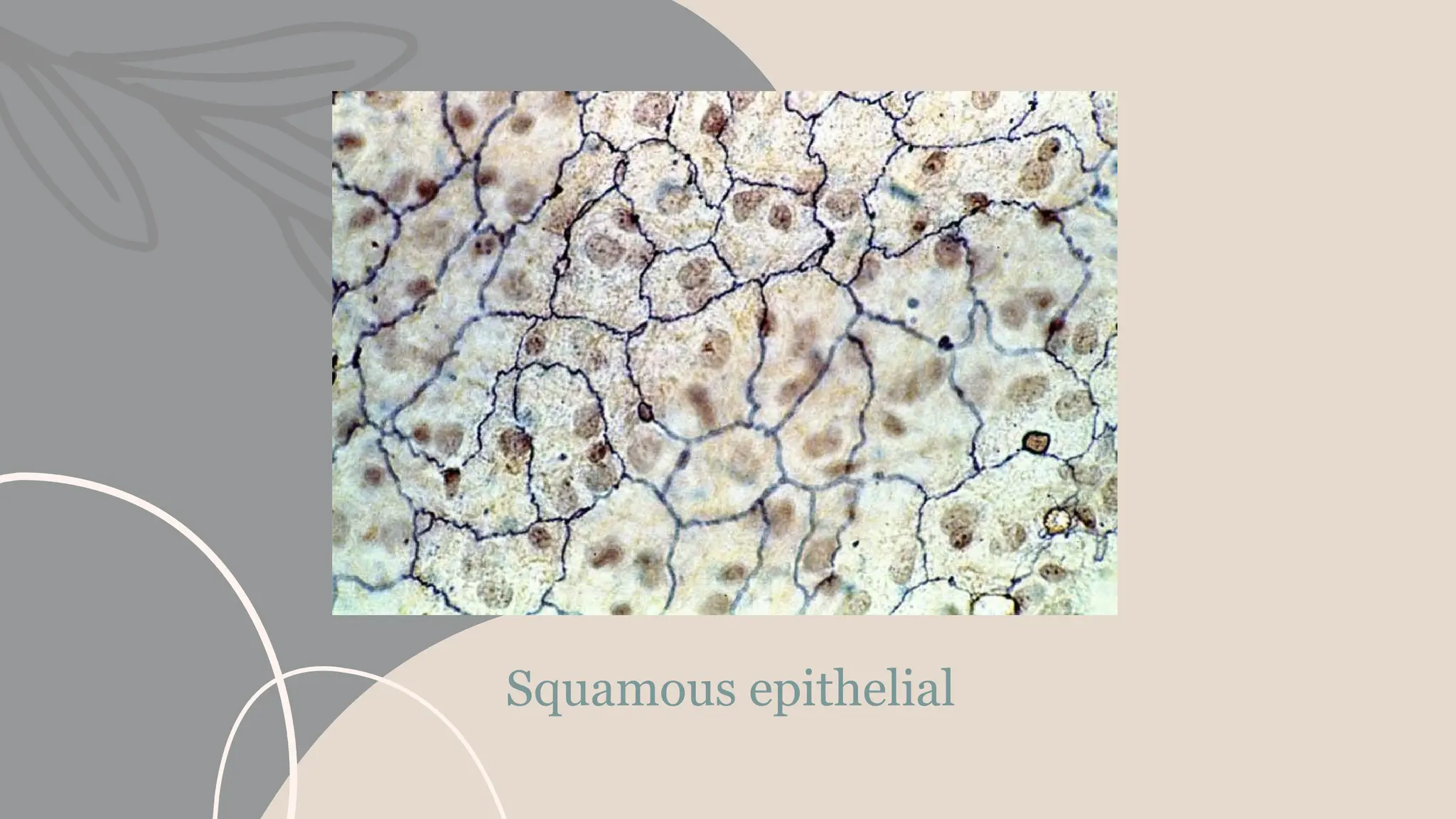
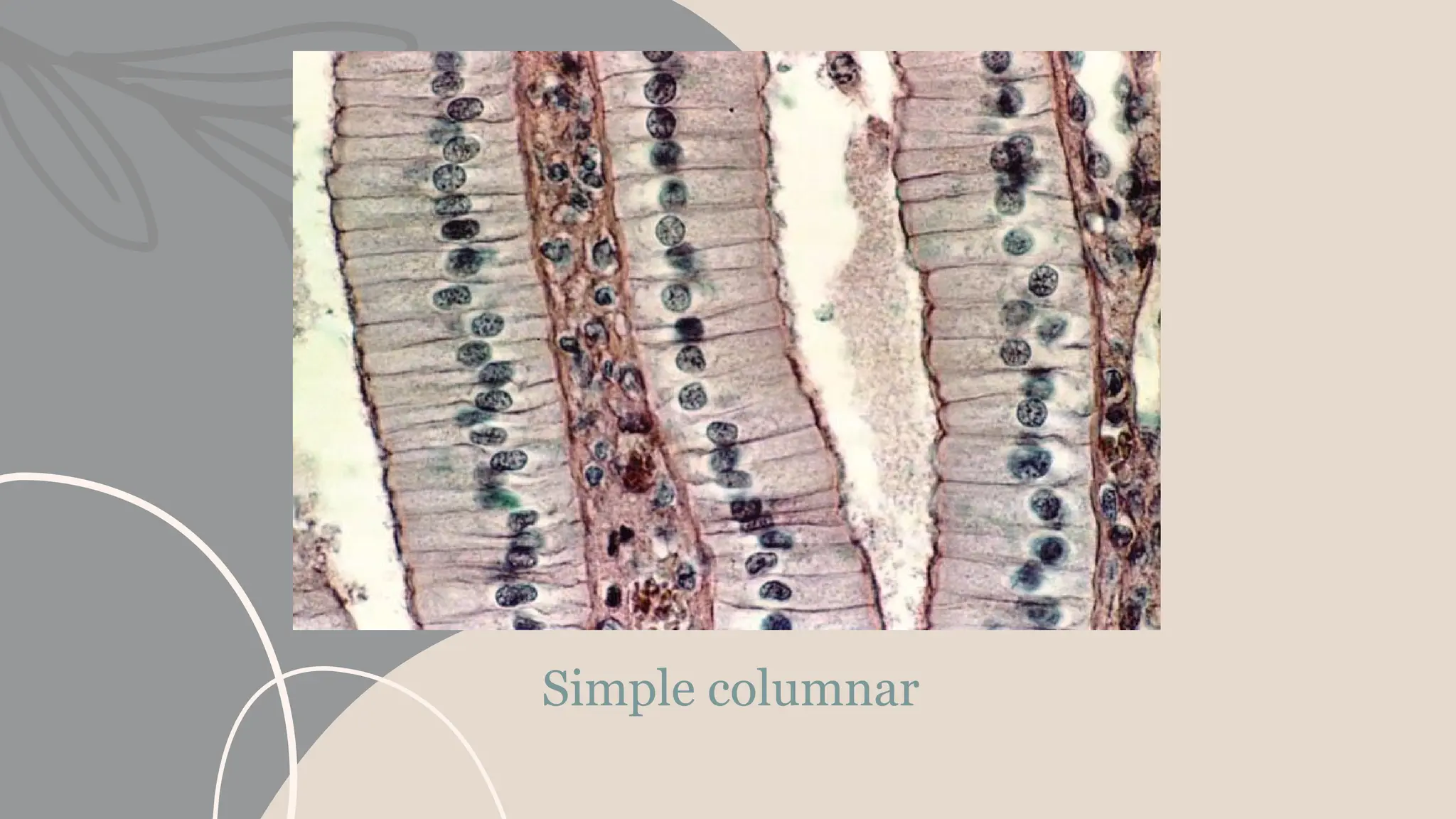
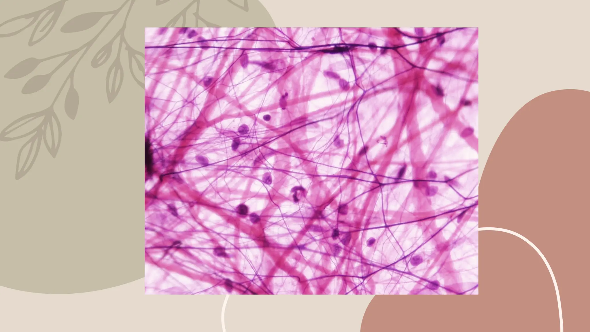
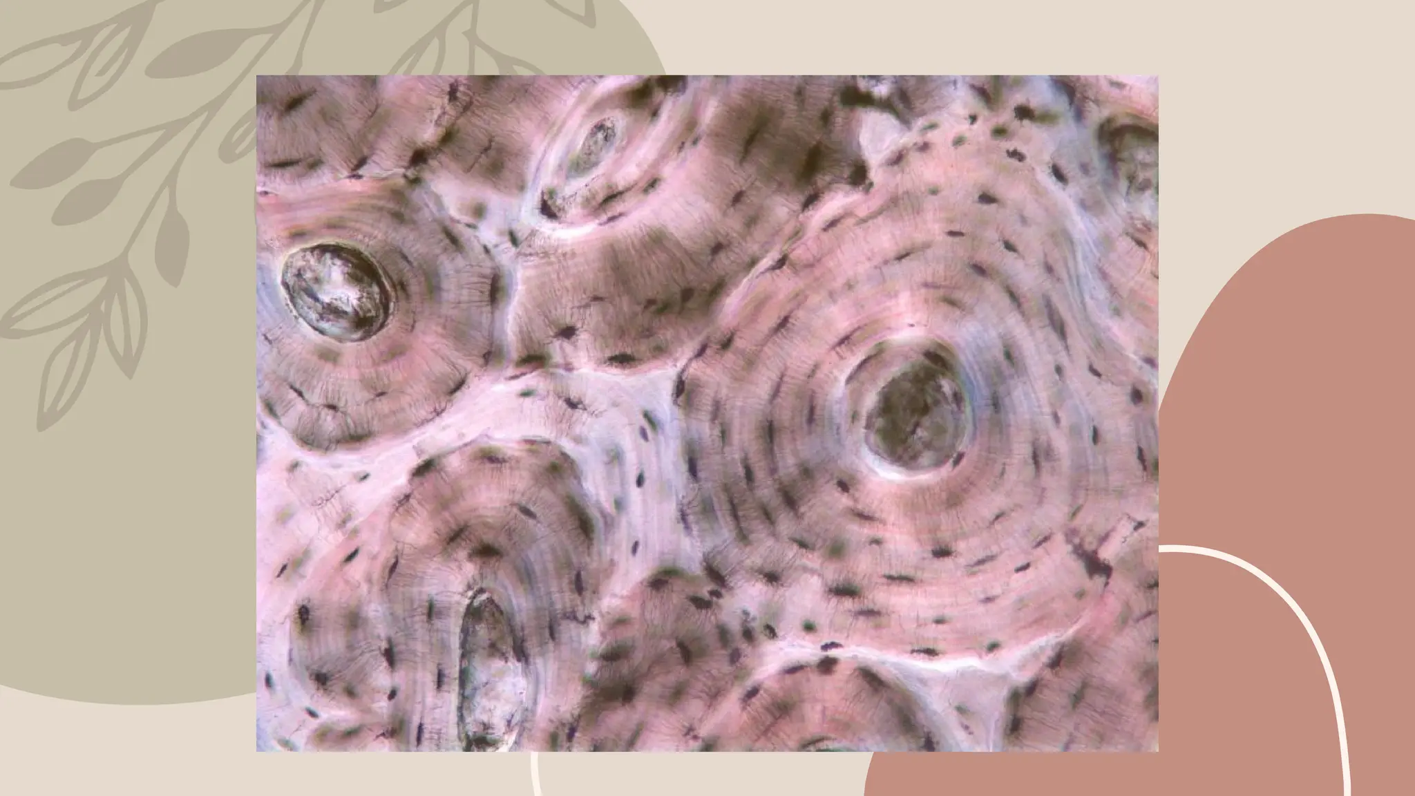
#10 simple squamous epithelium is a single layer of flat cells in contact with the basal lamina (one of the two layers of the basement membrane) of the epithelium.[1] This type of epithelium is often permeable and occurs where small molecules need to pass quickly through membranes via filtration or diffusion. Simple squamous epithelia are found in capillaries, alveoli, glomeruli, outer layer of skin and other tissues where rapid diffusion is required.[2] Cells are flat with flattened and oblong nuclei. It is also called pavement epithelium due to its tile-like appearance. This epithelium is associated with filtration and diffusion. This tissue is extremely thin, and forms a delicate lining. It offers very little protection
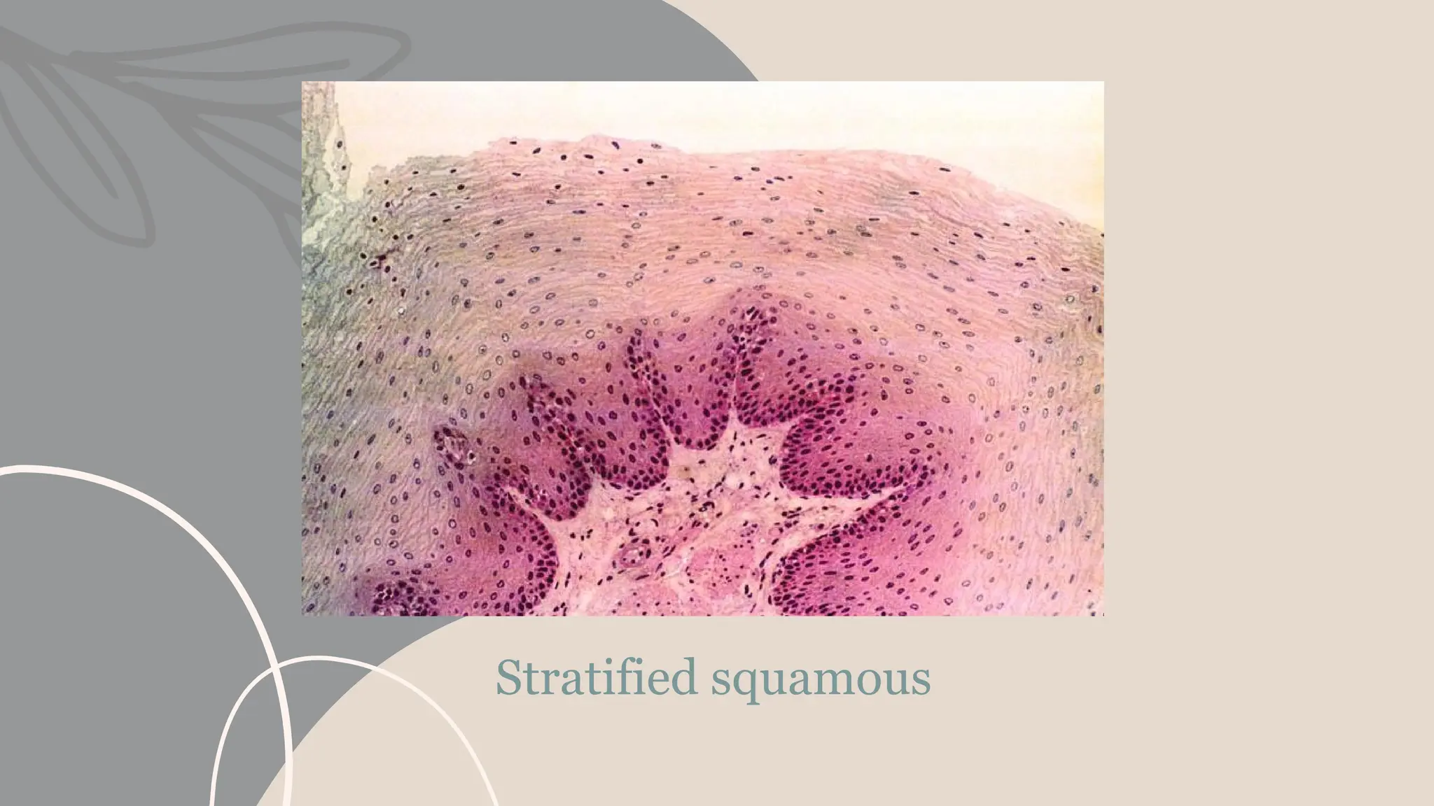
#11 A stratified squamous epithelium consists of squamous (flattened) epithelial cells arranged in layers upon a basal membrane. Only one layer is in contact with the basement membrane; the other layers adhere to one another to maintain structural integrity. Although this epithelium is referred to as squamous, many cells within the layers may not be flattened; this is due to the convention of naming epithelia according to the cell type at the surface. In the deeper layers, the cells may be columnar or cuboidal.[1] There are no intercellular spaces. This type of epithelium is well suited to areas in the body subject to constant abrasion, as the thickest layers can be sequentially sloughed off and replaced before the basement membrane is exposed. It forms the outermost layer of the skin and the inner lining of the mouth, esophagus and vagina.[2]
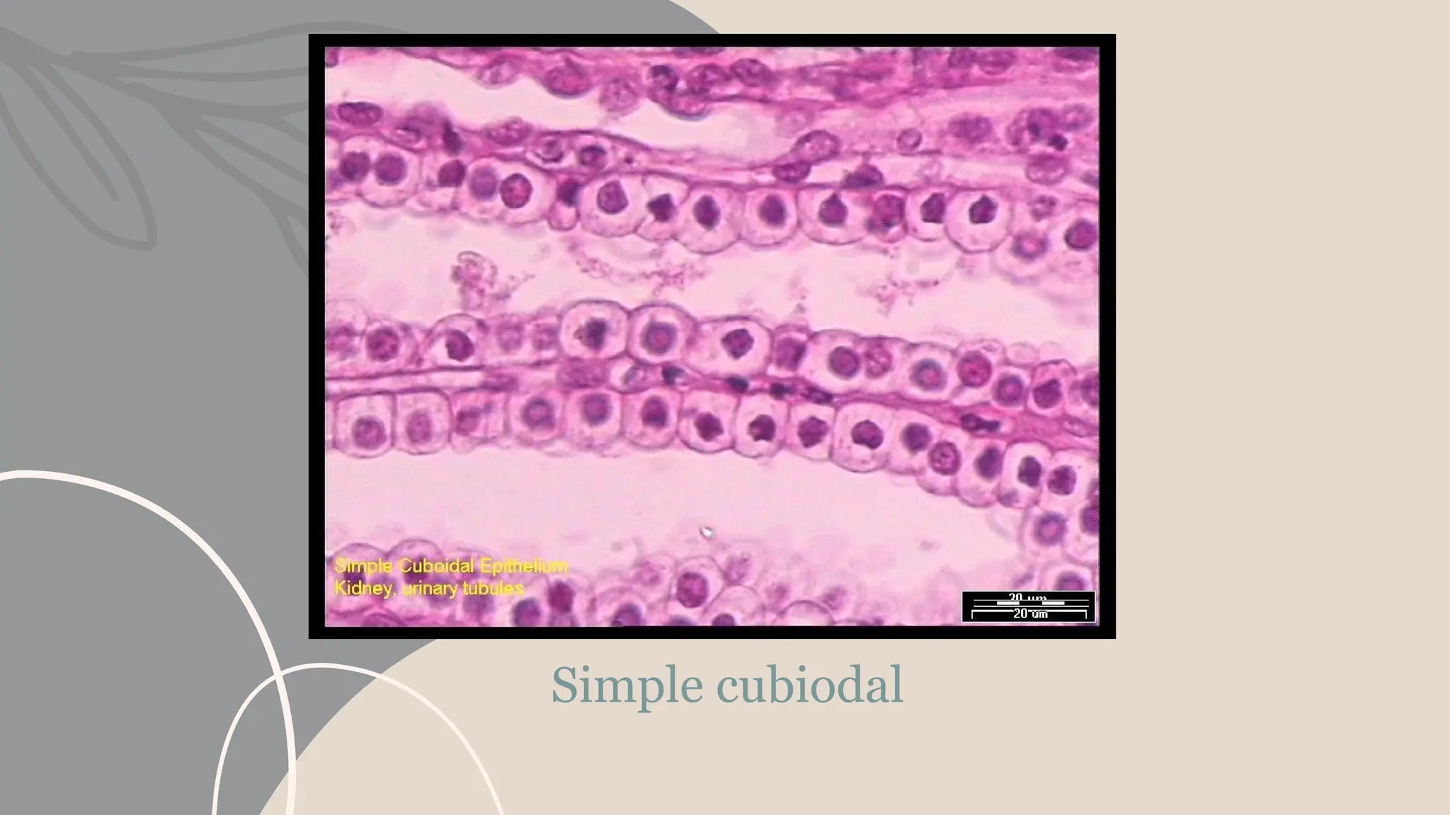
#12 Simple cuboidal epithelium is a type of epithelium that consists of a single layer of cuboidal (cube-like) cells. These cuboidal cells have large, spherical and central nuclei.
Simple cuboidal epithelia are found on the surface of ovaries, the lining of nephrons, the walls of the renal tubules, and parts of the eye and thyroid.
On these surfaces, the cells perform secretion and absorption.
#13 Ciliated epithelium: some simple epithelial tissue consists of cells that secrete mucus, a thick fluid secreted by specialized epithelial cells, known as goblet cells. Its function is to protect the underlying tissue. Mucous membranes usually possess small hair like projections on their surface, known as cilia. These are constantly moving and ‘waft’ the mucus along the surface. This type of tissue lines the ducts of the reproductive and respiratory tracts.
Squamous epthelial tissue of the skin also contains a tough, waterproof protein known as keratin and consequently the tissue is sometimes referred to as cornified or keratinized.
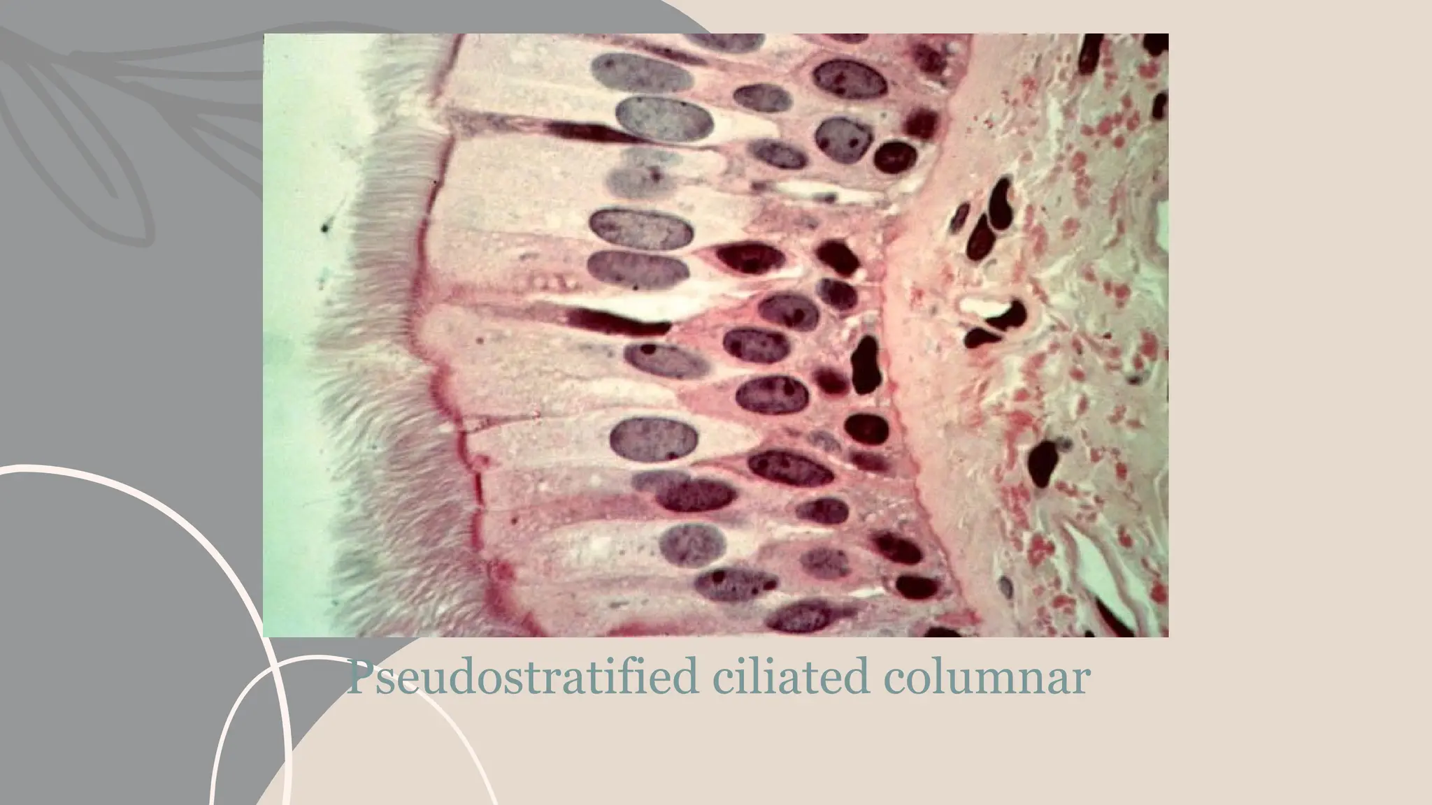
#14 A pseudostratified epithelium is a type of epithelium that, though comprising only a single layer of cells, has its cell nuclei positioned in a manner suggestive of stratified epithelia. As it rarely occurs as squamous or cuboidal epithelia, it is usually considered synonymous with the term pseudostratified columnar epithelium.
The term pseudostratified is derived from the appearance of this epithelium in section which conveys the erroneous (pseudo means almost or approaching) impression that there is more than one layer of cells, when in fact this is a true simple epithelium since all the cells rest on the basal lamina. The nucleous of these cells, however, are disposed at different levels, thus creating the illusion of cellular stratification. Not all ciliated cells extend to the luminal surface; such cells are capable of cell division providing replacements for cells lost or damaged.
Pseudostratified epithelia function in secretion or absorption. If a specimen looks stratified but has cilia, then it is a pseudostratified ciliated epithelium, since stratified epithelia do not have cilia.
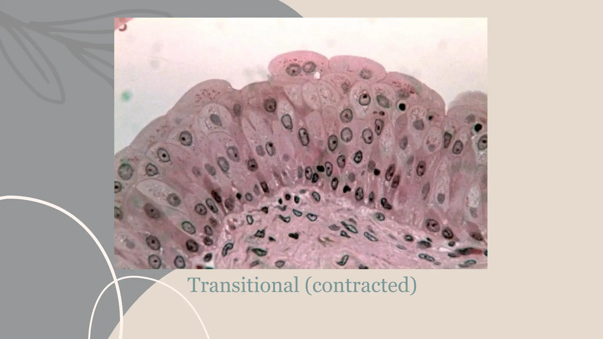
#15 Transitional epithelium is a type of stratified epithelium – tissue consisting of multiple layers of epithelial cells which can contract and expand as needed. It is so named because of this function in the transition of degree of distension. This tissue structure type is found in urothelium, including that of the renal pelvis, urinary bladder, the ureters, the superior urethra, and the prostatic and ejaculatory ducts of the prostate.