The document compares the scales of three large fish: the Arapaima gigas, Latimeria chalumnae, and Atractosteus spatula. It finds that each fish has unique scale structures corresponding to the attacks of their predators. The Arapaima gigas has flexible overlapping cycloid scales with a tough collagen base and hard mineralized surface, protecting it from piranha predators. The Latimeria chalumnae has overlapping elasmoid scales with a double-twisted collagen structure, providing energy dissipation against shark predators. The Atractosteus spatula has inflexible articulating ganoid scales with a hard enamel-like surface and tough dentine-like base, resisting powerful
In addition, it also provides non‐structural functions including lactate sequestration and vibration and tactile perception (Pina et al., 2016). The turtle shell is thus considered a multi‐functional system, with characteristics that likely reflect the product of multiple selective pressures and trade‐offs (Cover et al., 1967). Understanding the structural role of the shell in defense therefore requires detailed investigation(Acevedo et al.,2009).
fossil record - why it is incomplete - index fossils - principle of superposition - principle of fossil succession - lithostratigraphic units - biostratigraphic units - time stratigraphic units - facies fossils - correlation - biozones
Microfossils are very small remains of organisms 0.001 mm (1 micron) to 1 mm, that require magnification for study.
They are abundant, can be recovered from small samples.
Provide the main evidence for organic evolution through the time
They classified into two groups:
Organic-walled; Acritarchs, Dinoflagellate, Spores and Pollen grains … etc.
Foraminifera Each chamber interconnected by an opening (foramen) or several openings (foramina).
Known from Early Cambrian through to recent times, and has reached its acme during the Cenozoic.
Have a wide environmental range from terrestrial to deep sea and from polar to the tropical region.
Depending on the species, the shell may be made of organic compounds, sand grains and other particles cemented together, or from crystalline calcite.
Inorganic walled; Diatoms, Silicoflagellates, Ostracods, Conodonts, and Foraminifera
Grade 8 Integrated Science Chapter 16 Lesson 1 on fossils. This lesson gives detail about fossil, how they form, and the different types. The purpose of this lesson is for students to understand fossil and how they give us a record of our planet's past. Students should know the different types of fossils by the end of the lesson.
Fossils
What are the fossil types?
What Things Become Fossils?
How are Fossils Made?
What are the modes of fossil preservation for body fossils?
Turning to Stone
When Did They Live?
Fossilization
Taphonomy
What are some factors that can affect fossilization
How fossilization dependent upon the environment
This brief description of paleontological techniques will enhance reader's knowledge about fossil collection, tools, excavation, identification and techniques to study fossils.
Poster: Characterizing Ignition behavior through morphing to generic curvesEdward Blurock
The qualitative notion that ignition processes have similar behavior, even over an extensive range of starting conditions, is quantitatively demonstrated through the production of a single ’generic’ ignition curve. The key to the production of the generic curve is the recognition that the basic shapes of the species and temperature profiles occurring in the ignition process differ only in their ’timing’. By ’morphing’ the time scale, the profile shapes can be made to align. From the aligned profile shapes a generic or ’average’ profile can be derived. Synchronizing chemical events modifies the ignition progress times. In addition to fixing the ignition time to have the progress value of one, intermediate ignition events (such as selected profile maxima or inflection points) that occur before ignition are also aligned to have specific ’normalized’ times.
Presentation of Jessica Webb, Civil Society Specialist, Global Forest Watch, World Resource Institute. Delivered in the panel, titled "Addressing Extractive Challenge to Pursue Sustainable Development", organized by Publish What You Pay (PWYP) Indonesia in OGP Civil Society Day, OGP Global Summit on 27 October 2015 in Mexico.
March 2014 - Currency devaluation, limited effectFGV Brazil
A worsening external environment and the perception that the economy is deteriorating should keep the Brazilian real undervalued, but the recovery of industry will take much more than a devaluated currency.
The Brazilian Economy is one of the oldest publications for expert economic analysis of both the Brazilian and international economies. Through this publication, FGV’s Brazilian Institute of Economics and Finance (FGV/IBRE) compares different periods of the economy, assessing both macroeconomic considerations and scenarios related to finance, administration, marketing, management, insurance, statistics, and price indices.
For more information, and Brazilian economic index results, visit: http://bit.ly/1EA1Loz
Toelichting handboek ‘Verankeren van erfgoed in ruimtelijk beleid’ 5Onroerend Erfgoed
Presentatie 5 van vrijdag 19 februari 2016: toelichting van de case RUP Voorzieningenpool fase II Ravelijn in de stad Zoutleeuw, door Inge Gorissen, architect-stedenbouwkundige Creosum
February 2013 - No clear view of the futureFGV Brazil
After negotiating a path full of obstacles in 2012, mainly put up by the economic problems of the major world economies, Brazilian exporters have started the year hoping to recover the ground they lost last year, when foreign sales fell by 5.3% and the trade surplus plunged 34.7%. Exporters are not sure, however, that this time road conditions will be much better.
The Brazilian Economy is one of the oldest publications for expert economic analysis of both the Brazilian and international economies. Through this publication, FGV’s Brazilian Institute of Economics and Finance (FGV/IBRE) compares different periods of the economy, assessing both macroeconomic considerations and scenarios related to finance, administration, marketing, management, insurance, statistics, and price indices.
For more information, and Brazilian economic index results, visit: http://bit.ly/1EA1Loz
Toelichting handboek ‘Verankeren van erfgoed in ruimtelijk beleid’ 3Onroerend Erfgoed
Presentatie 3 van vrijdag 19 februari 2016: clusters, voorschriften voor het verankeren van erfgoed in het ruimtelijk beleid en de toepassing ervan, door Brecht Vandekerckhove, SUM
Biomimetic Materials in Our World: A Review.IOSR Journals
The study of biomineralization offers valuable and incredible insights into the scope and nature of material chemistry at the inorganic and organic surfaces. Biological systems (architecture) are replete with examples of organic supramolecular assemblies (double and triplet helices, multisubunit proteins, membrane-bound reaction centres, vesicle, tubules e. t. c.), some of which (collagen, cellulose and chitin) extend to microscopic dimensions in the form of hierarchical structure, There are ample opportunities of lessons from the biological (on growth and functional adaptation), and physical (properties and compositions) world. This review explores the field of biomimetic material chemistry as it relates to fibres with respect to their historical perspective, the use of the products of biomimetic material, the progressive efforts and a general overview. Conclusively, biomimetic materials research is indeed a rapidly growing and enormously promising field that needs to be explored.
Materials Technology for Engineers pre-test 1 notesmusadoto
Materials are probably more deep-seated in our culture than most of us realize. Transportation, housing, clothing, communication, recreation, and food production virtually every segment of our everyday lives is influenced to one degree or another by materials. Historically, the development and advancement of societies have been intimately tied to the members’ ability to produce and manipulate materials to fill their needs. In fact, early civilizations have been designated by the level of their materials development (i.e., Stone Age, Bronze Age). The earliest humans had access to only a very limited number of materials, those that occur naturally: stone, wood, clay, skins, and so on. With time they
discovered techniques for producing materials that had properties superior to those of the natural ones; these new materials included pottery and various metals. Furthermore, it was discovered that the properties of a material could be altered by heat treatments and by the addition of other substances. At this point, materials utilization was totally a selection process, that is, deciding from a given, rather limited set of materials the one that was best suited for an application by virtue of its characteristics. It was not until relatively recent times that scientists came to understand the relationships between the structural elements of materials and their properties. This knowledge acquired in the past 60 years or so, has empowered them to fashion, to a large degree, the characteristics of materials. Thus, tens of
thousands of different materials have evolved with rather specialized characteristics that meet the needs of our modern and complex society; these include metals, plastics, glasses, and fibers. The development of many technologies that make our existence so comfortable
has been intimately associated with the accessibility of suitable materials. An advancement
in the understanding of a material type is often the forerunner to the stepwise progression of a technology. For example, automobiles would not have been possible without the availability of inexpensive steel or some other comparable substitute. In our contemporary era, sophisticated electronic devices rely on components that are made from what are called semiconducting materials.
17) M-Here you see several examples of fossil mollusks- Mollusks are o.docxKeithldMSandersony
17) M.Here you see several examples of fossil mollusks. Mollusks are one of the most successful animal groups and include animals such as clams, snails, octopus, and squid. They have a large muscular foot and often secrete protective shells. M1. Examine the two fossil bivalves (clams). Both of these animals live by filtering food out of the water while sitting either on the seafloor or buried in the sediment. Why might burrowing help a clam? If a clam was sedentary and sitting exposed on the sea floor, what adaptations would be useful? M2. Examine the three fossil snails. Snails can fill many different roles within an ecosystem including scavenger, predator, herbivore, and even parasite. Which of these three shells is most likely to be a predator? Why did you come to that conclusion? M3. Now look at the two fossil ammonites, which are an extinct group of mollusk. These are relatives of squid and octopi that had elaborate shells with internal chambers, which can be seen in the specimen preserved as a mold. Examine the shape of the internal chambers, what function could the internal chambers serve? (make a hypothesis).
18) Sample N. Here are examples of Lophophorates including brachiopods (shells) and colonial bryozoans (stick-like). These are both animals that were extremely abundant in ancient oceans, but are much less common today. They are grouped together based on sharing a soft-tissue tentacled structure surrounding their mouth called a lophophore. Students often confuse these organisms with other animals (clams or coral), what features can you see that separate them from these groups?
19) Sample O. Arthropods (insects, spiders, crabs, and others) are the most diverse group of animals on the planet today. They have a distinctive exoskeleton and segmented bodies. Their exoskeletons are composed of chiton, which is an organic material rather than a mineral (protein). How would a skeleton made of protein alter their chance to become a fossil? Examine these examples of fossil trilobites, which are an extinct group of marine arthropods. They are common fossils in ancient rocks compared to other arthropods. Why might that be?
20) Sample P. These are examples of fossil echinoderms (sea stars, sea urchins, and relatives). Echinoderms have an internal skeleton, a variation on 5-fold symmetry, and a water vascular system with tube feet (used for locomotion and feeding). P1: These are examples of crinoids (feather stars or sea lilies). These animals are often confused with plants, but they are animals that filter the sea water with delicate arms. Think of them as a starfish atop a long stem. Crinoids were extremely abundant in the past, but are rare in the ocean today. Their stems could range in length from a few inches to over 100 feet! Why might a long stem benefit a filter feeding marine organism? P2: These are two examples of fossil echinoids, including sea urchin and a sea biscuit. Examine the sea urchin and notice it is covered with bumps wher.
Nanoscale science (or nanoscience) researches the phenomena, properties, and responses of materials at atomic, molecular, and macromolecular scales, and in general at sizes between 1 and 100 nm. In this scale, and especially below 5 nm, the properties of matter differ significantly (i.e., quantum-scale effects play a key role) from that at a larger particulate scale (Nguyen et al., 2009).
Nanotechnology is then the design, the manipulation, the building, the production and application, by regulating the shape and size, the properties-responses and functionality of structures, and devices and systems of the order or less than 100 nm. Nanotechnology is considered an advancing and emerging technology due to the possibility to advance well-established products and to create new products with totally new characteristics and functions with enormous potential in a wide range of applications (Azzam, 1977).
2. years ago. The fish scales have been traditionally classified into four
groups: placoid, elasmoid, cosmoid and ganoid. Placoid scales are
denticles with a flattened rectangular base plate embedded in the fish
body, and spines which project from the posterior surface. They have a
core with pulp which is surrounded by dentine and an outer vitroden-
tine layer. Cosmoid scales are similar to placoid scales and likely
evolved from the fusion of them; they have dentine, vitrodentine and a
tissue complex known as cosmine with interconnected canals and flask-
shaped cavities, but lack a pulp core. These rigid rhombic scales are
now, unlike the other scale classifications, entirely extinct. Ganoid
scales are modified cosmoid scales which are also rhombic, rigid, and
jointed articulating scales of two layers. A thin mineral surface layer
called ganoine replaces the vitrodentine, and lies atop a bony founda-
tion which replaces the cosmine. Peg and socket joints often join
ganoid scales. Elasmoid scales likely evolved from ganoid scales and
are the most common among living vertebrates. They are thin and
imbricate, resembling shingles on a roof, and consist of a bony surface
and a fibrous layer beneath of collagen. There are two subcategories,
ctenoid and cycloid, the difference between being that ctenoid scales
have developed surface spines which are bony and grow from the body
of the scale to the surface and the cycloid scales have a smooth surface
(Sire and Huysseune, 2003; Helfman et al., 2009; Sire et al., 2009;
Vickaryous and Sire, 2009).
Each of these scales has unique features and provides protection
with a modest weight penalty. In order to learn from natural fish scales,
modern tools and techniques, such as electron microscopy, nano-
indentation, computer x-ray tomography and finite element analysis,
provide insight into the features at the nano to macro level and reveal a
variety of toughening mechanisms that make fish armors highly
effective. Early studies on fish scales of this nature include
Polypterus senegalus (Bruet et al., 2008; Song et al., 2011), Morone
saxatilis (Zhu et al., 2013), Arapaima gigas (Torres et al., 2008; Lin
et al., 2011) and Atractosteus spatula (Allison et al., 2013; Yang et al.,
2013).
One principal function of fish scales is to resist penetration from
predators. In particular, the manner in which they resist pressure by
teeth has been addressed by several researchers. Zhu et al. (2012)
performed penetration tests on a Morone saxatilis (striped bass) fish
scale using a steel stylet simulating a sharp tooth. They compared the
force-penetration response of whole fish scales and just the collagen
layer with that of the synthetic polymers and found marked differences.
By analyzing the penetration sequence in the bony surface and
collagenous foundation, they classified it into three stages: stage I
represents a linear relationship between force and penetration distance
due to flexing of the scale and penetrating into the surface bone layer.
Stage II begins with a small force drop associated with the crack
opening in the bone layer and radiating from the penetration point
which finally propagates into the collagen layer. In stage III the force-
displacement curve plateaus as the stylet punctures the collagen layer.
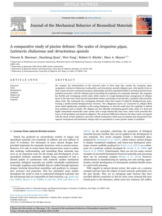
In similar vein, Vernerey and Barthelat (2010) described the load
redistribution mechanisms (proportional to scale size) from penetra-
tion on the overlapped fish scales (Fig. 1a). They also described
relationships between scale density, ratio of angular attachment
stiffness to bending stiffness, and a variety of other properties which
are key to the scales’ protective function (Fig. 1b) and vary according to
the environment in which the fish lives. If escaping from a predator is
critical, the structure of fish scale is “designed” in terms of flexibility
(strain stiffening) and lightness; if the protection from a predator is
more important, higher resistance to fracture and average bending
stiffness are design criteria. Similarly, characterizing the structure,
quantifying the mechanical parameters and understanding the salient
mechanisms can provide insight to understand the environment in
which the fish exist, the types of predators the fish may have faced, and
even aids in understanding the evolution of the fish.
In this review, we focus on the three resilient fish (Fig. 2) whose
highly effective dermal armors have protected them survive for millions
of years, namely Arapaima gigas (arapaima) (Torres et al., 2008; Lin
et al., 2011; Zimmermann et al., 2013), Latimeria chalumnae (coela-
canth) (Roux, 1942; Smith et al., 1972), and Atractosteus spatula
(alligator gar) (Allison et al., 2013; Yang et al., 2013), with an emphasis
on the structure-mechanical property relationships in their respective
armored scales. It has been established that the properties of fish scales
may vary with the head, body, and tail having unique traits (Marino
Cugno Garrano et al., 2012; Murcia et al., 2015), but this case there is
no knowledge of what specific part of the animal they are from; this
may lead to some experimental variation. The arapaima is a huge fish
which lives in the Amazon and grows upwards of 3 m in length and 200
kgf in weight (National Geographic, 2016a). Its fossil records have
remained the same for at least 23 million years, and it evolved in order
to peacefully cohabitate with the piranha, a predator with famously
sharp tricuspid teeth with a tooth tip radius of 13 μm. Despite its
ferocity, the piranha possesses a relatively small bite force, estimated
by Meyers et al. (2012) as 20 N. The coelacanth is another large fish
which lives at up to 700 m deep in the Indian ocean (specimens have
been found in the Madagascar and Indonesian coasts), and grows to
2 m in length and 90 kgf in weight. It has existed for 400 million years,
and was thought to have gone extinct with the dinosaurs until
rediscovered in 1938 (Smith, 1939). Shark bite marks found on
coelacanths suggest that the shark is one possible predator (Fricke
et al., 1991). Shark teeth are nearly as sharp as the piranha's, with a
16 μm tooth edge radius, but different types of sharks possess biting
forces ranging between 1–2400 N (Huber et al., 2009; Mara et al.,
2010; Ferrara et al., 2011). The bite force of sharks is surprisingly low,
and this is understandable because they do not possess bones and only
cartilage. Finally, the alligator gar is a third large fish which lives in the
brackish waters around the Gulf of Mexico and grows up to 3 m and
140 kgf (National Geographic, 2016b). The gar has existed in its
current form for roughly 100 million years, and must protect itself
from alligators. The alligator has 80 teeth although it may generate
3000 over its life (Potts, 1998), but these teeth which are not
Fig. 1. (a) Scales redistribute an applied load in a region proportional to the scale size. This also leads to a greater penetration resistance. (b) Plot of attachment to scale stiffness ratio as
a function of scale density illustrates several relationships between scale properties and features of the response such as average bending stiffness, resistance to fracture, and mass
density (from Vernerey and Barthelat (2010)).
V.R. Sherman et al. Journal of the mechanical behavior of biomedical materials xx (xxxx) xxxx–xxxx
2
3. particularly sharp, have a tip radius of the order of ~80–130 μm for
juveniles up to 3 mm for adults. This lack of sharpness, however, is
compensated by powerful jaws capable of bite forces of 10 N to 10 kN,
depending on the size (Erickson et al., 2003; Erickson et al., 2004).
2. Arapaima gigas
2.1. Structure of arapaima scales
As shown in Fig. 3, the arapaima fish has elasmoid scales (cycloid as
the sub-classification) which are composed of a hard and stiff outer
mineral layer and a tough lamellar Bouligand-like collagen structure
beneath. The scales are overlapped and arranged with a degree of
imbrication (exposed length/total length) equal to 0.4 and aspect ratio
(length/thickness) of 50 (Yang et al., 2014). The average number of
scale layers covering the fish's body is three, and the darker parts of the
scale are exposed while the light parts are overlapped by surrounding
scales (Fig. 3). The exposed scale is covered by a thick mineral layer
(~1 mm in adults) with ridges on the surface; in the embedded portion
of the scale the mineral layer is thinner (~0.5 mm). Beneath the
mineral, collagenous lamellae display a Bouligand-type structure,
involving large (~45–90°) angles between adjacent layers. Individual
lamella consists of parallel and straight collagen fibrils, although there
is significant misorientation between the fibril directions in adjacent
lamellae. Fibrils can be imaged by transmission electron microscopy
(TEM), as shown in Fig. 3, where the misorientation between the two
visible lamellae is visible. The cross-section of fibrils in layer 1 of
collagen is elliptical and the periodicity of the banding in collagen
fibrils in layer 2 is ~50 nm, i.e., less than the 67 nm characteristic d
period of collagen. This indicates that the fibrils are at an angle to the
surface, which can be calculated as θ=arccos(50/67)=~40°, and that
the included angle between the two layers is less than 90°.
2.2. Mechanical response of arapaima scales
Owing to the large overlap area over the entire fish, the flexibility of
each individual scale is a requirement for the mobility of the arapaima.
As such, its scales are flexible in all directions, a characteristic of all
elasmoid scale classifications as compared to ganoid scales, such as
those of the alligator gar. When these scales are tested in tension in the
longitudinal and transverse directions (Fig. 4a), with the mineral layer
either intact or removed, there is a substantial decrease in the strength
with the mineral layer present. This decrease is associated with the
mineral's inability to carry tensile load due to the presence or formation
of microcracks, i.e., the outer mineral layers merely increase the cross-
sectional area and correspondingly decrease the stress. Although the
arapaima fish scale is flexible in all directions, the scale in the
longitudinal direction, i.e., along its length, is stiffer (0.5 vs. 0.2 GPa)
and stronger (25–50 vs. 15–20 MPa) than in the transverse direction
(Yang et al., 2014). The stronger and stiffer direction of the scale aligns
with a possible exotendon function of the scales, where scales store
energy for more efficient swimming (Brainerd, 1994). The more
compliant direction allows the scales to accommodate movement as
they conform to the curvature of the fish body. However, as previously
addressed, the specific location of the scales on the fish is unknown and
this effect may vary throughout the fish.
Fig. 2. Ruthless predators and their prey. (a) The piranha is an infamously ferocious fish which cohabitates with the arapaima in the Amazon; the arapaima grows up to 3 m in length
and 180 kgf in weight. (b) The shark is suspected to be the main predator of the coelacanth, which can grow to over 2 m in length and 90 kgf in weight. (c) The alligator, known for its
massive jaw strength and powerful attacks, cohabitates the Mississippi basin with the alligator gar, a fish which grows up to 3 m in length and 140 kgf in weight.
Fig. 3. Structure of the scale of Arapaima gigas. The arapaima's elasmoid scale has a hierarchal structure which is designed to defend against piranha attacks. The underlying shape of
the scales can be seen in the overlapping arrangement schema of five scales. The exposed portion of the scale is dark and consists of mineral ridges atop a Bouligand -type lamellar base
of collagen, creating the cross-section shown. The white part of the scale is covered by surrounding scales, and consists of the same Bouligand layers as beneath the mineral ridges. These
collagen layers are made of sheets of oriented collagen fibrils, roughly 200 nm in diameter and with some degree of mineralization; two layers of differing orientations are observable in
the TEM micrograph (adapted from Zimmermann et al. (2013) and Yang et al. (2014)).
V.R. Sherman et al. Journal of the mechanical behavior of biomedical materials xx (xxxx) xxxx–xxxx
3
4. Scanning electron microscopy (SEM) shows that the lamellar angles
are large and variable (Fig. 4b), and are arranged in a Bouligand-type
structure, which is highly resistant to crack propagation. Under tensile
loading, some lamellae reorient as the deformation is applied so that
the lamellae close to the tensile axis can more effectively carry the
applied load. This reorientation has been estimated theoretically by
considering the elastic stretching, strain-rate sensitivity and interfi-
brillar sliding of the lamellae (Zimmermann et al., 2013; Yang et al.,
2014):
⎡
⎣
⎢
⎢
⎢
⎢
⎛
⎝
⎜
⎞
⎠
⎟
⎤
⎦
⎥
⎥
⎥
⎥
Ψ Ψ
Ψ ε
C
− = arccos
cos ( + 2)
2 +
,t
ε
ε
ε
E
1 0
0
̇
̇
t
f0
(1)
where Ψ1 is the predicted angle between the lamella and the tensile
axis (positive values correspond to rotation towards the tensile axis),
Ψ0 is the initial value of Ψ before the application of the load, εt is the
sum of the strain due to the elastic stretching of fibrils and rotation
with the effect of interfibrillar shear, ε̇ is the strain rate, ε ̇0 is a reference
strain rate, Ef is the modulus of a fibril, and C is an experimentally
measured constant. Predictions from Eq. (1) for the angular rotation
shown in Fig. 4c indicate that all orientations will rotate towards the
tensile axis, with the most significant reorientation occurring for Ψ
angles between 10° and 30°. Corresponding experimental measure-
ments of the scales, under tensile loading with real time small angle x-
ray scattering (SAXS) imaging, provide a detailed observation and
analysis of the mechanisms in Fig. 4d. SAXS results indicate that under
tensile loading, the collagen fibrils oriented to within ~15–30° of the
loading direction tend to rotate towards the tensile axis, whereas fibrils
in the 61–90° range rotate away from the tensile axis. Owing to the
large angles between layers, as the collagen fibrils oriented close to the
loading direction rotate towards it, the adjacent layers, which are far
from the tensile axis are subjected to tension perpendicular to the
fibrils, causing gaps to open. In addition, interfacial shear and
sympathetic lamellar rotation by adjacent layers contribute to these
orientations, on average, rotating away from the tensile direction (Yang
et al., 2014).
2.3. Arapaima failure prevention strategies
The arapaima can effectively defend against piranha attacks due to
the highly mineralized, hard surface layer of its scale, and a Bouligand-
type structure of the layer beneath. The mineral layer resists the
penetration by a tooth, and the Bouligand foundation provides
strength and toughness to accommodate the deformation. The damage
evolution in the scale, and its corresponding toughness, have been
examined by tensile testing notched specimens of the scale in situ in
the SEM (Yang et al., 2014). Fig. 5 shows a sequence of images
captured during such tests: at the beginning of the tensile test, fibrils at
the notch tip start to delaminate in the vicinity of the notch tip
(Fig. 5a). Under increasing load, the collagen fibrils become stretched,
with some fracturing close to the notch tip, while other fibrils away
from the tip become curved due to the geometry of notch-opening and
the rotating from the layers beneath (shown in Fig. 5b). Before fracture,
more collagen fibrils delaminate (labeled as 1 in Fig. 5c), some fracture
(labeled as 2), bend or buckle (labeled as 3), but other layers remain
intact and carry the load (labeled as 4 in Fig. 5c,d). Similarly, Dastjerdi
Fig. 4. Mechanical response and lamellar rotation of the arapaima scale. (a) The tensile response of the arapaima scale is characterized by elastic and plastic regions (Yang et al., 2014).
The tensile response of the scales shows that they are weaker when the mineral layer is not removed, due to the fact that the mineral is brittle and microcracks defeat its strength. (b) The
variation in lamellar orientation is not consistent between layers. Three orientations of adjacent layers are shown; two at acute angles and one at an obtuse angle. (c) The lamellar
reorientation of these layers can be predicted mathematically based on their initial orientation using Eq. (1). The plotted results show that under load, all layers should reorient towards
the tensile axis, but those layers with the strongest tendency are those oriented roughly 10–30° from the tensile axis. (d) The lamellar reorientation predicted from (c) is measured
experimentally using SAXS. (from Yang et al. (2014) and Zimmermann et al. (2013)).
V.R. Sherman et al. Journal of the mechanical behavior of biomedical materials xx (xxxx) xxxx–xxxx
4
5. and Barthelat (2015) measured the fracture toughness of striped bass
scales (also an elasmoid scale) and found them to be among nature's
toughest materials (Jc=15–18 kJ m−2
). The scales negate the effect of
cracks as they effectively delocalize into wide process zones with the
partially detached collagen fibers engulfing the crack front.
To further assess the effectiveness of arapaima scales, Meyers et al.
(2012) examined how an actual piranha tooth can penetrate the
arapaima scale. With the tooth mounted on the upper fixture of the
testing machine so that it could be compressed into hydrated arapaima
scales, it fractured before complete perforation of the scales occurred
(Fig. 6). The images show the sequence of events (for the tooth and the
scale) during the test, with a picture of the failed tooth as an inset to the
force-displacement plot. Microscopically, the lamellar base of the scales
was observed and shown to serve as a resilient and tough base which
acts synergistically with the hard and stiff outer mineral layer to
prevent penetration from the tooth. Hence, this highly effective armor
allows the passive arapaima to cohabitate peacefully with the piranha,
one of the most vicious and feared fish in the Amazonian waters.
3. Latimeria chalumnae
3.1. Structure of coelacanth scales
The coelacanth has an ancient type of elasmoid scales, which were
present several times during the fish evolution. (Fig. 7). Similar to
cycloid scales, these scales have a dark region with a rough and more
mineralized exposed surface, and an overlapped (embedded) region of
which the surface is light and smoother. The individual scales have an
elliptical shape with various sizes corresponding to the size of the fish
and they are typically ~10–35 mm in size for a 1 m fish. The degree of
imbrication is ~0.34 and the aspect ratio is ~55, similar to the
arapaima. These scales provide protection by means of a highly
mineralized outer layer with unique “double-twisted” Bouligand foun-
dation (Giraud et al., 1978). The surface of embedded region is shown
in Fig. 7b. From the center of the scale circular annular ridges with
various spacings can be observed. Additional ridges radiating from the
center are initially perpendicular to the annuli and then orient towards
the dorsal-lateral direction. These ridges, shown in Fig. 7c, have a
spacing of approximately 30 µm. On the surface of the exposed region,
denticles protrude from the scale towards the direction of the fish tail
(Fig. 7d). The mineral layer is needed for protection, although Sudo
Fig. 5. In situ SEM of a crack arrested in arapaima scales under tensile loading. (a) An initially notched scale is loaded in tension in the direction indicated. (b) As stretching continues,
multiple layers become apparent as lamellae stretch, reorient, bend, and buckle. (c) Lamellar delamination occurs as an energy absorbing mechanism. (d) The crack is fully arrested by
reorientation, bending and stretching of the layers (from Yang et al. (2014)).
Fig. 6. Piranha tooth penetration of the arapaima scale. During an indentation
experiment, the piranha tooth attempts to fully penetrate a single arapaima scale. A
sequence of images on the right shows a time evolution of the penetration. The protective
mineral and lamellar Bouligand structure base causes tooth fracture before the scale can
be fully penetrated. The fracture of the tooth is indicated by the drop in force as seen in
the force vs. displacement measurement (identified by black arrows) and the broken
tooth is pictured as an inset within the graph (from Meyers et al. (2012)).
V.R. Sherman et al. Journal of the mechanical behavior of biomedical materials xx (xxxx) xxxx–xxxx
5
6. et al. (2002) studied the scales of the Sebastes inermis (rockfish), which
also has ctenoid scales, and showed the surface roughness aligns with
the direction of the water current and serves to channel flow for a
hydrodynamic advantage.
Beneath the rough and hard exposed surface, the majority of the
scale thickness is composed of collagenous lamellae, identified as
isopedine (Goodrich, 1907; Ørvig, 1957; Smith et al., 1972; Giraud
et al., 1978). Isopedine is also present in the ganoid scales of Senegal
bichir (Bruet et al., 2008). The laminated structure of the coelacanth
scale is shown in Fig. 7e. The lamellae in the isopedine have two
superimposed and interpenetrating Bouligand structures with a re-
markably regular arrangement in which the parallel fibers in any one
lamella lie at a roughly 90° angle to the fibers in adjacent lamellae
(Fig. 7i) (Smith et al., 1972; Giraud et al., 1978). The architecture of
the coelacanth scale differs from the arapaima scales, as they have
struts with less ordered collagen fibrils connecting the lamellae and
filling the gaps between collagen bundles. Fig. 7f shows holes in the
scale cross-section which indicate that the collagenous bundles “pull-
out” when the sample is fractured. Transmission electron microscopy
of these scales (Fig. 7g) shows aligned collagen fibrils along the
lamellae and less organized fibrils in the struts, forming a continuous
network. The orientation of lamellae can be readily identified from the
oblique slice shown in Fig. 7h. The fibril directions are marked and
show that the orientations of the collagen lamellae in adjacent layers
are nearly orthogonal, while successive bilayers are characterized by a
clockwise rotation of ~30° (when observing from the top of the figure
down). This arrangement, first described by Giraud et al. (1978) and
subsequently termed “double twisted”, is illustrated schematically in
Fig. 7i, which shows the collagen orientation in each odd layer and each
even layer (including the rotation between the layers), and how the
combination of the two Bouligand structures forms the double twisted
structure.
3.2. Mechanical response of coelacanth scales
The pseudo-orthogonal ‘plywood’ structure of the isopedine of the
coelacanth scales leads to in-plane isotropy, with no significant
difference between the mechanical responses along longitudinal and
Fig. 7. Overview of the scale of coelacanth. (a) The entire body of coelacanth is covered by elasmoid scales. (b) The scales are oblong and the complete surface of the scale is comprised of
stacked layers, which originate at the intersection of the exposed and covered portions of the scale. A ridge at the edge of each oval layer transitions to the layer beneath. (c) Each layer
has comb-like ridges which radiate from the center of the scale. (d) The exposed portion of the scale has denticles which angle towards the tail of the fish. (e) Cross-sectional view of the
scale shows the internal collagenous lamellae. (f) SEM and (g) TEM of adjacent lamellae the collagenous struts connecting them. (h) An oblique cross-section shows the change in
orientation of adjacent layers. (i) The progression of layers and orientations shown schematically (from Giraud et al. (1978)).
V.R. Sherman et al. Journal of the mechanical behavior of biomedical materials xx (xxxx) xxxx–xxxx
6
7. transverse directions. Tensile stress-strain curves, shown in Fig. 8a and
b, reveal a Young's modulus in the longitudinal direction of ~210 MPa
with a tensile strength of ~50 MPa, as compared to respective values of
~250 MPa and ~50 MPa along transverse direction. This in-plane
‘isotropic’ mechanical response is substantially different from the
mechanical response of arapaima scales, which have higher strength
and stiffness in the longitudinal direction. The work-of-fracture in
coelacanth scales (area under the stress-strain curve) before complete
fracture is about 10 MJ m−3
in both longitudinal and transverse
directions. This is much higher than the work-of-fracture in arapaima
scales, which is 1–2 MJ m−3
. This indicates that the role of collagen
struts between the lamellae is important and provides additional
deformation ability to the structure, contributing significantly to the
toughness of these scales.
3.3. Coelacanth failure prevention strategies
Fig. 9a and b shows the extension of a crack in a pre-notched
Fig. 8. Mechanical response of the coelacanth scale. Comparison of the tensile stress as a function of strain for the coelacanth scales in (a) the longitudinal and (b) the transverse
direction. The in-plane isotropy results from the periodic relationship between lamellae; the lamellae are oriented as two interpenetrating Bouligand structures (ABAB) where the A and
B orientations are perpendicular to one another and subsequent AB pairs are twisted by ~30° with respect to the previous pair. This structure promotes strength as well as isotropy; the
relatively strong scales have an ultimate tensile strength between 40 and 50 MPa.
Fig. 9. Crack arrested by the collagen fibrils and EDX image of outer layer of coelacanth scale. (a) A notched tensile test is paused to illustrate the opening of the scale during tensile
crack propagation. (b) Expanded view of crack tip demonstrating extensive bridging of the crack by collagen fibers and blunting of the crack tip. Individual lamellae are not visible, but a
large amount of collagen fiber pullout and complete lamellar delamination are the key energy absorbing features. (c) SEM of the cross-section of the scale used for energy dispersive x-ray
analysis (EDX). (d) EDX shows high mineral content of outer surface as well as a small amount of mineral distributed in the scale indicated by the blue features. The black area
corresponds to low mineral content.
V.R. Sherman et al. Journal of the mechanical behavior of biomedical materials xx (xxxx) xxxx–xxxx
7
8. coelacanth scale. As the crack propagates, the collagen fibers form
bridges in front of the main crack front and delocalize failure. Thus, the
crack tip becomes blunted by collagen fibrillary bridging, similar to that
shown in striped bass scales (Vernerey and Barthelat, 2014). Fig. 9c
shows the highly mineralized surface layer (top); element mapping of
calcium in Fig. 9d indicates that this outer layer of the scale has much
higher mineralization than the inner layer.
To examine how these scales defend against an attack by the
coelacanth's predator, the shark, a shark's tooth is attached to a load
frame and penetrated through two scales, with fish flesh placed
underneath to mimic the coelacanth body. The force vs. displacement
curve in Fig. 10 shows two drops as the tooth penetrates through the
two scales. The penetration of one coelacanth scale occurs at approxi-
mately 15 N (the first drop in the curve) and 25 N for the subsequent
scale, which is inferior to the arapaima scale that can withstand loads
in excess of 100 N.1
As potential predators of the coelacanth, sharks
with ~ 70 teeth (depending on the species) may have a bite force of up
to 2400 N (blacktip shark: 420 N; horn shark: 200 N; hammerhead
shark: 2400 N; bullshark: 1000 N as summarized by Mara et al.
(2010)). The bite force is distributed across a number of teeth, although
in some cases (especially with larger sharks) the scales would be
expected to suffer penetration. The coelacanth scales would success-
fully defend from many smaller sharks, but inevitably the fish would
fall victim to the more powerful sharks in the ocean. For this reason,
the fish's ability to remain hidden (Fricke et al., 1991) is crucial to its
survival.
4. Atractosteus spatula
4.1. Structure of alligator gar scales
The alligator gar scales provide protection from alligators as well as
from self-predation. Sharp teeth are not a major concern, as in the case
for other fish, but the gar's defense must be effective in resisting the
powerful bite force and impact of ambush predators. The fish has
ganoid scales characterized by a hard enamel-like mineral layer and a
dentine-like foundation consisting of a bony composite of collagen and
mineral. In the living fish, the scales are not exposed but are covered by
a layer of skin (Daget et al., 2001). Fig. 11a shows the scales without
the outer skin layer. The ganoine layer (white in image, green in micro-
computed tomography (μ-CT) scan in Fig. 11b) covers most of the
surface of the scale, while the overlapped region is the bony foundation
(yellow in image, red in μ-CT). Fig. 11a shows the shapes and
organization of the gar scales, where three types of scales are required
to provide full coverage of the fish. Type I is the most common type of
scales and cover most of the gar. Type II scales are found where rows of
Type I scales converge, and Type III scales are found at the extremities
of the fish. The edges of the Types I and III scales may have serrated
ridges which serve to cut predators as the fish thrashes and are shown
by the arrow in Fig. 11b. The scales on the fish form an imbricated
array; the reported aspect ratio is 8.66 and the degree of imbrication is
0.78 (Yang et al., 2013) which is much larger than for the arapaima
(0.4) and coelacanth (0.34) and is a result of a small overlap region.
This highlights a major difference between the gar and most other fish:
it is protected principally by only one layer of scales, although this is
also the case for the ganoid scales of the Senegal bichir (Bruet et al.,
2008). In the overlap regions the thickness of the scale is reduced so
that adjacent scales fit to produce a constant thickness. These
chamfered overlapping edges provide protection against penetration
when the tooth tip impinges on the boundary. The advantage of a small
overlap area is to minimize weak interfaces at the junctions of scales,
while still permitting the mobility of the fish, as ganoid scales are much
stiffer than the scales of either the arapaima or the coelacanth and do
not readily flex during fish movement. The ganoine, shown in Fig. 11c,
consists of distinct layers of decussated mineral in a crossed pattern,
while in the bony layer, shown in Fig. 11d, hollow tubules and fibrils
(the head of a single fibril is shown in the inserted picture) are present
which run throughout the thickness of the scale (Fig. 11e).
4.2. Mechanical response of alligator gar scales
The main predator of the alligator gar is the alligator whose bite
force can range from 10 N to 10 kN (Erickson et al., 2003; Erickson
et al., 2004). Both the bite force and the radius of the tooth tip increase
with the mass of the predator. Thus the natural design of alligator gar
scale is to have stiff and strong fish scales with minimal overlap, where
connective tissue ensures the linkage as well as flexibility. The principal
function of the rigid scales is to resist the high forces by delocalizing
them. Fig. 12 shows compressive and tensile stress-strain curves of the
bony region of the gar scale. Results for the orientation perpendicular
to the scale surface (orientation A) are in black, with blue and red lines
for the orientation parallel to the surface plane of the scale, but
perpendicular (orientation B) and parallel to the serrated edge
(orientation C), respectively. The average compressive strength and
failure strain perpendicular to the surface are 300 MPa and 0.3,
respectively, and the two in-plane directions are 210 MPa and
240 MPa with maximum strain values of 0.17 and 0.25, respectively.
The ultimate strength and failure strain perpendicular to the scale are
significantly different from these properties in the two in-plane
directions, specifically because the tubules and fibrils run normal to
the scale surface, or from the interior to outer surface of the scale. In a
previous study, Yang et al. (2013) reported moduli for these scales:
5.4–6.0 GPa for dry scales and 4.8–5.5 GPa for wet ones (obtained by
compression results). In contrast, the elastic moduli measured using
nanoindentation showed far less variation: 10.0 GPa when wet and
10.9 to 13.2 GPa when dry. The latter tests probe a far smaller region of
the scale which is likely to be uniform, whereas the compressive tests
on ~2-mm sized cubes of bulk material sample a much larger volume of
the scale and probe its heterogeneous nature with flaws and pores, all
of which lead to a reduction of stiffness. The ultimate strength and
failure strain perpendicular to the scale are significantly different from
these properties in the two in-plane directions, specifically because the
tubules and fibrils run in the direction normal to the scale surface, or
Fig. 10. Penetration of a coelacanth scale by a shark tooth. Two overlapped scales are
penetrated in the exposed area by a shark tooth. The arrangements of the coelacanth
scales are such that the covered portion of a second scale exists immediately below
exposed surfaces. The force displacement diagram indicates that penetration and damage
of the scale is possible due to the shark's bite force; the two drops in load correspond to
visible penetration of the shark tooth through two scales.
1
Penetration distances measured in the testing machine are a function of both the
actual scale penetration and compression of the foundation beneath; as such, these
distances cannot be compared across the three fish.
V.R. Sherman et al. Journal of the mechanical behavior of biomedical materials xx (xxxx) xxxx–xxxx
8
9. from the interior to outer surface of the scale. However, there appears
to be no structural features which would lead to dissimilar responses in
the plane of the scale.
The difference between the properties of the scale when wet or dry
can also be seen by the tensile data for the longitudinal direction
(Fig. 12b). Interestingly, both wet and dry scales show similar stiffness
and strength. Typically both free convection dehydration and dehydra-
tion through the introduction of a polar solvent result in an increase in
inter-peptide hydrogen bonds which, although weaker in strength,
result in stiffening and strengthening due to the increase in quantity
(Murcia et al., 2016). However, this effect may be interrupted by high
amounts of mineralization, which is why the strength and stiffness in
Fig. 11. Structure of the scales of Atractosteus spatula. (a) The protective armor is formed of three principal types of scales, shown as type I, II, III. The outer surface of the scale is
covered with a layer of dermis. Below and in direct contact with the dermis, some portions of the scale are covered with ganoine, a white colored enamel-like mineral. The darker areas
around the ganoine are a bone-like composite of protein and mineral; these areas correspond to the portions of the scales that are directly overlapped by surrounding scales. (b) These
two regions are easily visualized using micro-computed tomography; in the scan shown the green is indicative of the dense ganoine mineral, while the red indicates the bony composite.
(c) The two regions and their interface are shown. A saw tooth interface is observed, as well as clearly defined mineral layers. The surface of the ganoine is characterized by small rounded
reliefs called tubercles. (d) The bony region of the scale has hollow tubules and collagen fibrils which run throughout. (e) The arrangement of these is demonstrated by a schematic which
shows the ganoine (outer layer) and the bony inner layer with tubules (hollow) and collagen (green) dispersed throughout. (For interpretation of the references to color in this figure
legend, the reader is referred to the web version of this article.)
Fig. 12. Mechanical response of alligator gar scale. (a) The compressive response of the bony region of the alligator gar scale. Differences between orientations are due to the orientation
of mineral and tubules in the scale. (b) The tensile response of the alligator gar scale. Dry scales have a linear response while hydrated scales have a bilinear response due to the plasticity
induced by the presence of water; in the dry state, hydrogen cross-linking creates strong bonds between all components, while in the hydrated state weaker hydrogen bonds are formed
with water molecules which allow plasticity as these bonds are broken and reformed (from Yang et al. (2013)). (For interpretation of the references to color in this figure legend, the
reader is referred to the web version of this article.)
V.R. Sherman et al. Journal of the mechanical behavior of biomedical materials xx (xxxx) xxxx–xxxx
9
10. the highly mineralized alligator gar scale are similar when wet and
when dry. The critical difference is the post-yield behavior in the wet
scales which display extensive plasticity, due to the breaking and
reforming of hydrogen bonds between water molecules and collagen
fibrils (Maciel et al., 1996). Such plasticity is clearly important to the
mechanical properties, especially the toughness of the gar scale. In
tension, the dry scales do not exhibit significant plasticity. However,
the compressive tests show a bi-linear response. This may be associated
with a much greater difficulty in opening cracks in compression.
Fig. 12b shows the curves in tension, with the absence of plasticity.
Dry and wet fish scales are toughened by differing mechanisms which
are addressed in the following section.
4.3. Alligator gar failure prevention strategies
The alligator gar has a variety of refined features and strategies
which aid in protection against attack, as desribed in detail by Sherman
et al. (2016). First, the outer mineral layer (Fig. 13) has an arrange-
ment of crystals which greatly contributes to the toughness of the
ganoine. Fig. 13a shows cracks which initiate in the ganoine layer
under compressive load yet arrest within the layer; this prevents cracks
from propagating into the bony layer to cause failure of the scale. The
mineral bundles in ganoine, observed from both a fractured surface
(Fig. 13b) and polished and etched surface (Fig. 13c), are shown to be
twisted and interlocked with other bundles, in a manner similar to that
seen in tooth enamel (Bajaj and Arola, 2009). This decussation, or
cross-plied arrangement of crystals, guides the cracks along a tortuous
path through the twists and turns of the mineral, requiring significantly
more energy to propagate.
Another important feature of the alligator gar scales are the tubules
in the inner layer that are oriented nearly perpendicular to the scale
surface. These tubules provide channels for vascular flow and the
delivery of nutrients. Fig. 14a shows a single tubule and the fracture
surface (Fig. 14b) shows the tubules and collagen fibers in the
structure, confirming that they are nearly parallel. The tubules can
cause crack meandering when the scale is dry (Fig. 14d) which would
increase the toughness of the scale by up to a factor of two for in-plane
deflections. However, the scales of the fish are not dry and this effect
does not appear to occur when the scales are hydrated (Fig. 14c). This
begs the question as to whether the tubules contribute to the in vivo
fracture resistance of the alligator gar scale. The water molecules can
act as a plasticizer and enhance plastic deformation and ductility in the
wet scale, which we believe is the principal toughening mechanism in
the presence of water; this is shown in Fig. 12b. Finite element
Fig. 13. Failure aversion of the alligator gar scale by mineral decussation. (a) Compressive failure of the mineral layer (the separation between mineral and bone is shown by the red
line). (b,c) The arrest of incipient cracking is due in part to the enamel-like weaving of the mineral, called decussation, which the ganoine exhibits. This decussation is observed on a
mineral fracture surface (b) and by use of an etchant (c). Both show the woven structure with mineral texture protruding towards the outer scale surface, which appears to be even more
extreme than the weave of tooth enamel. The etchant also clearly reveals distinct mineral layers which are indistinguishable in a fracture surface.
V.R. Sherman et al. Journal of the mechanical behavior of biomedical materials xx (xxxx) xxxx–xxxx
10
11. modeling was performed in COMSOL. The constitutive equations used
in the FEM analysis are based on the two responses given in Fig. 12b,
and dry scales were modeled as a linear elastic solid while wet scales
were modeled as a elastoplastic solid. These results confirm that the
plasticity in the wet scale acts to markedly diminish the stress
concentrations around the tubules by almost a factor of two, making
the stress distribution far more even throughout the scale. This allows
increased strain values to be achieved, and enhances the absorption of
strain energy (Fig. 14e and f).
The effectiveness of the gar scale array is demonstrated by experi-
ments involving the penetration of an alligator tooth shown in Fig. 15.
A foam pad was placed beneath the scales to simulate the flesh of the
fish. Application of pressure caused adjacent scales to hinge on one
another in order to resist from penetration. Small concentric cracks
occurred in the ganoine layer, but were arrested and did not propagate
or cause failure. Eventually, at a force of ~500 N, the alligator tooth
failed, illustrating the capability of the gar scale to withstand the high
forces from the predatory action of an alligator. Tooth failure under the
alligator's biting force is because in an actual bite, the force of up to
10 kN is distributed among the ~80 teeth in an alligator's mouth. The
radius of the adult tooth tip is much larger (~up to 3 mm) and the load
at which the tooth fracture would be significantly increased.
5. Comparison of the three fish scales
The arapaima, coelacanth and alligator gar scale have different
predators, and comparing the mechanical properties with the structur-
al characterization as well as the toughening mechanisms which were
described in the previous sections provides insights on the armor
suited for different applications. Key features of the three types of
scales being compared are presented in Table 1. The relative hardness
and stiffness of the three fish scales, calculated using micro- and nano-
indentation measurements, are plotted in Fig. 16. Although the
hardness and stiffness of these scales are different for each fish because
of their specific protective requirements, they are all designed with the
concept of a hard and stiff outer surface with a relatively soft and tough
foundation to accommodate excessive damage. Beyond this common
design principle, each scale has a specialized structure which corre-
sponds to the specific mechanisms providing a competitive advantage
over their predators, which have been discussed above. These mechan-
isms of protection against predators have clearly been successful as
they have enabled these fish to survive for millions of years.
To compare the three fish scales and their defense capability,
predator tooth sharpness, predator bite force as well as the modulus
of the fish scales are summarized in Fig. 17. To give a clear comparison,
Fig. 14. Failure aversion of the alligator gar scale by tubule effects. (a) High magnification view of tubule and surrounding collagen fibril heads. (b) Fracture along tubule and fibril
direction shows parallel and regularly spaced fibrils. (c) When wet, tubules have little influence on the crack propagation. (d) When dry, the tubules cause crack meandering as the crack
deflects from tubule to tubule. (e and f) Finite element simulations of an applied load of 70 MPa applied to dry (linear elastic) and wet (elastoplastic) scales. Contour lines are drawn at
77 MPa. (e) The plasticity of the hydrated tensile response leads to a decrease in the stress around tubules and more homogenous stress distribution. This decrease in stress corresponds
with an increase in localized strain. (f) The dry scale experiences significantly higher stresses around tubules, and inhomogeneous stress distribution with clear directions of high stress
which the cracks follow, causing crack meandering.
V.R. Sherman et al. Journal of the mechanical behavior of biomedical materials xx (xxxx) xxxx–xxxx
11
12. values are normalized by maximum values. It is clear that minimal
overlap (and corresponding large degree of imbrication) of systems
with stiff scales, such as alligator gar, can defend against a predator
with a large bite force but a large radius of the teeth tips. However,
facing predators with sharp teeth (small tip radius) requires more
flexible scales with large overlap regions (consistent to a low degree of
imbrication).
6. Bioinspired flexible armor
Inspiration from nature in the design of armor has existed since the
days of the Roman Empire. The lorica squamata mimicked lizard skin
and provided protection to the soldiers while ensuring mobility.
Indeed, the name squama signifies “scales” in Latin. The plates of
Lorica squamata were 0.5–1 mm thick and had dimensions of 15–
25 mm across, similar to the arapaima and coelacanth. These scales
provided the best protection, superior to Lorica segmentatata (metal
lamellar hoops associated with Roman legionnaires in movies), and
Lorica hamata (chain mail). Lorica squamata was superior because
the majority of the body was covered by two plates in which the kinetic
energy of projectiles was transferred laterally.
Researchers are currently using new techniques to mimic the
overlap system of the fish scales as well as specification of the aspect
ratio and imbrication in order to tune the flexibility and protection
function of future bioinspired armor. Rudykh et al. (2015) investigated
the resistance to penetration of a microstructured elasmoid scale-
inspired armor. Indentation and bending tests on bioinspired 3-D
printed structures, shown in Fig. 18a and b, optimized protection
against penetration and flexibility, amplifying penetration resistance by
a factor of 50 while reducing flexibility by less than a factor of 5. This
was achieved by identifying and separately analyzing the mechanisms
which govern flexibility and penetration resistance. Similar efforts by
Funk et al. (2015) have led to the creation of a synthetic “fish skin” for
the production of soft materials through a combination of a mesh or
dermis-like layer and rigid scales. The assembled product is not specific
to any particular classification of fish scale, but incorporates compo-
nents that are key to the armors mechanical response. The resulting
assembly, shown in Fig. 18c and d, is flexible, lightweight, transparent,
and robust under mechanical load. It is claimed to have a potential
application as a thin protective coating for soft materials. It is also
important to understand how a stiff plate performs on a soft substrate;
Martini and Barthelat (2016a) showed that small plates may fail by
tilting due to a localized force, and emphasized the importance of
avoiding this dangerous failure mode which drastically reduces the
effectiveness of stiff armor plates. Additionally, Martini and Barthelat
(2016b) produce a bioinspired armor shown in Fig. 18e and f, made of
ceramic tiles and a soft substrate and capable of a large degree of
flexibility and also resistant to penetration.
Another unique biological feature learned from the arapaima scale
is that of the concept of “flexible ceramics”. This refers to the ability of
the collagenous foundation of the scales to flex without damaging the
mineralized surface (Meyers et al., 2012), as shown in Fig. 18g and h,
which allows arrangements of larger scales to retain flexibility. The
tensile strains at the bottom of the mineralized ridges are considerably
lower than those encountered if the mineralized layer had a homo-
geneous thickness. Correspondingly, localized cracking occurs at the
bottom of the ridges but is much less damaging. Rudykh and Boyce
(2014) similarly demonstrated that super flexible composites may be
produced with an elasmoid scale type arrangement, using a large
volume fraction of the stiff phase in order to promote protection with
little compromise in flexibility.
Recent studies have targeted the design of armor plates inspired
from the architecture of the alligator gar scale. These armored plates, 3-
D printed with ABS and machined using zirconia, simplify the
geometry of the gar scale into an easily manufactured shape which
retains its primary features to allow for exceptional flexibility and
penetration resistance with little compromise between the two. To
achieve this, an exceptionally hard and inflexible articulating unit
hinges on adjacent units through matching radii of curvature using
arrays of additively manufactured ABS and machined zirconia tiles in
Fig. 15. Penetration of an alligator gar scale array by an alligator tooth. As pressure is applied, the scales hinge at their interfaces in order to absorb and conform to the flexing
(overlapped scales redistribute penetration load over a larger area). As the test continues, the tooth breaks at 500 N, which corresponds to the maximum force in the plot. The scales are
sufficiently strong to resist the high applied force from one tooth (biting force distributed to ~80 teeth) and defeat the attempted penetration by an alligator tooth. The inset shows the
fractured tooth after an “attempted attack”.
Table 1
Comparison of the properties of arapaima, coelacanth, and alligator gar scales.
Fish Aspect ratio Imbrication degree Mineral content Structure Ultimate tensile strength Tensile modulus Nano-indentation modulus
Arapaima 50 0.4 43% Bouligand 15–40 MPa 200–500 MPa 1.3 GPa to 0.5 GPa
Coelacanth 55 0.34 53% Double-twisted Bouligand 50 MPa 200 MPa 0.5 GPa to 0.2 GPa
Alligator gar 8.66 0.78 65% Bone and enamel 90–115 MPa 2 GPa 3.7 GPa and 0.75 GPa
V.R. Sherman et al. Journal of the mechanical behavior of biomedical materials xx (xxxx) xxxx–xxxx
12
13. the image of gar scales (Fig. 19). Fig. 19a–c shows the initial
production of ABS scales, and their ability to flex and retain coverage.
Fig. 19d is the next iteration of the model with zirconia tiles mounted
on Kevlar in order to retain the crucial flexibility with much improved
protection.
7. Conclusions
In this paper, we have reviewed the armored scales of three large
fish: the Arapaima gigas (arapaima), Latimeria chalumnae (coela-
canth) and Atractosteus spatula (alligator gar). Each of these fish
utilize a different class of scales for protection, respectively cycloid,
ctenoid, and ganoid. These finely tuned dermal armors have protected
these fish for millions of years, and barring direct or indirect human
intervention, will likely continue to do so. Like most armors, these
scales are designed with a hard outer surface to resist penetration and a
tough inner foundation to accommodate excessive strains. However,
each type of scale owes its effectiveness to specific features which are
related to the fish's main predators:
• The elasmoid (cycloid) scale of the arapaima enables flexibility in
spite of a highly mineralized exposed surface and substantial overlap
to effectively resist the penetration of piranha teeth. The ridges on
the surface enable the mineral to effectively flex, minimizing the
tensile stresses acting on it.
• The elasmoid (ctenoid) scale of the coelacanth has a much lower
stiffness but higher ultimate strength (40–60 MPa) than that of the
arapaima (30 MPa), with a considerably higher work-of-fracture (9–
10 MJ m−3
vs. 1–1.5 MJ m−3
). Although the coelacanth scale uses
similar mechanisms to the arapaima, the interfibrillar collagen
struts between the collagen bundles in the structure contribute
significantly to the energy dissipation.
• The ganoid scale of the alligator gar resists the extreme bite forces of
its predators by having a highly mineralized, tough and strong
foundation beneath a hard and stiff ganoine outer layer. The wet gar
scales dissipate energy as the water molecules act as a plasticizer to
promote ductility while in the dry scales the tubules provide
toughening through crack deflection and meandering.
Fig. 16. Nanoindentation and microindentation of the cross-sections of scales. (a) Nanoindentation of the arapaima scale shows that the surface mineral has a hardness of ~1.3 GPa;
the hardness decays with decreasing mineral to a value of ~0.5 GPa in the Bouligand-type foundation. (b) The coelacanth scale has a nanoindentation hardness of ~0.5 GPa in the
mineral layer which, similar to the arapaima, continuously decreases to ~0.2 GPa as mineral content decreases. (c) The alligator gar scale has a nanoindentation hardness of ~3.7 GPa in
the mineralized ganoine outer surface; a sharp transition between the outer surface and the boney base causes an immediate hardness decrease to ~0.75 GPa. (d) Hardness vs.
normalized depth plots of across each of the three scales reveal harder outer surfaces and softer foundations.
Fig. 17. Comparison of normalized predator tooth sharpness, predator bite force, and prey scale modulus. The high imbrication of stiff alligator gar scales can defend against predators
with large bite forces and dull teeth (large tooth tip radius), while the more flexible scales of the coelacanth and arapaima can defend against sharp teeth (small tooth tip radius).
V.R. Sherman et al. Journal of the mechanical behavior of biomedical materials xx (xxxx) xxxx–xxxx
13
14. Each type of scale has unique and fascinating features in its nano-,
micro-, and meso-structure which lead to its capacity to prevent failure
when under attack by either sharp teeth or crushing force of predators.
If placed in alternative environments, each fish would be likely to suffer
as the minimally overlapped gar scales may provide regions where a
piranha's sharp teeth can penetrate into the fish's connective tissue and
flesh, while the cycloid scales of the arapaima may not resist the
powerful bite of the alligator. The understanding of these dermal
armors and their effectiveness in protecting these three fish may inspire
the production of novel designs for flexible body armor which provides
superior safety and protection from physical threats.
Acknowledgements
This work was funded by the Multi-University Research Initiative
Grant no. AFOSR-FA9550-15-1-0009 from the Air Force Office of
Scientific Research to the University of California, Riverside, specifi-
cally through subcontracts to the University of California, San Diego
and the University of California, Berkeley. We gratefully acknowledge
Dianne and Gary Ulery for generously providing a supply of alligator
gar scales. Prof. P. Hastings and Mr. H.J. Walker from Scripps Institute
of Oceanography generously provided us the scales of preserved
coelacanths. Discussions with Prof. J. McKittrick, Dr. B. Gludovatz,
and Dr. E.A. Zimmerman were helpful in the development of our ideas.
Fig. 18. Bioinspired armor designs. (a) and (b) Microstructured elasmoid scale armor produced by Rudykh et al. (2015) amplifies penetration resistance by a factor of 50 while reducing
flexibility by less than a factor of 5. (c) and (d) A synthetic “fish skin” developed by Funk et al. (2015) combines a mesh or dermis-like layer and rigid protective scales. (e) and (f) Martini
and Barthelat (2016b) produced the this armor by stretching a soft substrate and affixing ceramic tiles, which leads to imbrication upon release. (g) and (h) Schematic of the flexing of the
arapaima scale, which illustrates the concept of a “flexible ceramic”. Ridges minimize the tensile strains in the mineralized layer, preventing large cracks from forming during flexing.
From Meyers et al. (2012).
V.R. Sherman et al. Journal of the mechanical behavior of biomedical materials xx (xxxx) xxxx–xxxx
14
15. We thank Mr. E. Hahn for the help revising the paper as well as
contributions to sample preparation and microscopy, and Mr. E.
Zamborsky at ARGEN for the manufacturing of ceramic scales.
References
Allison, P.G., Chandler, M.Q., Rodriguez, R.I., Williams, B.A., Moser, R.D., Weiss, C.A.,
Poda, A.R., Lafferty, B.J., Kennedy, A.J., Seiter, J.M., Hodo, W.D., Cook, R.F., 2013.
Mechanical properties and structure of the biological multilayered material system,
Atractosteus spatula scales. Acta Biomater. 9 (2), 5289–5296.
Bajaj, D., Arola, D.D., 2009. On the R-curve behavior of human tooth enamel.
Biomaterials 30 (23–24), 4037–4046.
Brainerd, E.L., 1994. Mechanical design of polypterid fish integument for energy-storage
during recoil aspiration. J. Zool. 232, 7–19.
Bruet, B.J.F., Song, J.H., Boyce, M.C., Ortiz, C., 2008. Materials design principles of
ancient fish armour. Nat. Mater. 7 (9), 748–756.
Chen, P.Y., McKittrick, J., Meyers, M.A., 2012. Biological materials: functional
adaptations and bioinspired designs. Progress. Mater. Sci. 57 (8), 1492–1704.
Chintapalli, R.K., Mirkhalaf, M., Dastjerdi, A.K., Barthelat, F., 2014. Fabrication, testing
and modeling of a new flexible armor inspired from natural fish scales and
osteoderms. Bioinspir. Biomimetics 9 (3), 036005.
Daget, J., Gayet, M., Meunier, F.J., Sire, J.Y., 2001. Major discoveries on the dermal
skeleton of fossil and Recent polypteriforms: a review. Fish Fisheries 2 (2), 113–124.
Dastjerdi, A.K., Barthelat, F., 2015. Teleost fish scales amongst the toughest collagenous
materials. J. Mech. Behav. Biomed. Mater. 52, 95–107.
Deville, S., Saiz, E., Nalla, R.K., Tomsia, A.P., 2006. Freezing as a path to build complex
composites. Science 311 (5760), 515–518.
Erickson, G.M., Lappin, A.K., Vliet, K.A., 2003. The ontogeny of bite-force performance
in American alligator (Alligator mississippiensis). J. Zool. 260, 317–327.
Erickson, G.M., Lappin, A.K., Parker, T., Vliet, K.A., 2004. Comparison of bite-force
performance between long-term captive and wild American alligators (Alligator
mississippiensis). J. Zool. 262, 21–28.
Ferrara, T.L., Clausen, P., Huber, D.R., McHenry, C.R., Peddemors, V., Wroe, S., 2011.
Mechanics of biting in great white and sandtiger sharks. J. Biomech. 44 (3),
430–435.
Fricke, H., Schauer, J., Hissmann, K., Kasang, L., Plante, R., 1991. Coelacanth Latimeria
Chalumnae aggregates in caves: first observations on their resting habitat and social
behavior. Environ. Biol. Fishes 30 (3), 281–285.
Funk, N., Vera, M., Szewciw, L.J., Barthelat, F., Stoykovich, M.P., Vernerey, F.J., 2015.
Bioinspired fabrication and characterization of a synthetic fish skin for the protection
of soft. Mater. ACS Appl. Mater. Interfaces 7 (10), 5972–5983.
Giraud, M.M., Castanet, J., Meunier, F.J., Bouligand, Y., 1978. The fibrous structure of
coelacanth scales: a twisted ‘plywood’. Tissue cell 10 (4), 671–686.
Goodrich, E.S., 1907. On the scales of fish, living and extinct, and their importance in
classification. Proc. Zool. Soc. Lond. 77, 751–774.
Helfman, G.S., Collette, B.B., Facey, D.E., 2009. The Diversity Of Fishes-Biology,
Evolution, and Ecology. Wiley-Blackwell, Oxford, United Kingdom.
Huber, D.R., Claes, J.M., Mallefet, J., Herrel, A., 2009. Is extreme bite performance
associated with extreme morphologies in sharks? Physiol. Biochem. Zool. 82 (1),
20–28.
Ji, B.H., Gao, H.J., 2010. Mechanical principles of biological nanocomposites. Annu. Rev.
Mater. Res. Vol 40 (40), 77–100.
Lin, Y.S., Wei, C.T., Olevsky, E.A., Meyers, M.A., 2011. Mechanical properties and the
laminate structure of Arapaima gigas scales. J. Mech. Behav. Biomed. Mater. 4 (7),
1145–1156.
Maciel, K.T., Carvalho, R.M., Ringle, R.D., Preston, C.D., Pashley, D.H., 1996. The effects
of acetone, ethanol, HEMA, and air on the stiffness of human decalcified dentin
matrix. J. Dent. Res. 75 (11), 1851–1858.
Mara, K.R., Motta, P.J., Huber, D.R., 2010. Bite force and performance in the
Durophagous Bonnethead Shark, Sphyrna tiburo. J. Exp. Zool. A Ecol. Genet.
Physiol. 313 (2), 95–105.
Marino Cugno Garrano, A., La Rosa, G., Zhang, D., Niu, L.N., Tay, F.R., Majd, H., Arola,
D., 2012. On the mechanical behavior of scales from Cyprinus carpio. J. Mech.
Behav. Biomed. Mater. 7, 17–29.
Martini, R., Barthelat, F., 2016a. Stability of hard plates on soft substrates and
application to the design of bioinspired segmented armor. J. Mech. Phys. Solids 92,
195–209.
Martini, R., Barthelat, F., 2016b. Stretch-and-release fabrication, testing and
optimization of a flexible ceramic armor inspired from fish scales. Bioinspir. Biomim.
11 (6), 066001.
Meyers, M.A., Chen, P.Y., Lin, A.Y.M., Seki, Y., 2008. Biological materials: structure and
mechanical properties. Progress. Mater. Sci. 53 (1), 1–206.
Meyers, M.A., Lin, Y.S., Olevsky, E.A., Chen, P.Y., 2012. Battle in the Amazon: arapaima
versus Piranha. Adv. Eng. Mater. 14 (5), B279–B288.
Munch, E., Launey, M.E., Alsem, D.H., Saiz, E., Tomsia, A.P., Ritchie, R.O., 2008. Tough,
bio-inspired hybrid materials. Science 322 (5907), 1516–1520.
Murcia, S., McConville, M., Li, G.H., Ossa, A., Arola, D., 2015. Temperature effects on the
fracture resistance of scales from Cyprinus carpio. Acta Biomater. 14, 154–163.
Murcia, S., Li, G.H., Yahyazadehfar, M., Sasser, M., Ossa, A., Arola, D., 2016. Effects of
polar solvents on the mechanical behavior of fish scales. Mater. Sci. Eng. C – Mater.
Biol. Appl. 61, 23–31.
National Geographic "Arapaima." From 〈http://environment.nationalgeographic.com/
environment/freshwater/arapaima/〉 (Retrieved 22.08.16).
National Geographic, 2016b. 〈http://environment.nationalgeographic.com/
environment/freshwater/alligator-gar/〉
Ørvig, T., 1957. Remarks on the vertebrate fauna of the Lower Upper Devonian of
Escuminac Bay, P.Q., Canada, with special reference to the Porolepiform
Crossopterygians. Ark. Zool. (10).
Porter, M.M., Yeh, M., Strawson, J., Goehring, T., Lujan, S., Siripasopsotorn, P., Meyers,
M.A., McKittrick, J., 2012. Magnetic freeze casting inspired by nature. Mater. Sci.
Eng. A 556, 741–750.
Potts, S., 1998. The American Alligator. Capstone books, Mankato, Minnesota.
Roux, G., 1942. The microscopic anatomy of the Latimeria scale. S. Afr. J. Med. Sci. 7,
1–18.
Rudykh, S., Boyce, M.C., 2014. Analysis of elasmoid fish imbricated layered scale-tissue
systems and their bio-inspired analogues at finite strains and bending. IMA J. Appl.
Math. 79 (5), 830–847.
Fig. 19. 3D printed ABS and machined zirconia scales. (a) through (c) 3-D printed ABS and (d) machined zirconia scales were inspired from an idealized and symmetric ganoid scale
geometry. They are designed to utilize key features of the alligator gar scales which lead to the scales’ flexibility and effectiveness. This is achieved through matching radii of curvature on
all sides of the scale which allows for hinge-like action between scales, as well as sufficient overlap to prevent gaps from forming between scales as they hinge. ABS scales were produced
in order to refine the geometry and as a proof of concept, while zirconia scales were produced due to their strength and toughness as a technical ceramic.
V.R. Sherman et al. Journal of the mechanical behavior of biomedical materials xx (xxxx) xxxx–xxxx
15
16. Rudykh, S., Ortiz, C., Boyce, M.C., 2015. Flexibility and protection by design: imbricated
hybrid microstructures of bio-inspired armor. Soft Matter 11 (13), 2547–2554.
Sacks, M.S., Sun, W., 2003. Multiaxial mechanical behavior of biological materials. Annu.
Rev. Biomed. Eng. 5, 251–284.
Sherman, V.R., Yaraghi, N.A., Kisailus, D., Meyers, M.A., 2016. Microstructural and
geometric influences in the protective scales of Atractosteus spatula. J. R. Soc.
Interface, In Press.
Sire, J.Y., Huysseune, A., 2003. Formation of dermal skeletal and dental tissues in fish: a
comparative and evolutionary approach. Biol. Rev. 78 (2), 219–249.
Sire, J.Y., Donoghue, P.C.J., Vickaryous, M.K., 2009. Origin and evolution of the
integumentary skeleton in non-tetrapod vertebrates. J. Anat. 214 (4), 409–440.
Smith, J.L.B., 1939. A living fish of Mesozoic type. Nature 143, 455–456.
Smith, M.M., Miller, W.A., Hobdell, M.H., 1972. Structure of scales of Latimeria-
Chalumnae. J. Zool. 167 (Aug), 501–509.
Song, J., Ortiz, C., Boyce, M.C., 2011. Threat-protection mechanics of an armored fish. J.
Mech. Behav. Biomed. Mater. 4 (5), 699–712.
Sudo, S., Tsuyuki, K., Ito, Y., Ikohagi, T., 2002. A study on the surface shape of fish scales.
JSME Int. J. Ser. C – Mech. Syst. Mach. Elem. Manuf. 45 (4), 1100–1105.
Torres, F.G., Troncoso, O.P., Nakamatsu, J., Grande, C.J., Gomez, C.M., 2008.
Characterization of the nanocomposite laminate structure occurring in fish scales
from Arapaima Gigas. Mater. Sci. Eng. C 28 (8), 1276–1283.
Vernerey, F.J., Barthelat, F., 2010. On the mechanics of fishscale structures. Int. J Solids
Struct. 47 (17), 2268–2275.
Vernerey, F.J., Barthelat, F., 2014. Skin and scales of teleost fish: Simple structure but
high performance and multiple functions. J. Mech. Phys. Solids 68, 66–76.
Vickaryous, M.K., Sire, J.Y., 2009. The integumentary skeleton of tetrapods: origin,
evolution, and development. J. Anat. 214 (4), 441–464.
Wegst, U.G.K., Bai, H., Saiz, E., Tomsia, A.P., Ritchie, R.O., 2015. Bioinspired structural
materials. Nat. Mater. 14 (1), 23–36.
Yang, W., Gludovatz, B., Zimmermann, E.A., Bale, H.A., Ritchie, R.O., Meyers, M.A.,
2013. Structure and fracture resistance of alligator gar (Atractosteus spatula)
armored fish scales. Acta Biomater. 9 (4), 5876–5889.
Yang, W., Sherman, V.R., Gludovatz, B., Mackey, M., Zimmermann, E.A., Chang, E.H.,
Schaible, E., Qin, Z., Buehler, M.J., Ritchie, R.O., Meyers, M.A., 2014. Protective role
of Arapaima gigas fish scales: structure and mechanical behavior. Acta Biomater. 10
(8), 3599–3614.
Zhu, D.J., Szewciw, L., Vernerey, F., Barthelat, F., 2013. Puncture resistance of the scaled
skin from striped bass: collective mechanisms and inspiration for new flexible armor
designs. J. Mech. Behav. Biomed. Mater. 24, 30–40.
Zhu, D.J., Ortega, C.F., Motamedi, R., Szewciw, L., Vernerey, F., Barthelat, F., 2012.
Structure and mechanical performance of a “Modern” Fish Scale. Adv. Eng. Mater.
14 (4), B185–B194.
Zimmermann, E.A., Gludovatz, B., Schaible, E., Dave, N.K.N., Yang, W., Meyers, M.A.,
Ritchie, R.O., 2013. Mechanical adaptability of the Bouligand-type structure in
natural dermal armour. Nat. Commun. 4, 2634.
V.R. Sherman et al. Journal of the mechanical behavior of biomedical materials xx (xxxx) xxxx–xxxx
16