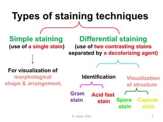
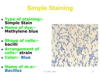
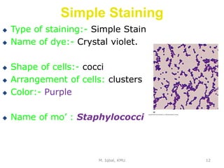
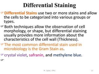
Simple and Gram Staining
Microbiology
Unit II
Muhammad Iqbal
Lecturer
Khyber Medical University
M. Iqbal, KMU 2
Staining of Bacteria
 Bacteria are almost colorless and transparent
 A staining technique is often applied to the
cells to color them.
Their shape and size can be easily determined
under the microscope.
M. Iqbal, KMU 3
Types of staining techniques
Simple staining
(use of a single stain)
Differential staining
(use of two contrasting stains
separated by a decolorizing agent)
For visualization of
morphological
shape & arrangement.
Identification Visualization
of structure
Gram
stain
Acid fast
stain Spore
stain
Capsule
stain
M. Iqbal, KMU 4
Smear Preparation:
Preparation and Fixation of Bacteria
for Staining.
Objective:
To kill the microorganism & fix them
to the slide to prevent them from
being washed out during the process
of staining.
M. Iqbal, KMU 5
Smear preparation
S Fixation
M. Iqbal, KMU 6
M. Iqbal, KMU 7
 Staining:
 Staining is an auxiliary technique used
in microscopy to enhance contrast in
the microscopic image. Stains are used
to highlight cell structures. Different
types of staining methods are used to
study cell structures and differentiate
one bacterium from another.
Simple Staining is the use of single
basic dye to color the bacterial cells
e.g. methylene blue, crystal violet, or
safranin.
M. Iqbal, KMU 8
Simple Staining
All bacteria take the color of the dye.
 Objective:-
To show the morphological
shapes and arrangement of
bacterial cells.
M. Iqbal, KMU 9
Basic Shapes of Bacteria
Cocci Bacilli
M. Iqbal, KMU 10
Arrangements
Cocci
Irregular Clusters Chains or Pairs
Tetrads
Staphylococci Micrococci Streptococci
M. Iqbal, KMU 11
Simple Staining
 Type of staining:-
Simple Stain
 Name of dye:-
Methylene blue
 Shape of cells:-
bacilli
 Arrangement of
cells:- strain
 Color:- Blue
 Name of m.o:-
Bacillus
M. Iqbal, KMU 12
Simple Staining
 Type of staining:- Simple Stain
 Name of dye:- Crystal violet.
 Shape of cells:- cocci
 Arrangement of cells: clusters
 Color:- Purple
 Name of mo’ : Staphylococci
Differential Staining
Differential Stains use two or more stains and allow
the cells to be categorized into various groups or
types.
Both techniques allow the observation of cell
morphology, or shape, but differential staining
usually provides more information about the
characteristics of the cell wall (Thickness).
The most common differential stain used in
microbiology is the Gram Stain as.
crystal violet, safranin, and methylene blue.

M. Iqbal, KMU 13
M. Iqbal, KMU 14
M. Iqbal, KMU 15
Gram Stain:
 It is the most
important differential
stain used in
bacteriology because
 it classifies bacteria
into two major groups:
a) Gram positive:
Appears violet after
Gram’s stain
b) Gram negative:
Appears pink after
Gram’s stain
Crystal violet
↓
Iodine
↓
Alcohol
↓
Safranin
M. Iqbal, KMU 16
M. Iqbal, KMU
17
Gram +ve
S. aureus
Gram –ve
E. coli
Step 1: Crystal Violet
(30—60 sec)
Step 2: Iodine
(mordant)
(30 sec)
Step 3: Decolorization
(Alcohol)– 5 sec
Step 4: Safranin
(30—60 sec)
Gram-positive bacteria
• Have a thick peptidoglycan layer surrounds the cell.
• The stain gets trapped into this layer and the
bacteria turned purple.
• Retain the color of the primary stain (crystal violet)
after decolorization with alcohol
Gram-negative bacteria
• have a thin peptidoglycan layer that does not retain
crystal violet stain.
• Instead, it has a thick lipid layer which is dissolved
easily upon decolorization with Alcohol.
• Therefore, cells will be counterstained with safranin
and turned pink.
M. Iqbal, KMU 18
Gram Stain
• Materials:-
• Cultures of Staphylococcus aureus,
Bacillus subtilis, E.coli
• Gram stain:
Crystal violet (primary stain)
Iodine (mordant)
Alcohol (decolorizing agent)
Safranin (counter stain)
M. Iqbal, KMU 19
Results
M. Iqbal, KMU 20
Shape: Cocci
Arrangment: irregular clusters
Colour: Violet
Gram’s reaction: Gram’s +ve
Name of microorganism: Staphylococci
Results
M. Iqbal, KMU 21
Shape: Bacilli
Arrangment: Chains
Colour: Violet
Gram’s reaction: Gram +ve
Name of microorganism: Bacillus
Results
M. Iqbal, KMU
22
Shape: Rods
Arrangment: Single
Colour: pink
Gram’s reaction: Gram –ve
Name of microorganism: Gram negative bacilli

4)Microbial Control Methods Unit-II 2.pptx

  • 1.
    Simple and GramStaining Microbiology Unit II Muhammad Iqbal Lecturer Khyber Medical University
  • 2.
    M. Iqbal, KMU2 Staining of Bacteria  Bacteria are almost colorless and transparent  A staining technique is often applied to the cells to color them. Their shape and size can be easily determined under the microscope.
  • 3.
    M. Iqbal, KMU3 Types of staining techniques Simple staining (use of a single stain) Differential staining (use of two contrasting stains separated by a decolorizing agent) For visualization of morphological shape & arrangement. Identification Visualization of structure Gram stain Acid fast stain Spore stain Capsule stain
  • 4.
    M. Iqbal, KMU4 Smear Preparation: Preparation and Fixation of Bacteria for Staining. Objective: To kill the microorganism & fix them to the slide to prevent them from being washed out during the process of staining.
  • 5.
    M. Iqbal, KMU5 Smear preparation S Fixation
  • 6.
  • 7.
    M. Iqbal, KMU7  Staining:  Staining is an auxiliary technique used in microscopy to enhance contrast in the microscopic image. Stains are used to highlight cell structures. Different types of staining methods are used to study cell structures and differentiate one bacterium from another. Simple Staining is the use of single basic dye to color the bacterial cells e.g. methylene blue, crystal violet, or safranin.
  • 8.
    M. Iqbal, KMU8 Simple Staining All bacteria take the color of the dye.  Objective:- To show the morphological shapes and arrangement of bacterial cells.
  • 9.
    M. Iqbal, KMU9 Basic Shapes of Bacteria Cocci Bacilli
  • 10.
    M. Iqbal, KMU10 Arrangements Cocci Irregular Clusters Chains or Pairs Tetrads Staphylococci Micrococci Streptococci
  • 11.
    M. Iqbal, KMU11 Simple Staining  Type of staining:- Simple Stain  Name of dye:- Methylene blue  Shape of cells:- bacilli  Arrangement of cells:- strain  Color:- Blue  Name of m.o:- Bacillus
  • 12.
    M. Iqbal, KMU12 Simple Staining  Type of staining:- Simple Stain  Name of dye:- Crystal violet.  Shape of cells:- cocci  Arrangement of cells: clusters  Color:- Purple  Name of mo’ : Staphylococci
  • 13.
    Differential Staining Differential Stainsuse two or more stains and allow the cells to be categorized into various groups or types. Both techniques allow the observation of cell morphology, or shape, but differential staining usually provides more information about the characteristics of the cell wall (Thickness). The most common differential stain used in microbiology is the Gram Stain as. crystal violet, safranin, and methylene blue.  M. Iqbal, KMU 13
  • 14.
  • 15.
    M. Iqbal, KMU15 Gram Stain:  It is the most important differential stain used in bacteriology because  it classifies bacteria into two major groups: a) Gram positive: Appears violet after Gram’s stain b) Gram negative: Appears pink after Gram’s stain
  • 16.
  • 17.
    M. Iqbal, KMU 17 Gram+ve S. aureus Gram –ve E. coli Step 1: Crystal Violet (30—60 sec) Step 2: Iodine (mordant) (30 sec) Step 3: Decolorization (Alcohol)– 5 sec Step 4: Safranin (30—60 sec)
  • 18.
    Gram-positive bacteria • Havea thick peptidoglycan layer surrounds the cell. • The stain gets trapped into this layer and the bacteria turned purple. • Retain the color of the primary stain (crystal violet) after decolorization with alcohol Gram-negative bacteria • have a thin peptidoglycan layer that does not retain crystal violet stain. • Instead, it has a thick lipid layer which is dissolved easily upon decolorization with Alcohol. • Therefore, cells will be counterstained with safranin and turned pink. M. Iqbal, KMU 18
  • 19.
    Gram Stain • Materials:- •Cultures of Staphylococcus aureus, Bacillus subtilis, E.coli • Gram stain: Crystal violet (primary stain) Iodine (mordant) Alcohol (decolorizing agent) Safranin (counter stain) M. Iqbal, KMU 19
  • 20.
    Results M. Iqbal, KMU20 Shape: Cocci Arrangment: irregular clusters Colour: Violet Gram’s reaction: Gram’s +ve Name of microorganism: Staphylococci
  • 21.
    Results M. Iqbal, KMU21 Shape: Bacilli Arrangment: Chains Colour: Violet Gram’s reaction: Gram +ve Name of microorganism: Bacillus
  • 22.
    Results M. Iqbal, KMU 22 Shape:Rods Arrangment: Single Colour: pink Gram’s reaction: Gram –ve Name of microorganism: Gram negative bacilli