4 u1.0-b978-1-4160-4224-2..50045-4..docpdf
•
2 likes•1,014 views

This document discusses anemia and pregnancy. It defines anemia and notes that 20-60% of pregnant women will be anemic at some point during their pregnancy. The key issues in evaluating anemia are determining the morphology of the anemia and the reticulocyte count to classify the anemia and identify any underlying diseases or mechanisms. A complete blood count and reticulocyte count provide information to classify the anemia as microcytic, normocytic, or macrocytic and determine if the bone marrow is hyperproliferative or hypoproliferative. Additional tests may then be used to confirm the diagnosis. Normal hematologic changes in pregnancy include an increase in blood volume through hemodil

Recommended
More Related Content
What's hot
What's hot (20)
Similar to 4 u1.0-b978-1-4160-4224-2..50045-4..docpdf
Similar to 4 u1.0-b978-1-4160-4224-2..50045-4..docpdf (20)
More from Loveis1able Khumpuangdee
More from Loveis1able Khumpuangdee (20)
Recently uploaded
Recently uploaded (20)
4 u1.0-b978-1-4160-4224-2..50045-4..docpdf
- 1. Chapter 42 Anemia and Pregnancy Sarah J. Kilpatrick, MD, PhD Anemia is defined as a hemoglobin (Hb) value less than the lower limit tion of anemia provides an exhaustive catalog of diagnoses, it does not of normal that is not explained by the state of hydration. This defini- lend itself to a systematic investigation of an individual patient. Rather, tion has physiologic validity, in that it is the amount of Hb per unit when the patient is anemic one wants to know (1) the morphology of volume of blood that determines the oxygen-carrying capacity of the anemia and (2) the reticulocyte count. Determining the answers blood. The normal Hb level for the adult female is 14.0 ± 2.0 g/dL.1 to these questions allows one to make a first approximation of a spe- Based on this normal value, 20% to 60% of prenatal patients will be cific diagnosis and to answer the following questions: found to be anemic at some time during their pregnancy. In one study, 32% of women presenting at less than 7 weeks’ gestation had an Hb 1. What is the mechanism of the anemia? value lower than 12 g/dL, suggesting that the prevalence of anemia is 2. Is there an underlying disease? high.2 Because of the normal hemodilution that occurs during preg- 3. What is appropriate treatment? nancy, the Centers for Disease Control and Prevention defines anemia in pregnancy as an Hb concentration lower than 11 g/dL in the first The CBC and the reticulocyte count provide the answers to the and third trimesters, or lower than 10.5 g/dL in the second trimester.3 first two questions. These data allow a morphologic classification of Some women with mild anemia will be missed using this definition. the anemia and indicate whether the marrow is hyperproliferative or hypoproliferative. The patient’s Hb value is determined by converting the pigment to cyanmethemoglobin and quantitating the amount Clinical Presentation spectrophotometrically. The remainder of the values are obtained by flow cytometry with an electronic cell counter. Symptoms caused by anemia are those resulting from tissue hypoxia, Based on the size of the red blood cells (RBCs), anemia can be the cardiovascular system’s attempts to compensate for the anemia, or classified as microcytic, normocytic, or macrocytic. The appearance of an underlying disease. Tissue hypoxia produces fatigue, lightheaded- the RBCs may also provide a clue to the mechanism of the anemia. For ness, weakness, and exertional dyspnea. Cardiovascular compensation example, hypochromic microcytic cells associated with a low reticulo- leads to a hyperdynamic circulation, with attendant symptoms of pal- cyte count suggests iron deficiency, thalassemia trait, sideroblastic pitations and tachycardia. Clinical conditions commonly associated anemia, or lead poisoning. Oval macrocytes combined with a low with anemia in pregnancy include multiple pregnancy, trophoblastic reticulocyte count and hypersegmented polymorphonuclear leuko- disease, chronic renal disease, chronic liver disease, and chronic infec- cytes suggest megaloblastic anemia (vitamin B12 or folate deficiency). tion. In obstetric patients, however, anemia is most commonly discov- Oval microcytes and an elevated reticulocyte count are characteristic ered not because of symptoms but because a complete blood count of hereditary spherocytosis. Various poikilocytes, such as sickle cells, (CBC) is obtained as part of routine laboratory evaluation, either at acanthocytes, target cells, and schistocytes, suggest sickle cell disease, the initial prenatal visit or at repeat screening at 24 to 28 weeks’ acanthocytosis, Hb C disease, and mechanical RBC destruction, gestation. respectively. Although the CBC is an excellent first step in the approxi- mate diagnosis of anemia, additional studies are usually necessary to confirm the diagnosis. Table 42-2 lists laboratory studies frequently Evaluation of Anemia used in the evaluation of an anemic patient. Serum Hb and serum haptoglobin levels are useful in defining Anemia is not a diagnosis; rather, like fever or edema, it is a sign. The intravascular hemolysis. If serum haptoglobin is absent or low in con- key issue in the evaluation of anemia is to define the underlying mech- junction with an elevated serum Hb, the presence of intravascular anism or pathologic process. Although a mild anemia caused by iron hemolysis is established. Further studies are necessary to rule in or deficiency during pregnancy is of little consequence to either the rule out specific causes of intravascular hemolysis, such as severe mother or the fetus, a similarly mild anemia caused by carcinoma of autoimmune hemolytic anemia (direct Coombs test), paroxysmal the colon has grave implications. One must also keep in mind the nocturnal hemoglobinuria (PNH) (osmotic fragility), and hemoglo- genetic implications of many anemias, such as the hemoglobinopathies binopathies including sickle cell disease and thalassemia major (Hb and hereditary spherocytosis. electrophoresis). Table 42-1 presents a classification of anemia based on the patho- Total bilirubin is elevated modestly in hemolytic anemia (rarely in physiologic mechanism involved. Although a mechanistic classifica- excess of 4 mg/dL). The increase results predominantly from an
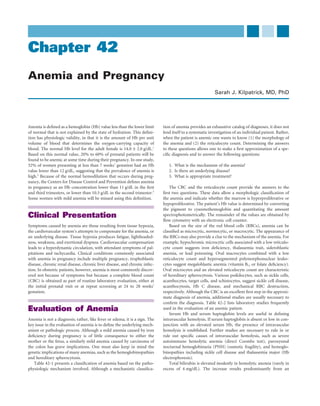
- 2. 870 CHAPTER 42 Anemia and Pregnancy TABLE 42-1 ANEMIA CLASSIFIED BY TABLE 42-2 LABORATORY STUDIES USEFUL IN PATHOPHYSIOLOGIC MECHANISM EVALUATION OF ANEMIA I. Dilutional (expansion of the plasma volume) Laboratory Study Reference Range A. Pregnancy B. Hyperglobulinemia Red blood cell (RBC) count 4.0-5.2 × 10¹²/L C. Massive splenomegaly Mean corpuscular volume (MCV) 80-100 μm3 II. Decreased RBC production Mean corpuscular hemoglobin 31-36 g/dL A. Bone marrow failure concentration (MCHC) 1. Decreased building blocks or stimulation Reticulocyte count 48-152 × 109/L (0.5-1.5%) a. Iron, protein Serum (free) hemoglobin 1.0-5 mg/dL b. Chronic infection, chronic renal disease Serum haptoglobin 30-200 mg/dL 2. Decreased erythron Total bilirubin 0.1-1.2 mg/dL a. Hypoplasia (hereditary, drugs, radiation, toxins) Direct Coombs test Negative b. Marrow replacement (tumor, fibrosis, infection) Hb electrophoresis >98% A B. Ineffective production <3.5% A2 1. Megaloblastic (vitamin B12 and folate deficiency, <2% F myelodysplasia, erythroleukemia) Serum ferritin >20 μg/L 2. Normoblastic (refractory anemia, thalassemia) Plasma iron 33-102 μg/dL III. Increased RBC loss Plasma total iron-binding capacity 194-372 μg/dL A. Acute hemorrhage Transferrin saturation 16-60% B. Hemolysis Folate level 1. Intrinsic RBC disorders Serum >20 μg/L a. Hereditary Red blood cells 165 ng/mL (1) Hemoglobinopathies Serum vitamin B12 190-950 ng/L (2) RBC enzyme deficiency Anti-intrinsic factor antibody (AIF) Negative (3) Membrane defects (4) Porphyrias b. Acquired (1) Paroxysmal nocturnal hemoglobinuria (2) Lead poisoning must be discontinued for 24 to 48 hours before these studies are carried 2. Extrinsic RBC disorders out. In iron deficiency, the FEP increases approximately fivefold. Iron a. Immune is transported in the plasma bound to transferrin. In the iron-deficient b. Mechanical state, the plasma iron decreases, the TIBC increases, and the percent c. Infection of iron saturation decreases. In contrast, with chronic infection, both d. Chemical agents the plasma iron and the TIBC are decreased, but the percent saturation e. Burns remains normal. f. Hypersplenism Serum folate, RBC folate, and serum vitamin B12 levels are useful g. Liver disease in defining the cause of macrocytic anemia. Because the RBC folate RBC, red blood cell. more accurately reflects the body’s folate stores, many laboratories no longer offer the serum folate determination. The presence of serum intrinsic factor antibodies is specific for pernicious anemia. However, increase in the indirect fraction. However, significant hemolysis can they are undetectable in approximately 40% of cases, so the absence occur without an elevation in the bilirubin. Therefore, the bilirubin of these antibodies does not rule out a diagnosis of pernicious level is helpful only when it is elevated. anemia. The direct Coombs test uses anti-human immunoglobulin to detect Although a bone marrow aspiration or biopsy can add much useful immunoglobulins attached to the surface of RBCs. A positive test information, it is rarely done today in pregnant anemic women. In indicates an immune cause for a hemolytic anemia. In such cases, it is addition to providing a ratio of myeloid to erythroid production important to search for underlying causes for autoimmunity, such as (normal, approximately 3 : 1), it provides a measure of iron stores, connective tissue disease, lymphoma, carcinoma, and sarcoidosis. The allows a differential count of myeloid and erythroid precursors, pro- diagnosis and management of glucose-6-phosphate dehydrogenase vides evidence of infiltration with neoplasm, and allows histologic and (G6PD) deficiency and of the various hemoglobinopathies are dis- bacteriologic confirmation of infection. cussed later in this chapter. The free erythrocyte protoporphyrin (FEP),4 plasma iron, plasma total iron-binding capacity (TIBC),5 and serum ferritin level6,7 are useful in establishing a diagnosis of iron deficiency. Protoporphyrin is Normal Hematologic Events generated in the penultimate step of heme synthesis, with iron subse- quently incorporated into protoporphyrin to create heme. Iron defi- Associated with Pregnancy ciency causes elevated FEP. Serum ferritin correlates closely with body iron stores, and many investigators support the use of serum ferritin Blood Volume Changes as the best single test in patients with anemia to make a diagnosis of During pregnancy, there is normally a 36% increase in the blood iron deficiency anemia.6,8 A ferritin level of 12 μg/L or lower is consis- volume, the maximum being reached at 34 weeks’ gestation.11 The tent with iron deficiency anemia. Plasma iron and serum ferritin levels plasma volume increases by 47%, but the RBC mass increases only by are both increased after ingestion of iron.9,10 Therefore, iron therapy 17%; the latter reaches its maximum at term. As shown in Figure 42-1,
- 3. CHAPTER 42 Anemia and Pregnancy 871 6 TABLE 42-3 IRON REQUIREMENTS FOR PREGNANCY 5 Required for Average (mg) Range (mg) Volume (I) 4 External iron loss 170 150-200 3 Expansion of red blood cell 450 200-600 2 mass Fetal iron 270 200-370 1 Iron in placenta and cord 90 30-170 Blood loss at delivery 150 90-310 0 Total requirement 980 580-1340 0 10 20 30 40 50 Gestational age Requirement less red blood 840 440-1050 cell expansion Blood volume RBC volume Plasma volume Hematocrit FIGURE 42-1 Hematologic changes during pregnancy. RBC, red were receiving iron supplementation than in those who were not.16 The blood cell. (Redrawn from Peck TM, Arias F: Hematologic changes usual sequence of events in regard to iron deficiency is an absence of associated with pregnancy. Clin Obstet 22:785, 1979.) iron in the marrow followed by the development of abnormal plasma iron studies (transferrin, ferritin, or FEP). The RBCs first become microcytic, then hypochromic. Finally, anemia develops. this disparity produces a relative hemodilution throughout pregnancy, Most women enter pregnancy with marginal iron stores. Pregnancy which reaches its maximum between 28 and 34 weeks. Although this places a large demand on iron balance that cannot be met with the dilutional effect lowers the Hb, hematocrit (Hct), and RBC count, it usual diet. In the absence of supplementation, iron deficiency develops. causes no change in the mean corpuscular volume or in the mean Supplementation with 60 mg of elemental iron per day during the corpuscular Hb concentration. Therefore, serial evaluation of these second and third trimesters meets the daily requirement. The Institute two indices is useful in differentiating dilutional anemia from progres- of Medicine recommends that supplementation be offered only to sive iron deficiency anemia during pregnancy: In the former, the women whose serum ferritin level is less than 20 μg/L.17 Although this indices do not change; in the latter, they decrease progressively. is a valid recommendation scientifically, the high cost of the screening limits its applicability. The fetal compartment preferentially obtains iron, folate, and Iron Kinetics vitamin B12 from—and at the expense of—the mother.18-20 Maternal The classic study by Scott and Pritchard shows that iron stores in iron is transferred to the fetus via serum transferrin. Transferrin binds healthy women are marginal at best.12 These authors evaluated iron to receptors in placental syncytiotrophoblast, where the iron is released stores in the bone marrow of healthy, white college students who and subsequently binds to ferritin in placenta cells. It is then trans- had never been pregnant and had never donated blood. Approximately ferred to apotransferrin, which enters into the fetal circulation as holo- two thirds had minimal iron stores. In another study, Pritchard transferrin. If maternal iron status is low, placental transferrin receptors and colleagues demonstrated that almost 50% of healthy primigra- increase to facilitate more uptake of iron by the placenta.21 vidas had minimal iron stores in the marrow during the first trimester.13 The major reason for poor iron stores is thought to be menstrual Folate loss. Data from Monsen and associates indicated that the usual men- Folic acid, a water-soluble vitamin, is widely available in the diet. strual loss is 25 to 30 mL of whole blood.14 This is equivalent to 12 to Dietary folates are, in fact, a family of compounds that appear as poly- 15 mg of elemental iron, because each milliliter of blood contains glutamates. In humans, the only source of folate is the diet, and absorp- 0.5 mg of iron. To meet the iron loss for menses alone, a woman must tion occurs primarily in the proximal jejunum. Before folate can be absorb 1.5 to 2.0 mg of elemental iron from her diet each day. Because absorbed, it must be reduced to the monoglutamate form.22 Pancreatic only 10% of dietary iron is usually absorbed, and the average diet conjugases within the intestine are responsible for this process. The contains only 6 mg/1000 kilocalories, a woman’s iron balance is pre- activity of conjugase is decreased by use of anticonvulsants, oral con- carious at best. traceptives, alcohol, or sulfa drugs.23 Therefore, in addition to an abso- Pregnancy presents substantial demands on iron balance, exceeding lute diminution in dietary intake, the combination of increased need that saved by 9 months of amenorrhea.13 Table 42-3 lists the iron (e.g., multiple pregnancy, hemolytic anemia) and decreased absorption requirements for pregnancy. If available iron stores are insufficient to can lead to folate deficiency.20,24,25 meet the demands of pregnancy, iron-deficient erythropoiesis results. The folate requirement of 50 μg/day for a nonpregnant woman In a prospective study of 35 nonanemic women, ferritin levels were increases to 300 to 500 μg/day during pregnancy.13,26 Because adequate measured before and during pregnancy to determine the relationship folate intake before and during the first weeks of pregnancy reduces of iron stores to developing anemia.15 Approximately 60% of the the occurrence of neural tube defects, all women considering preg- women with a ferritin concentration less than 20 μg/L before preg- nancy should consume 400 μg of folate per day.27 One study estimated nancy were anemic by 20 weeks’ gestation, compared with 25% of that a folate supplementation of 400 μg/day would reduce the risk of women with normal prepregnancy ferritin levels. Fenton and col- neural tube defects by 36%, and that 5 mg/day would reduce the risk leagues used serum ferritin levels to evaluate iron stores in pregnant by 85%.28 If a previous pregnancy was complicated by a neural tube women and found significantly higher ferritin levels in women who defect, the mother’s intake of folate should be 4 mg/day in the next
- 4. 872 CHAPTER 42 Anemia and Pregnancy pregnancy, starting at least 4 weeks before conception and continuing (hemolysis, elevated liver enzymes, low platelets) and thrombotic through the first 3 months of pregnancy.27,29 When folate depletion thrombocytopenic purpura—and in association with prosthetic heart occurs, the usual sequence of events is a decreased serum folate, hyper- valves. Other types of poikilocytes identified include sickle cells, target segmentation of polymorphonuclear leukocytes, a decrease in RBC cells, stomatocytes, ovalocytes, spherocytes, elliptocytes, and folate, the appearance of ovalocytes in the blood, development of an acanthocytes. abnormal marrow, and, finally, anemia.22 The Coombs test differentiates immune from nonimmune causes of hemolysis. Immune hemolysis is related to alloantibodies, drug- induced antibodies, and autoantibodies. Nonimmune causes of he- Vitamin B12 molysis include various hemoglobinopathies, hereditary disorders of Vitamin B12, also abundantly available in the diet, is bound to animal the RBC membrane (spherocytosis and elliptocytosis), hereditary defi- protein. Absorption requires hydrochloric acid and pepsin to free the ciency of an RBC enzyme, and the porphyrias. Acquired nonimmune cobalamin molecule from protein. Intrinsic factor is also essential for hemolysis is caused by either PNH or lead poisoning. absorption. After absorption, transport occurs via binding to transco- Bone marrow examination is essential for the evaluation of patients balamin II. Most of the vitamin B12 is stored in the liver, and individuals who have hypoproliferative anemias with normal iron studies. If eryth- typically have a 2- to 3-year store available.5 ropoiesis is megaloblastic, folate or vitamin B12 deficiency is a likely cause. If it is sideroblastic, both acquired and hereditary forms of sid- eroblastic anemia must be considered. Finally, if erythropoiesis is normoblastic, etiologic mechanisms fall Morphologic Classification into two major categories. The first category has myeloid-to-erythroid production ratios greater than 4 : 1 and includes aplasia, bone marrow of Anemia infiltration, effects of chronic diseases, and endocrine disorders such as hypothyroidism and hypopituitarism. In the second category, there Microcytic Anemia is ineffective erythropoiesis, usually associated with a myeloid to ery- The microcytic anemias are characterized by abnormal Hb synthesis throid production ratio lower than 2 : 1. with normal RBC production. A logical progression of diagnostic steps requires, first, that iron deficiency anemia be ruled out. If iron defi- ciency anemia is diagnosed rather than ruled out, it is important to Macrocytic Anemia consider gastrointestinal bleeding as the cause, although it is rare in Macrocytic anemia is associated with either (1) an increased rate of pregnant women. This can be accomplished by testing the stool for the RBC production and release of less than fully mature RBCs or (2) presence of occult blood with guaiac or other equally sensitive reagent. disorders of impaired DNA synthesis. Early use of a bone marrow If a microcytic anemia is not the result of iron deficiency, one must examination is helpful in pointing the investigation in the correct seek another cause, such as hemoglobinopathy, chronic infection, or direction. If maturation is megaloblastic, abnormal serum vitamin B12 one of the sideroblastic anemias. For this purpose, the following tests and RBC folate levels allow a diagnosis of vitamin B12 or folate defi- should be considered: ciency. If a diagnosis of folate deficiency is confirmed, the various causes of decreased deconjugation of the polyglutamate and malab- Hb electrophoresis sorption must be considered. If anti-intrinsic factor antibodies are Plasma iron and TIBC present, a diagnosis of pernicious anemia is ensured. If anti-intrinsic FEP factor antibodies are absent, a Schilling test is required to differentiate DNA probing for α-genes between pernicious anemia and a small-bowel malabsorption syn- Bone marrow examination drome. The Schilling test is performed by oral loading with cobalt 58—labeled cobalamin. Urinary excretion of cobalamin measured As noted, iron deficiency anemia is associated with decreased serum over 24 hours is then assessed. If abnormal excretion is noted (<10%), iron, increased TIBC (>400 μg/dL), reduced serum ferritin concentra- the test is repeated with 58Co-labeled cobalamin bound to intrinsic tion (<30 μg/L), and elevated FEP. Anemia of chronic disorders is factor. If pernicious anemia is present, excretion will normalize; if associated with decreased serum iron and elevated FEP but paradoxi- malabsorption is the cause, excretion will remain reduced. cally normal or increased ferritin and decreased TIBC. If the serum iron and TIBC are normal or increased and FEP is normal, the patient usually has thalassemia or a sideroblastic anemia. Hb electrophoresis and DNA probes are used to define the thalassemias, and ring sidero- Anemia and Perinatal blasts are present in the bone marrow of individuals with hereditary or acquired sideroblastic anemia. Morbidity and Mortality Fetal Effects of Maternal Anemia Normocytic Anemia Although it has been traditionally taught that significant maternal Because of the diverse nature of normocytic anemia, it is the most anemia is associated with suboptimal fetal outcome, data supporting difficult type to evaluate. The reticulocyte count varies according to this concept are scarce. Most recent studies, including a meta-analysis, whether RBC production is increased, normal, or decreased. If eryth- reported a significant relationship between anemia early in pregnancy ropoiesis is increased, one must differentiate between hemorrhage and and preterm delivery but no significant association with small-for- an increased rate of destruction. The blood smear may reveal a type gestational-age (SGA) neonates.30-33 Earlier studies reporting on mater- of RBC shape that can be virtually diagnostic. Fragmented cells are nal anemia and poor fetal outcome produced conflicting data.34-37 seen in microangiopathic hemolysis—as in the HELLP syndrome Studies in sheep showed that fetal oxygen consumption is maintained
- 5. CHAPTER 42 Anemia and Pregnancy 873 until the maternal Hct was reduced by more than 50%.38 Therefore, maternal anemia needs to be severe to affect the fetus. Elevated Hb early in pregnancy has also been associated with poor Specific Anemias perinatal outcome, including stillbirth and SGA neonates.39,40 In a case- control study, women with stillbirths had a significantly higher inci- Space does not allow a detailed discussion of the diagnosis and treat- dence of Hb higher than 14.5 g/dL than did control women without ment of literally hundreds of different anemias. Instead, a scheme of stillbirths.40 Although the mechanism for this association is unknown, diagnostic studies that are useful in evaluating any anemia and a dis- the authors hypothesized that high Hb may be a marker of inadequate cussion of specific anemias that are commonly seen during pregnancy plasma-volume expansion and hence reduced blood flow to the are presented. intervillous space. An alternate hypothesis was offered by Sagen and associates, who believed that these data were evidence of chronic hypoxia.39 Iron Deficiency Anemia In Africa, Asia, and Latin America, the relative risk of maternal Iron deficiency is the cause of 75% of all anemias in pregnancy, and mortality with severe anemia (Hb < 4.7 g/dL) was significantly elevated its prevalence may be as high as 47%.48-50 Clinical symptoms include at 3.5.41 In a case series of 130 women with a Hb less than 5 g/dL in easy fatigue, lethargy, and headache. Pica, which may involve the inges- the third trimester in India, more than half had a preterm delivery, tion of clay, dirt, ice, or starch, is a classic manifestation of iron defi- and more than 25% had postpartum hemorrhage, sepsis, or a stillbirth. ciency and was significantly associated in one study with lower maternal Further, eight women died.42 However, these studies and others are Hb but not with adverse pregnancy outcomes.51 Clinical findings fraught with confounders, including hemorrhage at delivery and lack include pallor, glossitis, and cheilitis. Koilonychia has been associated of access to care, so it is difficult to know whether the key factor associ- with iron deficiency anemia but is a rare finding. The laboratory char- ated with these deaths was the baseline anemia. Therefore, although acteristics of iron deficiency anemia are a microcytic, hypochromic profound maternal anemia can have adverse effects on the fetus, the anemia with evidence of depleted iron stores, low plasma iron, high margin of safety appears to be large. It may be that the prevalence of TIBC, low serum ferritin, and/or elevated FEP. If a bone marrow severe anemia is too low in industrialized countries to see consistent examination is performed, stainable iron is found to be markedly associations with poor fetal or maternal outcomes. depleted or absent. Although iron supplementation has not been Although there is no association between maternal third-trimester shown to alter perinatal outcome, the Centers for Disease Control and Hb and cord-blood Hb, maternal Hb and/or ferritin were significantly Prevention strongly recommends screening and treatment of iron defi- associated with cord-blood ferritin.21,43 In several studies, the umbilical ciency anemia in pregnancy.6,52,53 The rationale is that treatment main- cord-blood ferritin levels of infants whose mothers were iron deficient tains maternal iron stores and may be beneficial for neonatal iron were reduced, compared with those of infants whose mothers were not stores.6 iron deficient.16,44 Yet, infants whose ferritin levels were low were not The specific treatment is oral iron, most commonly ferrous sulfate, anemic and had normal iron kinetics, and their serum ferritin values 325 mg one to three times daily. Other iron preparations are more were not in the iron-deficient range. In a study of newborns of women expensive and do not offer any advantage over ferrous sulfate if equal with severe folate deficiency, Pritchard and colleagues found normal amounts of elemental iron are given. Reticulocytosis should be observed neonatal levels of folate.13 Even more interesting, Colomer found that after 7 to 10 days of therapy, and the Hb can rise by as much as 1 g/wk 1-year-old infants had a 5.7-fold increased risk of anemia if their in severely anemic individuals. Absorption from the gastrointestinal mothers were anemic at delivery, compared with nonanemic mothers, tract can be enhanced by the administration of 500 mg of ascorbic acid even after the data were controlled for feeding practices and socioeco- with each dose of iron. Gastrointestinal side effects associated with iron nomic status.45 In another interesting trial, iron supplementation in therapy include nausea, vomiting, abdominal cramps, diarrhea, and women who were not anemic in the first trimester was associated with constipation. These symptoms correspond to the dose of elemental a significant reduction in the incidence of birth weight less than 2500 g iron ingested; if symptoms are troublesome, the dose of iron should and in incidence of SGA neonates.46 This trial randomized nonanemic be reduced. Ferrous sulfate syrup (300 mg/5 mL) is an effective way of women to receive 30 mg ferrous sulfate or placebo by 20 weeks’ gesta- tailoring the dose to the patient’s tolerance. Once the anemia has tion. Almost 17% of the placebo-treated women but only 4% of the resolved, the patient should continue to receive iron therapy for an iron-treated women had low-birth-weight neonates, and 18% and 7%, additional 6 months to replace iron stores. respectively, had babies that were SGA. These provocative results, indi- Parenteral administration of iron is rarely indicated and should be cating that improved iron reserves enhance fetal growth independent reserved for patients with a malabsorption syndrome and those who of anemia status, may be generalizable only to populations with a high refuse to take oral iron and are significantly anemic (Hb < 8.5 g/dL).54 incidence of smoking, because 36% to 40% of the women in each There are currently three parenteral forms of iron approved for use in group smoked. However, another randomized trial of routine versus the United States: iron dextran, sodium ferric gluconate, and iron indicated (Hb < 10 g/dL) treatment with iron revealed no difference sucrose.55 These are usually given intramuscularly or intravascularly, in perinatal outcome or long-term outcome, including subsequent and, because severe anaphylaxis can occur in 1% of patients, a test dose pregnancies.47 should be administered first. In the absence of any reaction, daily injec- tions of 2 mL (100 mg) can be administered until the full dose is reached. Iron dextran contains 50 mg of iron per milliliter and comes Genetic Implications in 2-mL ampules. The required dose of iron dextran needed to correct Many of the hemolytic anemias are inherited as either autosomal- anemia and replenish stores can be calculated as follows56: dominant or recessive traits. Therefore, once the correct diagnosis has been made, the genetic implications should be thoroughly discussed 1. Milligrams of Fe needed = Hb deficit (in g/dL) × lean body wt with the patient and her partner. If appropriate, the discussion should (lb) + 1000, where the Hb deficit is calculated for women as 12 include antenatal diagnosis. minus the patient’s Hb value
- 6. 874 CHAPTER 42 Anemia and Pregnancy 2. Milliliters of iron dextran needed = mg of Fe needed ÷ The most common causes of vitamin B12 deficiency are autoim- 50 mg/mL mune inhibition of intrinsic factor production (pernicious anemia), inadequate production of intrinsic factor after gastrectomy, and the Iron sucrose and sodium ferric gluconate preparations appear to presence of a malabsorption syndrome. The morphologic features of have fewer adverse events, including anaphylaxis, in part because of B12 deficiency are similar to those of folate deficiency. In this instance, lower molecular weights.55-57 In a recent study, oral iron was compared the serum vitamin B12 level is low and the folate level is normal. with intravenous iron sucrose in a randomized trial of pregnant Because ineffective erythropoiesis is a prominent feature, evidence of women with Hb values of 8 to 10 g/dL.58 The increase in Hb (2 g/dL) low-grade hemolysis may be present (increased bilirubin and decreased was the same in both groups at day 30. In another, partially random- haptoglobin). The measurement of anti-intrinsic factor antibodies is ized study comparing oral iron with intramuscular iron dextran in useful. Treatment consists of an intramuscular dose of 1000 μg (1 mg) anemic women, there was no significant difference in term Hb levels of vitamin B12 every day for 1 week, then 1 mg every week for 4 weeks, but a significant increase in ferritin in the group treated with intra- and then 1 mg every month for the remainder of the patient’s life in muscular iron, compared with orally treated women.59 In contrast, cases of pernicious anemia. A prompt reticulocyte response is antici- another randomized trial comparing oral to intravenous iron sucrose pated after 3 to 5 days of therapy. found that the latter was associated with a significantly larger increase in Hb at 2 and 4 weeks and at delivery.60 However, at delivery, there was less than 1 mg/dL difference between the mean Hb values in the Hereditary Spherocytosis two groups. The authors reported no serious side effects in either and Elliptocytosis group. Spherocytosis is the most common form of inherited hemolytic Subcutaneous erythropoietin with or without oral iron therapy or anemia. The inheritance is autosomal-dominant with variable pene- intravenous iron sucrose has been used successfully to treat severe iron trance. Hereditary spherocytosis (HS) is characterized by a structural deficiency anemia in pregnancy, with no significant risks to the defect in the erythrocyte membrane caused by several different molec- mother.61-63 In one study in women for whom oral iron therapy had ular defects in the membrane proteins, including spectrin deficiency failed and who had an Hb value lower than 8.5 g/dL, the addition of and ankyrin deficinecy.66 The classic characteristic is an increased RBC erythropoietin to oral iron was associated with normalization of Hb osmotic fragility. The prevalence of the disorder is 2-3/10,000, which in 2 weeks in 73% of the women.62 Darbepoetin alfa, which has a longer implies around 1000 pregnancies annually in women with spherocy- half-life than erythropoietin, has also been used to successfully treat tosis. A hemolytic crisis can be precipitated by many conditions, such anemia after renal transplantation in a pregnant patient.64 as infection, trauma, and pregnancy itself.67 A relationship between increased hemolysis and increased maternal blood volume and splenic blood flow has been proposed. An alternative suggestion Megaloblastic Anemia is an increased osmotic fragility during the third trimester of Megaloblastic anemia is the second most common nutritional anemia pregnancy.68 seen during pregnancy. Most commonly, folate deficiency is the cause, The diagnosis is suspected on the basis of family history and find- but a deficiency in vitamin B12 must also be considered. The etiology ings in the CBC and reticulocyte count that suggest a hyperprolifera- of these anemias are poor nutrition and or decreased absorption. With tive anemia. Confirmation is obtained with the osmotic fragility test. the increase in pregnancies occurring after bariatric surgery, it is pos- Prenatal care of women with hereditary spherocytosis who have not sible that bariatric surgery may become a common cause of folate or had a splenectomy requires vigilance for hemolytic crisis and folate B12 deficiency in pregnancy in the United States.65 supplementation to ensure adequate marrow function.69 A hemolytic Patients with folate deficiency present with the typical symptoms crisis can be treated conservatively with replacement transfusions or of anemia plus roughness of the skin and glossitis. The CBC reveals a with splenectomy. Because splenectomy is mechanically difficult to macrocytic or normocytic, normochromic anemia with hypersegmen- accomplish during the third trimester of pregnancy, it is sometimes tation of the polymorphonuclear leukocytes. The reticulocyte count is preceded by delivery. In the absence of severe, untreated anemia, sphe- normal or low, and the white blood cell and platelet counts are fre- rocytosis does not contribute to perinatal morbidity or mortality. quently decreased. Bone marrow examination is not usually necessary Hereditary elliptocytosis, also inherited as an autosomal-dominant for diagnosis, but if it is done, megaloblastic erythropoiesis is noted. trait, is a milder hemolytic state also caused by a structural defect in The RBC folate level is decreased to less than 165 ng/mL (serum folate the RBC wall. The signs and symptoms are similar to those of sphero- to less than 2 μg/L), and the vitamin B12 level is normal. Treatment cytosis but are not as severe. Most cases detected during pregnancy consists of oral folic acid administered in a dose of 1 mg/day. Parenteral have been successfully treated with supportive therapy alone.70 folic acid may be indicated for individuals with malabsorption. A reticulocyte response should be seen in 48 to 72 hours, and the platelet count should normalize within a few days. The neutrophils normalize Autoimmune Hemolytic Anemia after 1 to 2 weeks. There are two major types of antibodies responsible for autoimmune In addition to anemia, women with vitamin B12 deficiency may also hemolytic anemia: warm-reactive and cold-reactive. Most warm- manifest neurologic defects relating to damage to the posterior columns reactive antibodies are of the immunoglobulin G (IgG) class and are of the spinal cord. It is critical that individuals with vitamin B12 defi- directed against some component of the Rh system on the surface of ciency not be treated with folic acid alone. Such treatment may well the RBC. In contrast, most cold-reactive antibodies are IgM; they are improve the anemia but has absolutely no salutary effect on the neu- usually anti-I or anti-i. Autoimmune hemolytic anemia with warm- ropathy and, in fact, may make it worse. As with folate deficiency, reactive antibodies is frequently seen in association with various hema- vitamin B12 deficiency is associated with dietary deficiency, an increased tologic malignancies (chronic lymphocytic leukemia, lymphoma), requirement, or both. Except in strict vegetarians who avoid all animal lupus erythematosus, viral infections, and drug ingestion. Penicillin products, dietary deficiency is rare. and α-methyldopa have been reported to cause autoimmune hemo-
- 7. CHAPTER 42 Anemia and Pregnancy 875 lytic anemia. Cold-reacting antibodies can be seen in association with enced metabolic disturbances or infections that precipitate an acute mycoplasmal infections, infectious mononucleosis, and lymphoreticu- hemolytic episode. Most affected African Americans carry a variant lar neoplasms. with these properties. Their hemolytic episodes are relatively mild. In a large number of cases, no specific inciting event can be identi- Greeks, Sardinians, and Sephardic Jews are more likely to carry G6PD fied.71 The diagnosis is suspected when a hyperproliferative, macro- Mediterranean, in which hemolysis is characteristically more severe cytic anemia is identified. The stained smear of peripheral blood often and favism (hemolysis induced by ingestion of fava beans) occurs. The reveals microcytes, polychromatophilia, poikilocytosis, and the pres- G6PD-deficient African-American population has not been reported ence of normoblasts. Leukocytosis is frequently seen and is a result of to experience favism. marrow hyperactivity. The critical study to confirm the diagnosis is a It is relatively unusual for a pregnant woman to experience severe positive direct Coombs test. There are several case reports in the litera- sequelae of G6PD deficiency. However, Silverstein and associates ture describing pregnancy-induced hemolytic anemia in which no reported Hct levels lower than 30% in 62% of 180 G6PD-deficient etiology could be discerned, the disease was diagnosed during preg- women.75 Prudence would argue against exposure of a known carrier nancy, and spontaneous remission occurred after delivery.72,73 The to precipitants of hemolysis. Sulfonamides, sulfones, some antimalari- most recent report described a woman who had hemolytic anemia als, nitrofurans, naphthalene, probenecid, para-aminosalicylic acid– diagnosed in three separate pregnancies that was not responsive to isoniazid, and nalidixic acid are among the medications and commonly either steroids or intravenous IgG therapy but resolved after delivery occurring environmental chemicals known to precipitate RBC destruc- in all pregnancies.72 tion in at-risk individuals. Treatment of autoimmune hemolytic anemia is directed toward One report suggested an increased incidence of low-birth-weight both the hemolytic process and the underlying disease. Blood trans- infants born to G6PD-deficient mothers, but no correction for the fusion, corticosteroid therapy, immunosuppression, and splenectomy effects of anemia or urinary tract infection was employed.76 Affected are the most frequently used measures. In cases with warm-reactive male infants born of carrier females have a higher incidence of neona- antibodies, corticosteroid should be tried initially, because approxi- tal hyperbilirubinemia, sometimes severe, than normal infants, and mately 80% of patients respond dramatically. Splenectomy is an careful observation of those at risk is strongly advised.77 The incidence effective form of treatment in approximately 60% of patients with of severe jaundice in G6PD-deficient newborn males is approximately warm-reactive antibodies. If the disease is refractory to both cortico- 5%, rising to 50% if there is a history of an icteric sibling. steroid therapy and splenectomy, a trial of immunosuppression If a hemolytic episode occurs during pregnancy because of G6PD is warranted. The treatment of cold-reactive antibodies depends on deficiency in a female heterozygote or the very rare homozygote, man- the severity of the hemolytic process. In patients with mild anemia, agement should include prompt discontinuation of any medication or avoidance of cold temperatures is all that is required. Corticosteroid other agent that may be responsible, treatment of any intercurrent therapy and splenectomy are usually not effective if the majority illness, and, if clinically indicated, transfusion support. In patients with of the RBC breakdown is intravascular. In patients with severe the variant common among African Americans, even in the male hemi- anemia, a trial of immunosuppression or plasmapheresis should be zygote, the G6PD activity of young RBCs is much higher than in RBCs considered. that have circulated for weeks and months. Old cells may be totally devoid of activity. Hence, the hemolytic episode, recognized early, is generally relatively mild and can be limited to the oldest population of Gluose-6-Phosphate circulating RBCs if the inciting agent is eliminated. A comprehensive Dehydrogenase Deficiency review of G6PD deficiency was published by Beutler.74 More than 20 different hereditary RBC enzyme defects have been described, most with an associated hemolytic anemia. Of these, only G6PD deficiency occurs with more than occasional frequency. The Aplastic and Hypoplastic Anemia genetic locus controlling G6PD synthesis is on the X chromosome, and Aplastic anemia is characterized by pancytopenia in the presence of a males with an abnormal gene may suffer hemolysis, especially if they hypocellular bone marrow. If it is left untreated, patients usually die are exposed to oxidant drugs that stress the pentose phosphate pathway from infection or bleeding. Three mechanisms have been postulated of the erythrocyte. Female heterozygotes are generally clinically unaf- to explain the development of aplastic anemia: (1) insufficient stem fected by similar exposure. The G6PD activity of the RBCs in hetero- cells resulting from an intrinsic defect or a reduction in number after zygous females is usually intermediate between the activity in exposure to a noxious agent, (2) the presence of a suppressor substance hemizygous males and that in normal subjects. However, some female that inhibits the maturation of the myeloid precursors, and (3) devel- carriers have normal G6PD activity, whereas others have activity that opment of an autoimmune reaction that causes death of the stem falls within the range seen in affected males. It has been proposed that cells. this is consistent with the Lyon hypothesis, that one of the two X Agents such as benzene, ionizing radiation, nitrogen mustard, anti- chromosomes of every female cell is randomly inactivated in early metabolites, antimitotic agents, certain antibiotics, and toxic chemicals embryonic life and continues to be inactive throughout all cell divi- predictably lead to marrow aplasia. In another category are agents such sions.74 Therefore, a few heterozygous women may be severely defi- as chloramphenicol, anticonvulsants, analgesics, and gold salts, which cient in G6PD activity, but most have sufficient activity to withstand induce aplasia only occasionally. Finally, hundreds of agents of various added stress on this critical metabolic pathway in erythrocytes. types have been implicated in several cases as causes of aplastic anemia. The ethnic groups in which variants of the deficiency occur with In about 50% of the cases, however, careful search does not reveal any greatest frequency are blacks, Mediterranean populations, Sephardic causative agent. and Asiatic Jews, and selected Asian populations. Of African-American Holly described eight patients with hypoplastic anemia detected males in the United States, 12% are reported to be deficient in G6PD during pregnancy that remitted spontaneously after delivery.78 The activity. Most affected individuals are hematologically normal unless bone marrow was described as hypocellular with an increase in mega- they have been exposed to certain drugs or chemicals or have experi- karyocytes. There are now many case reports and series of pregnancy-
- 8. 876 CHAPTER 42 Anemia and Pregnancy associated aplastic anemia, although they present a spectrum of clinical also been widely used with some benefit. However, the remission rate and bone marrow findings that makes it difficult to substantiate the with steroids is only 12%. existence of an aplastic anemia specifically related to pregnancy.79-84 Because of anecdotal reports of complete remission after pregnancy Many papers used the criteria delineated by Snyder and coworkers84 termination, it is tempting to consider therapeutic abortion. However, as evidence that the disease was pregnancy related: identification of the thorough examination of the available literature indicates that abor- disease after the onset of pregnancy; no other etiology of aplastic tion or premature termination of pregnancy is not associated with a anemia; decrease in all blood cell counts and in Hb; and hypoplastic more favorable outcome. The only reason to terminate pregnancy bone marrow. However, recovery from the aplastic anemia was not prematurely is inability to treat the patient satisfactorily during preg- universally documented after delivery, which raises the question of nancy with transfusion alone and a consequent need to proceed to whether it is truly pregnancy related.80,82 marrow transplantation. Patients with aplastic anemia seek medical attention because of symptoms related to profound anemia, bleeding, or infection. The CBC reveals pancytopenia with a hypoproliferative reticulocyte count. Paroxysmal Nocturnal Hemoglobinuria Examination of the bone marrow reveals hypoplasia with normoblas- PNH was named for its characteristic nighttime hemolysis with dark tic erythropoiesis. Severe aplastic anemia is fatal for more than 50% of early-morning urine. Hemolysis in PNH occurs as a result of a somatic affected patients.85 mutation in the phosphatidylinositol glycan class A (PIGA) gene on Bone marrow transplantation is now the treatment of choice, and the X chromosome. This enzyme mediates formation of phosphati- long-term survival of 50% to 70% of patients can be expected. Alterna- dylinositol anchors for various transmembrane proteins, including tives include antithymocyte globulin, immunosuppressive therapy, inhibitors of the complement proteins.93 These latter proteins nor- and other supportive therapy described later in this section.86 Survivors mally are present in the RBC and protect against complement activa- have had successful pregnancies after bone marrow transplantation.87-90 tion. Their reduction renders RBCs susceptible to intravascular The largest series examined pregnancy outcomes in 146 pregnancies hemolysis by complement. PNH usually begins insidiously, and there occurring after treatment for aplastic anemia in 41 women.89 The out- is no familial tendency. Considerable variability exists in severity of the comes in cases treated with total-body irradiation and bone marrow disease, and the classic presentation of hemoglobinuria is seen in only transplantation were compared with those in cases treated with high- 25% of patients. Exacerbations of the hemolytic process are precipi- dose chemotherapy and bone marrow transplantation. The data dem- tated by infection, menstruation, transfusion, surgery, and ingestion onstrated no increase in the incidence of congenital anomalies in of iron. infants. However, total-body irradiation was associated with an The most serious complications are marrow aplasia, thrombosis, increased risk of spontaneous abortion. Twenty-five percent of the and infection. Thrombosis accounts for 50% of deaths in nonpregnant pregnancies ended with a preterm delivery or delivery of a low-birth- patients and often involves intra-abdominal vessels, including Budd- weight infant. A more recent paper described pregnancy outcomes of Chiari syndrome resulting from hepatic vein thrombosis.94,95 Although 36 women with aplastic anemia who had been treated with immuno- anemia is the most prominent hematologic feature of PNH, leukope- suppression before their pregnancy.91 Only 11 of these women had nia and thrombocytopenia also occur frequently. The diagnosis is complete remission before they became pregnant, and 19% of the total based on tests including the sucrose hemolysis and acidified serum group had a relapse of their aplastic anemia during pregnancy that lysis tests, which demonstrate the sensitivity of the patient’s RBCs to required transfusion. Two women died, one of whom also had PNH, complement. and two women had eclampsia. The majority of the pregnancies There are two excellent reviews of PNH in pregnancy.93,96 A review resulted in live births, with a 14% prematurity rate. Several patients of 20 case reports and series encompassing 33 pregnancies in 24 women were treated with cyclosporine or corticosteroids during their revealed several interesting features. One third of these cases were pregnancy. diagnosed for the first time during pregnancy, and 12% of the preg- During pregnancy, supportive therapy remains the major objective, nancies were complicated by thromboembolism with three fourths of because bone marrow transplantation is still relatively contraindicated the patients having Budd-Chiari syndrome or hepatic vein thrombus.96 in pregnancy. In recent years, with modern supportive therapy, the Half of these women died. In addition, there were two maternal deaths maternal mortality rate has been 15% or less, and more than 90% of from infection, which means that the maternal mortality rate in this patients survive in remission.80,82 Treatment consists of maintenance summary of 24 women with PNH was 17%. In addition, 73% had of Hb levels through periodic transfusion, prevention and treatment anemia or hemolysis during pregnancy, and 27% developed thrombo- of infection, stimulation of hematopoiesis with androgens, splenec- cytopenia. In another review, the most common complication was tomy, intravenous immune globulin (IVIG), and intravenous ste- venous thrombosis.93 Although at least two pregnant or puerperal roids.92 Two case reports described successful pregnancies with a women developed a thromboembolism despite receiving thrombopro- combination of RBC and platelet transfusions, cyclosporine, human phylaxis, experts continue to recommend thromboprophylaxis.93,96 granulocyte colony-stimulating factor, high-dose intravenous predni- This may be particularly challenging if the patient develops thrombo- sone, and intravenous immunoglobulin.79,83 In a series of 14 women cytopenia. There is one case report of a successful labor epidural place- diagnosed during pregnancy, all of whom were treated with transfu- ment in a woman with PNH and a platelet count of 64,000/mL.97 sions only, there were no deaths, and 10 of the women had normal The optimal treatment of PNH is replacement of the abnormal pregnancy outcomes.82 The four abnormal outcomes were spontane- stem cells with cells capable of producing the normal cellular compo- ous abortion, preterm delivery, preeclampsia, and intrauterine growth nents. This has been accomplished by bone marrow transplantation. restriction (IUGR). Androgen therapy can be effective at stimulating The major therapeutic modalities during pregnancy are iron therapy, erythropoiesis; however, androgens are contraindicated during preg- transfusions, corticosteroids, and androgen treatment (if the fetus nancy unless the fetus is demonstrated to be male. Androgenic agents is male).93,98,99 Iron can be administered orally to replace the consider- commonly used include the anabolic steroids, oxymetholone, nandro- able amount lost in the urine. However, in patients with significant lone decanoate, or testosterone enanthate. Adrenocorticosteroids have iron deficiency, such treatment may lead to a burst of erythropoiesis,
- 9. CHAPTER 42 Anemia and Pregnancy 877 with delivery of a cohort of cells susceptible to the lytic action of Genotype complement. If a hemolytic episode follows iron therapy, it should be treated with either suppression of erythropoiesis by transfusion or Asian African suppression of hemolysis with corticosteroids. When acute hemolytic Phenotype Pattern Pattern episodes occur, treatment is aimed at diminishing hemolysis and pre- venting complications. α α α α Normal Hemoglobinopathies α α α α The hemoglobinopathies can be broadly divided into two general types. In the thalassemia syndromes, normal Hb is synthesized at an abnormally slow rate. In contrast, the structural hemoglobinopathies α α occur because of a specific change in the amino acid content of Hb. Heterozygous These structural changes may have either no effect or profound effects α-thalassemia 2 on the function of Hb, including instability of the molecule, reduced α α α α solubility, methemoglobinemia, and increased or decreased oxygen affinity. α Thalassemia Syndromes The thalassemia syndromes are named and classified by the type of α-Thalassemia 1 chain that is inadequately produced. The two most common types are α α α α-thalassemia and β-thalassemia, both of which affect the synthesis of Hb A. Reduced synthesis of γ or δ chains and combinations in which two or more globin chains are affected are relatively rare. In each instance, the thalassemia is a quantitative disorder of globin synthesis. Hb H disease a-Thalassemia α In patients with α-thalassemia, one or more structural genes are physi- cally absent from the genome. The various α-thalassemia genotypes are summarized in Figure 42-2. In blacks, the most common two-gene deletion state consists of one gene missing on each chromosome. In Asians, most often both genes are missing from the same chromosome. Homozygous In the homozygous stage, all four genes are deleted and no chains are hydrops fetalis produced. In such cases, the fetus is unable to synthesize normal Hb F or any adult hemoglobins. This deficiency results in high-output cardiac failure, hydrops fetalis, and stillbirth.100 The most severe form of α-thalassemia compatible with extrauter- FIGURE 42-2 Genotypes of the various α-thalassemia ine life is Hb H disease, which results from deletion of three α genes. syndromes. Hb H, hemoglobin H. In these patients, abnormally high quantities of both Hb H (β4) and Hb Barts (γ4) accumulate. Because Hb H precipitates within the RBC, the cell is removed by the reticuloendothelial system, leading to a β-globin gene is on chromosome 11. In homozygous β-thalassemia, moderately severe hemolytic anemia. In α-thalassemia minor (also α-chain production is unimpeded, and these highly unstable chains called α-thalassemia-1), two genes are deleted, resulting in a mild accumulate and eventually precipitate; markedly ineffective erythro- hypochromic, microcytic anemia that must be differentiated from iron poiesis and severe hemolysis result in a condition known as β- deficiency. A single gene deletion (α-thalassemia 2) is clinically unde- thalassemia major or Cooley anemia. There is variation in severity tectable and is called the “silent carrier” state. depending on whether homozygous for reduced (β+) or absent (β0) The diagnosis of α-thalassemia is presumptive by exclusion of iron β-globin synthesis (see Table 42-4). The fetus is protected from severe deficiency and β-thalassemia. Although α-thalassemia-1 minor does disease by α-chain production. However, this protection disappears not present a hazard to the adult, there are serious genetic implications rapidly after birth, with the affected infant becoming anemic by 3 to 6 when a mating of two individuals with the trait occurs. Under these months of age. The infant has splenomegaly and requires blood trans- circumstances, one must make a specific diagnosis by using restriction fusions every 3 to 4 weeks. Death typically occurs by the third decade endonuclease techniques or a DNA probe before undertaking antenatal of life and is usually secondary to myocardial hemochromatosis. diagnosis.101 Female infants surviving until puberty are usually amenorrheic and have severely impaired fertility.103,104 b-Thalassemia β-Thalassemia minor (also called β-thalassemia trait) results in a β-Thalassemia is autosomal-recessive and is more common in people variable degree of illness, depending on the rate of β-chain production. of Mediterranean, Middle Eastern, and Asian descent. The underpro- The characteristic findings include a relatively high RBC membrane duction of β-globulin chains is caused by point mutations with single rigidity, moderate to marked microcytosis, and a peripheral smear nucleotide substitution or oligonucleotide addition or deletion.102 The resembling that observed in iron deficiency. Hb electrophoresis char-
- 10. 878 CHAPTER 42 Anemia and Pregnancy TABLE 42-4 HEMATOLOGIC AND CLINICAL ASPECTS OF THE THALASSEMIA SYNDROMES Hemoglobin (Hb) Pattern* Condition Hb Level Hb A2 HB F Other Hb Clinical Severity Homozygotes α-Thalassemia ↓↓↓↓ 0 0 80% Hb Barts, remainder Hb Hydrops fetalis H and H Portland, some Hb A β+-Thalassemia ↓↓↓ Variable ↑↑ Some Hb A Moderately severe Cooley anemia β0-Thalassemia ↓↓↓↓ Variable ↑↑↑ No Hb A Severe Cooley anemia δβ0-Thalassemia ↓↓ 0 100% No Hb A Thalassemia intermedia Heterozygotes α-Thalassemia silent carrier N N N 1-2% Hb Barts in cord blood at birth N α-Thalassemia trait ↓ N N 5% Hb Barts in cord blood at birth Very mild Hb H disease ↓↓ N N 4-30% Hb H in adults; 25% Hb Barts in Thalassemia intermedia cord blood β+-Thalassemia ↓ to ↓↓ ↑ ↑ None Mild β0-Thalassemia ↓ to ↓↓ ↓ ↑↑↑ None Mild *Number of arrows indicates relative intensity of increase or decrease. ≠, increased; Ø, decreased; b+, reduced b-globin synthesis; b0, absent b-globin synthesis; db0, both d- and b-globin synthesis reduced or absent; N, normal. acteristically shows an elevation of Hb A2. β-Thalassemia trait does not TABLE 42-5 FREQUENCY OF THE MOST impair fertility, and the incidence of prematurity, low-birth-weight COMMON HEMOGLOBINOPATHIES infants, and infants of abnormal size for gestational age is identical to IN ADULT AFRICAN AMERICANS that in normal women.105,106 Nineteen women with β-thalassemia major or intermedia (e.g., the compound heterozygous state) were Hemoglobinopathy Abbreviated Name Frequency followed through 22 pregnancies; 21 viable infants were delivered.107 These patients all had intensive treatment, including transfusions and Sickle cell trait Hb SA 1 : 122 Sickle cell anemia Hb SS 1 : 708 iron-chelating agents, if necessary, before pregnancy or if their Hb Sickle cell–hemoglobin C Hb SC 1 : 757 concentration was greater than 7 g/dL. In addition, all women had a disease prepregnancy cardiac echocardiogram showing a left ventricular ejec- Hemoglobin C disease Hb CC 1 : 4790 tion fraction greater than 55%. These results suggest that women with Hemoglobin C trait Hb CA 1 : 41 well-managed, stable β-thalassemia can do very well during preg- Hemoglobin S–β-thalassemia Hb S-β-thal 1 : 1672 nancy.107 The clinical characteristics and hematologic findings of the Hemoglobin S-high F Hb S-HPFH 1 : 3412 various thalassemias are summarized in Table 42-4. Because of increased Asian immigration, the number of β- thalassemia cases in the United States has risen, so maternal screen- ing of appropriate women is important.108 In California, cases of The nomenclature and frequency of the most common hemoglobin- β-thalassemia major, Hb E/β-thalassemia, and other combined struc- opathies among African Americans are depicted in Table 42-5.114 Con- tural Hb abnormalities are more common than phenylketonuria or firmation of a diagnosis of a specific hemoglobinopathy requires galactosemia.108 A suggestion for easy antenatal maternal screening for identification of the abnormal Hb by means of Hb electrophoresis. α- and β-thalassemia is shown in Figure 42-3.105,109 Prenatal diagnosis, including preimplantation genetic diagnosis, is now available for β- Sickle Cell Trait thalassemia by polymerase chain reaction techniques of mutation Traditionally, women with sickle cell trait have been thought to do well detection on fetal blood or fetal DNA obtained from amniocentesis or during pregnancy and labor. However, new studies have reported con- chorionic villus sampling.102,110-113 flicting results about increased morbidities in women with sickle trait.115-117 A case-control study from Mississippi, in which women with or without the trait were matched for race, reported a significant Structural Hemoglobinopathies decrease in gestational age at birth (33 versus 35 weeks), lower mean To date, several hundred variants of α, β, γ, and δ chains have been birth weight, and an increased rate of fetal death (9.7% versus 3.5%) identified. Most differ from normal chains by only one amino acid. in the women with sickle cell trait.116 Of interest, 42% of the fetal
- 11. CHAPTER 42 Anemia and Pregnancy 879 CBC with RBC indices MCV 80 μm3 MCV 80 μm3 No thalassemia Hb electrophoresis, Fe studies* Hb A2 3.5% Hb A2 3.5% Hb A2 3.5% and Low Fe studies and NI Fe studies† NI Fe studies Probable -thalassemia Fe deficiency -Thalassemia Father evaluated Father evaluated MCV 80 μm3 MCV 80 μm3 Hb A2 3.5% Hb A2 3.5% No -thalassemia -Thalassemia -Thalassemia No -thalassemia Cord blood for Counseling Hb electrophoresis Hb Barts prenatal Dx when infant 1 yr FIGURE 42-3 Maternal screening for a- and b-thalassemia. *May be serum Fe, total Fe binding capacity. †Percent transferrin saturation >15 or ferritin >12 μg/L. CBC, complete blood count; Dx, diagnosis; Fe, iron; Hb, hemoglobin; NI, normal; MCV, mean corpuscular volume; RBC, red blood cell. deaths in the sickle cell trait group were early deaths (16 to 20 weeks). Sickle Cell Anemia In contrast, in a large cohort study of all African-American deliveries Patients with sickle cell anemia (SCA) suffer from lifelong complica- at one institution that compared those with and without maternal tions, in part as a result of the markedly shortened life span of their sickle cell trait, the trait was found to have a significant protective effect RBCs. Virtually all signs and symptoms of SCA are secondary to hemol- for preterm delivery at less than 32 weeks (0.9% versus 4.5%).115 This ysis, vaso-occlusive disease, or an increased susceptibility to infection protective effect was even more apparent in women with multiple (Table 42-6). Clinical manifestations may affect growth and develop- gestations, with 0% versus 22% delivering before 32 weeks. ment, with growth restriction and skeletal changes secondary to expan- Because there is an increased rate of urinary tract infection among sion of the marrow cavity. Painful crises may occur in the long bones, women with sickle cell trait, pregnant patients should be repeatedly abdomen, chest, or back. The cardiovascular manifestations are those screened for asymptomatic bacteriuria.118-120 Recently, in a large case- of a hyperdynamic circulation, and pulmonary signs may be secondary control study of women with or without sickle cell trait who were to either infection or vaso-occlusion. In addition to painful vaso-occlu- matched for race, age, gestational age, and entry into prenatal care, sive episodes, patients may exhibit hepatomegaly, signs and symptoms there was no significant difference in the incidence of positive urine of hepatitis, cholecystitis, and painful splenic infarcts. Genitourinary cultures (22% versus 19%).117 However, pyelonephritis was signifi- signs include an impairment in concentrating ability (hyposthenuria), cantly more common in the women with sickle cell trait (2.4% versus hematuria, and pyelonephritis. 0.7%). Another study suggested that the risk of preeclampsia Whether pregnancy in women with SCA is associated with more was increased to 25% in those with the trait, compared with 10% maternal complications is controversial. One comparison of pregnancy in a sickle-negative control group.121 These patients may become outcomes between women with and without SCA revealed no signifi- iron deficient, and iron supplementation during pregnancy is cant differences. Rates of maternal morbidity from SCA were the indicated. same during pregnancy as in the nonpregnant state.122 However,
- 12. 880 CHAPTER 42 Anemia and Pregnancy TABLE 42-6 CLINICAL MANIFESTATIONS OF cated.129,130 The goal of partial exchange transfusions is to keep the Hb SICKLE CELL ANEMIA A level higher than 50% and the Hct greater than 25%.131 A prospective, randomized study of 72 patients with SCA showed no significant dif- I. Growth and development ference in perinatal outcome between women who were treated with A. Retarded growth prophylactic transfusions and those who received transfusions only if B. Skeletal changes their Hb level fell to less than 6 g/dL or the Hct to less than 18%.127 C. Decreased life span However, this study did report a significant decrease in crises during II. Sickle cell crisis pregnancy, from 50% to 14%, in the group receiving prophylactic trans- A. Painful vaso-occlusive episodes: bones, abdomen, chest, and back fusions. Sixty-six patients with sickle cell–Hb C disease and 23 with III. Cardiovascular manifestations of hyperdynamic circulation sickle cell–β-thalassemia received transfusions for hematologic reasons A. Cardiomegaly only and experienced similar perinatal outcomes.127 However, the ben- B. Systolic murmurs efits attained must be balanced against a 25% incidence of alloimmu- C. Failure nization and 20% occurrence of delayed transfusion reaction. IV. Pulmonary signs Several studies documented no relationship between maternal A. Infection: pneumococcus, mycoplasma, hemophilus, anemia and risk for IUGR or perinatal death in women with SCA.124,132 salmonella The use of prophylactic transfusions should be individualized. An B. Vascular occlusion excellent review of SCA in nonpregnant individuals suggested that V. Abdominal involvement transfusion is indicated for symptomatic acute anemia, severe symp- A. Painful vaso-occlusive episodes B. Hepatomegaly tomatic chronic anemia, acute chest syndrome with hypoxia, and C. Hepatitis surgery with general anesthesia and may be useful for severe protracted D. Cholecystitis pain episodes.133 Most observers believe that the prepregnancy course E. Splenic infarction of a woman is a good index of how she will fare during pregnancy. VI. Bone and joint changes Although fetal outcomes are generally good in pregnancies compli- A. Bone marrow infarction cated by SCA, there continues to be an increased risk of prematurity B. Osteomyelitis: salmonella and IUGR.122,123,134,135 The most recent series showed an incidence of C. Arthritis IUGR and preterm delivery of 45% each, and both were significantly VII. Genitourinary signs more common in women with SCA than in the control group without A. Hyposthenuria B. Hematuria SCA.123 C. Pyelonephritis Serial ultrasound studies should be done throughout pregnancy to VIII. Neurologic manifestations confirm normal fetal growth. There are no prospective studies on the A. Vascular occlusion use of antepartum fetal testing in women with SCA, so this should be B. Convulsions instituted at the discretion of the physician. In addition, preimplanta- C. Hemorrhage tion genetic diagnosis with polymerase chain reaction assays is avail- D. Visual disturbances able for SCA.110 IX. Ocular manifestations In general, prenatal vitamins without iron should be given to A. Conjunctival vessel changes women who are receiving multiple transfusions. But all women with B. Vitreous hemorrhage SCA should have an additional folic acid supplement of 1.0 mg/day prescribed. The pneumococcal vaccine should be given if the patient has not had the vaccine within the past year. During labor and delivery, another study showed a significant increase in antepartum admissions, the patient must remain well oxygenated and hydrated. If an exchange preterm labor or preterm premature rupture of membranes, and post- transfusion protocol has been used and the Hb A level is greater than partum infection in women with versus those without SCA.123 In two 40%, painful crises are distinctly unusual.136 Finally, a recent retrospec- studies, there was no difference in the incidence of preeclampsia in tive study of 40 women with SCA reported that the initial prenatal women with and without SCA.123,124 Series examining maternal deaths white blood cell count was significantly higher in those women who have been too small to determine whether there is an increased risk subsequently developed SCA-related complications (11.2 × 10⁹/L) with SCA; however, pulmonary embolus or acute chest syndrome or during their pregnancy than in those who did not develop complica- both was the cause in 5 of 7 deaths.125 tions (8 × 10⁹/L).137 It is not known whether the frequency of painful crises in women with SCA changes with pregnancy. In one large study, the average Hemoglobin Sickle C Disease number of crises per patient per pregnancy was 1 to 2, and other studies Women who are doubly heterozygous for both the Hb S and the Hb have suggested that 20% to 50% of affected pregnant women had C genes are said to have Hb SC disease. Hb electrophoresis reveals crises.122,124,126,127 Treatment is largely symptomatic, with the major approximately 60% Hb C and 40% Hb S. Patients with Hb SC disease objectives being to end a painful crisis and to combat infection. Hydra- typically have a normal habitus, a healthy childhood, and a normal life tion, oxygen therapy, and pain management are the cornerstones of span. If a systematic screening program has not been used, the condi- managing a pain crisis. Acute chest syndrome is one of the most severe tion may first be detected in many women during the latter part of complications of SCA and can be very difficult to treat. It has been pregnancy, when a complication occurs. At the beginning of preg- reported to occur in up to 20% of pregnancies.122,124,128 Urinary tract nancy, most women are mildly anemic and splenomegaly is present. and pulmonary infections should be diagnosed promptly and treated Examination of a peripheral blood smear shows numerous target cells. vigorously with appropriate antibiotics. Transfusion therapy has been Hb electrophoresis ensures the correct diagnosis.138,139 used widely for years in the treatment of symptomatic patients. Partial During pregnancy, 40% to 60% of patients with Hb SC disease exchange transfusions or prophylactic transfusions have been advo- present as if they had SCA. In contrast to patients with SCA, those with
- 13. CHAPTER 42 Anemia and Pregnancy 881 Hb SC disease frequently experience rapid and severe anemic crises usually is discovered during a medical evaluation. Mild hemolytic resulting from splenic sequestration. These patients also have a greater anemia with an Hct in the range of 25% to 35% is characteristic. The tendency to experience bone marrow necrosis with the release of fat- RBCs show microspherocytes and characteristic targeting. No increased forming marrow emboli. The clinical manifestations of Hb SC disease morbidity or mortality is associated with pregnancy, and no specific are otherwise similar to those of SCA but milder, and the general therapy is indicated. management of symptomatic patients is identical. Considerations for the management of labor are the same as with SCA. In a recent report, Hemoglobin E Disease women with Hb SC disease had a significantly increased risk of ante- The recent immigration of Southeast Asians to the United States has partum admission, IUGR, and postpartum infection compared to resulted in an increase in the number of individuals with Hb E trait women without sickle disease, but these risks were only half as great and disease. The clinical and laboratory manifestations of the various as those of the women with SCA.123 Similarly, Serjeant and associates Hb E syndromes are outlined in Table 42-7.142,143 Most individuals have reported that the rates of miscarriage, live-born delivery, and newborn a mild microcytic anemia that is of no clinical significance, and no weight less than 2500 g in women with Hb SC disease were similar to treatment is necessary. However, patients who are homozygous for Hb those in women with normal Hb and significantly better than those in E have a greater degree of microcytosis and are frequently anemic. women with SCA.140 Rates of pain crises, acute chest syndrome, and Target cells are prominent. As with Hb C trait and disease, no specific urinary tract infections were similar in women with Hb SC disease and therapy is required, and reproductive outcome is normal. those with SCA.140 Hemoglobin S-b-Thalassemia Patients with Hb S-β-thalassemia are heterozygous for the sickle cell Anemias Associated with and the β-thalassemia genes, and in general about 10% of sickle cell Systemic Disease disease is caused by Hb S-β-thalassemia.111 In addition to decreased β-chain production, there is a variably increased production of Hb F The normal bone marrow has the capacity to increase its RBC produc- and Hb A2. Because of this variable production rate, Hb electrophoresis tion sixfold to eightfold in response to anemia. This compensatory reveals a spectrum of Hb concentrations. Hb S may account for 70% mechanism, which is responsible also for the increase in RBC mass in to 95% of the Hb present, with Hb F rarely exceeding 20%.141 Because normal pregnancy, is triggered by tissue hypoxia and mediated by of the thalassemia influence, the Hb S concentration exceeds the Hb A erythropoietin. The response may be absent or blunted in some cir- concentration. This is in sharp contrast to patients with sickle cell trait, cumstances, most commonly in chronic disorders. Chronic infections, in whom Hb A levels exceed the concentration of Hb S. rheumatoid arthritis, and other inflammatory states are characterized The diagnosis is made in an anemic patient by demonstrating by a mild normocytic, normochromic anemia (or sometimes a hypo- increased Hb A2 and Hb F levels in association with a level of Hb S chromic, microcytic anemia) with low serum iron concentration, low exceeding that of Hb A. The peripheral smear reveals hypochromia and transferrin level, inappropriately low reticulocyte count, and generous microcytosis with anisocytosis, poikilocytosis, basophilic stippling, and but poorly utilized stores of reticuloendothelial iron. Although the target cells. The clinical manifestations of this disorder parallel those of bone marrow is normally cellular, it does not respond appropriately SCA but are generally milder. Painful crises may occur; however, these to the mildly accelerated RBC destruction typical of chronic inflam- patients have a normal body habitus and frequently enjoy an uncom- mation. Studies thus far have not determined whether the defect in promised life span. The role of exchange transfusion should be similar erythropoiesis can be attributed to inadequate erythropoietin secre- to that in patients with SCA; that is, exchange transfusion should be tion. In the absence of pregnancy, the Hb concentration in these reserved for the woman who experiences painful crises or whose anemia chronic states is frequently 9 to 10 g/dL, and the Hct concentration is leads to an Hct lower than 25%. approximately 30%. The hydremia of pregnancy may lower these values somewhat. Hemoglobin C Trait and Disease A similar but frequently more complicated anemia accompanies Hb C trait is an asymptomatic trait without reproductive consequences. renal failure. Here, more often perhaps than in chronic inflammatory Target cells are found in the peripheral smear, but anemia is not states, blood loss and hemolysis are contributory factors, and the present. Hb C disease, the homozygous state, is a mild disorder that serum iron and transferrin changes noted earlier are less regular. In TABLE 42-7 VARIOUS GENOTYPES OF HEMOGLOBIN E AND THEIR PHENOTYPIC EXPRESSION* Hb Electrophoresis (%) Genotype Degree of Anemia MCV† A + A2 E F S Phenotype Expression A/E 0 ↓ 68 30 <2 0 None E/E 0 to + ↓↓ <4 94 <2 0 None E/α-thal + to ++ ↓ 50 15 35 0 None S/E ++ ↓ 0 40 0 60 None E/β+-thal ++ ↓↓ 10 60 30 0 Splenomegaly E/β0-thal +++ ↓↓ 0 60 40 0 Splenomegaly *Number of + symbols indicates relative severity of anemia. † Number of arrows indicates relative amount of decrease. Hb, hemoglobin; MCV, mean corpuscular volume.