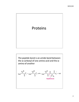
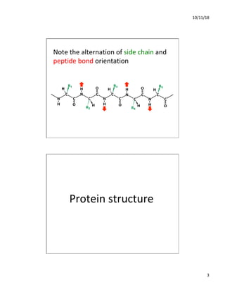
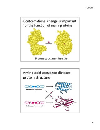
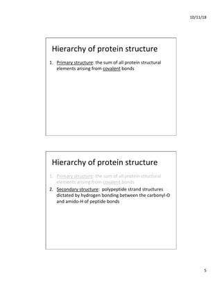
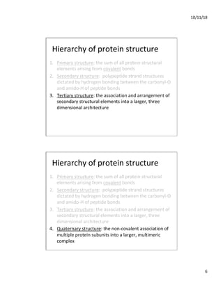
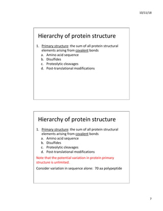
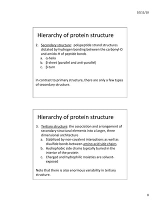
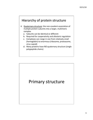
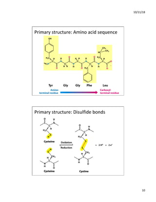
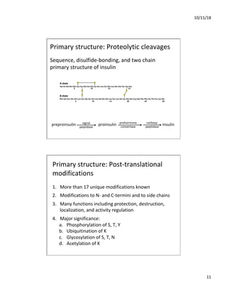
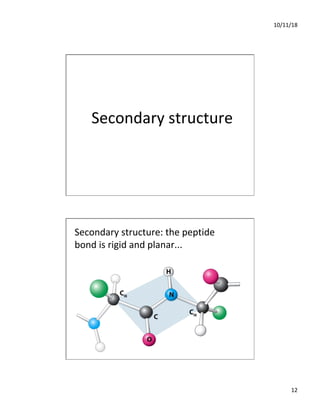
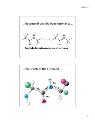
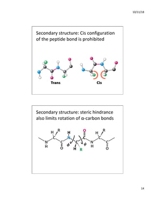
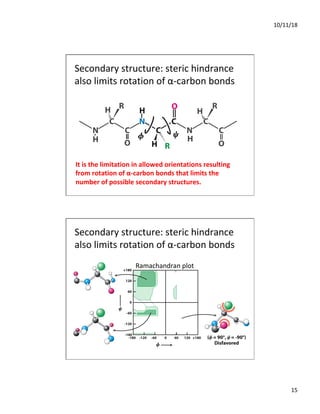
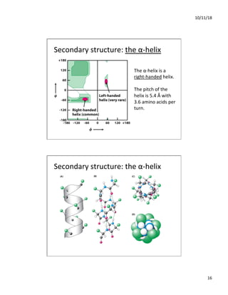
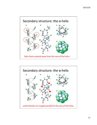
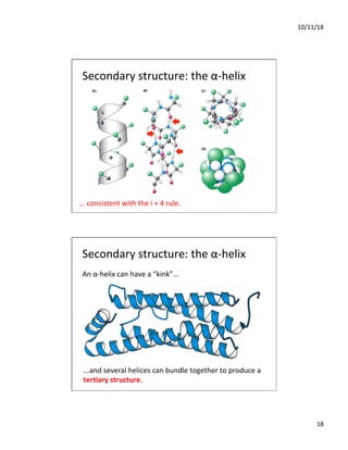
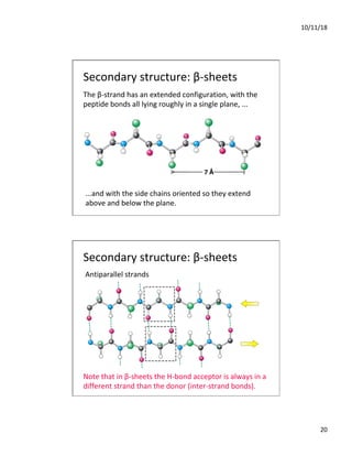
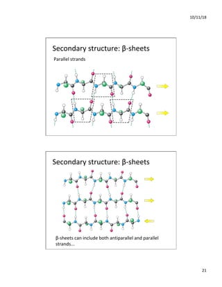
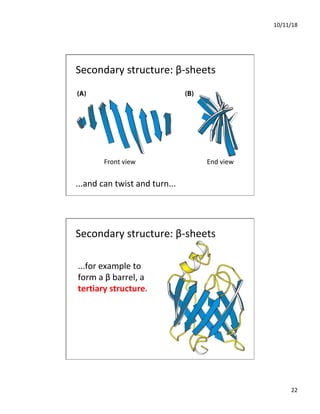
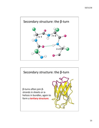
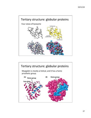
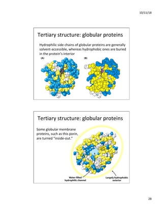
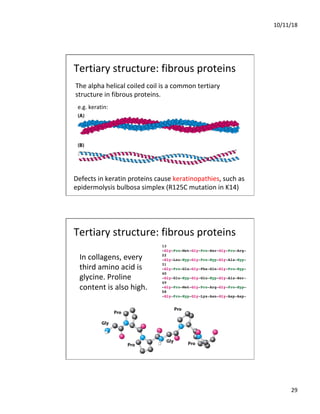
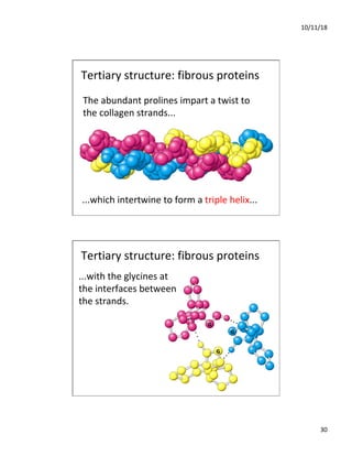
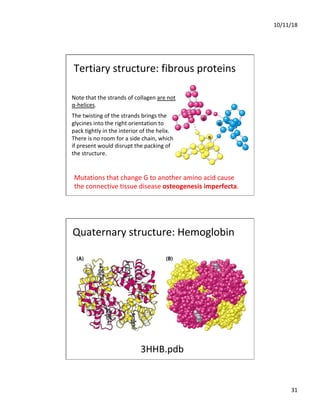
10/11/18
1
Biology of Cells & Tissues
Lecture 2: Protein Structure & Function I
Dan Hardy
Department of Cell Biology & Biochemistry
October 29, 2018
Lecture Plan
1. Hierarchy of protein structure
2. Elements of primary structure
3. Three types of secondary structure
4. Protein tertiary and quaternary structure
10/11/18
2
Proteins
The peptide bond is an amide bond between
the α-carboxyl of one amino acid and the α-
amino of another
10/11/18
3
Note the alternation of side chain and
peptide bond orientation
Protein structure
10/11/18
4
Conformational change is important
for the function of many proteins
Protein structure = function
Amino acid sequence dictates
protein structure
10/11/18
5
Hierarchy of protein structure
1. Primary structure: the sum of all protein structural
elements arising from covalent bonds
Hierarchy of protein structure
1. Primary structure: the sum of all protein structural
elements arising from covalent bonds
2. Secondary structure: polypeptide strand structures
dictated by hydrogen bonding between the carbonyl-O
and amido-H of peptide bonds
10/11/18
6
Hierarchy of protein structure
1. Primary structure: the sum of all protein structural
elements arising from covalent bonds
2. Secondary structure: polypeptide strand structures
dictated by hydrogen bonding between the carbonyl-O
and amido-H of peptide bonds
3. Tertiary structure: the association and arrangement of
secondary structural elements into a larger, three
dimensional architecture
4. Quaternary structure: the non-covalent association of
multiple protein subunits into a larger, multimeric
complex
10/11/18
7
Hierarchy of protein structure
1. Primary structure: the sum of all protein structural
elements arising from covalent bonds
a. Amino acid sequence
b. Disulfides
c. Proteolytic cleavages
d. Post-translational modifications
Hierarchy of protein structure
1. Primary structure: the sum of all protein structural
elements arising from covalent bonds
a. Amino acid sequence
b. Disulfides
c. Proteolytic cleavages
d. Post-translational modifications
Note that the potential variation in protein primary
structure is unlimited.
Consider variation in sequence alone: 70 aa polypeptide
10/11/18
8
Hierarchy of protein structure
2. Secondary structure: polypeptide strand structures
dictated by hydrogen bonding between the carbonyl-O
and amido-H of peptide bonds
a. α-helix
b. β-sheet (parallel and anti-parallel)
c. β-turn
In contrast to primary structure, there are only a few types
of secondary structure.
Hierarchy of protein structure
3. Tertiary structure: the association and arrangement of
secondary structural elements into a larger, three
dimensional architecture
a. Stabilized by non-covalent interactions as well as
disulfide bonds between amino acid side chains
b. Hydrophobic side chains typically buried in the
interior of the protein
c. Charged and hydrophilic moieties are solvent-
exposed
Note that there is also enormous variability in tertiary
structure.
10/11/18
9
Hierarchy of protein structure
4. Quaternary structure: the non-covalent association of
multiple protein subunits into a larger, multimeric
complex
a. Subunits can be identical or different
b. Required for cooperativity and allosteric regulation
c. Complexes can range in size from relatively small
(hemoglobin) to enormous (ribosome, proteasome,
virus capsid)
d. Many proteins have NO quaternary structure (single
polypeptide chains)
Primary structure
10/11/18
10
Primary structure: Amino acid sequence
Primary structure: Disulfide bonds
10/11/18
11
Sequence, disulfide-bonding, and two chain
primary structure of insulin
Primary structure: Proteolytic cleavages
preproinsulin proinsulin insulin
signal
peptidase
prohormone
convertase
carboxy-
peptidase
Primary structure: Post-translational
modifications
1. More than 17 unique modifications known
2. Modifications to N- and C-termini and to side chains
3. Many functions including protection, destruction,
localization, and activity regulation
4. Major significance:
a. Phosphorylation of S, T, Y
b. Ubiquitination of K
c. Glycosylation of S, T, N
d. Acetylation of K
10/11/18
12
Secondary structure
Secondary structure: the peptide
bond is rigid and planar...
10/11/18
13
...because of peptide bond resonance...
...that shortens the C-N bond.
10/11/18
14
Secondary structure: Cis configuration
of the peptide bond is prohibited
Secondary structure: steric hindrance
also limits rotation of α-carbon bonds
10/11/18
15
Secondary structure: steric hindrance
also limits rotation of α-carbon bonds
It is the limitation in allowed orientations resulting
from rotation of α-carbon bonds that limits the
number of possible secondary structures.
Secondary structure: steric hindrance
also limits rotation of α-carbon bonds
Ramachandran plot
10/11/18
16
Secondary structure: the α-helix
The α-helix is a
right-handed helix.
The pitch of the
helix is 5.4 Å with
3.6 amino acids per
turn.
Secondary structure: the α-helix
10/11/18
17
Secondary structure: the α-helix
Side chains extend away from the axis of the helix...
Secondary structure: the α-helix
..and H-bonds run roughly parallel to the axis of the helix...
10/11/18
18
Secondary structure: the α-helix
... consistent with the i + 4 rule.
Secondary structure: the α-helix
An α-helix can have a “kink”...
...and several helices can bundle together to produce a
tertiary structure.
10/11/18
19
Question
If a lipid bilayer is about 30 Å thick, how many
contiguous hydrophobic amino acids would be
required to form a membrane-spanning α-helix?
A. 5
B. 10
C. 15
D. 20
E. α-helices cannot span lipid bilayers
Secondary structure: β-sheets
10/11/18
20
Secondary structure: β-sheets
The β-strand has an extended configuration, with the
peptide bonds all lying roughly in a single plane, ...
...and with the side chains oriented so they extend
above and below the plane.
Secondary structure: β-sheets
Antiparallel strands
Note that in β-sheets the H-bond acceptor is always in a
different strand than the donor (inter-strand bonds).
10/11/18
21
Secondary structure: β-sheets
Parallel strands
Secondary structure: β-sheets
β-sheets can include both antiparallel and parallel
strands...
10/11/18
22
Secondary structure: β-sheets
...and can twist and turn...
Front view End view
Secondary structure: β-sheets
...for example to
form a β barrel, a
tertiary structure.
10/11/18
23
Secondary structure: the β-turn
β-turns often join β-
strands in sheets or α-
helices in bundles, again to
form a tertiary structure.
10/11/18
24
What determines the secondary
structure a polypeptide will form?
The peptide VDLLKN
in an α-helix of one
protein and in a β-
strand of another
Short sequences can form different secondary structures...
...and there is only a modest preference for certain
amino acids in secondary structures.
10/11/18
25
Pro and Gly (“kinky” and “flexible”) are most likely to be
in β-turns, and unlikely to be in α-helices or β-sheets,
but otherwise correlations are not very strong.
Algorithms for predicting secondary structure from
sequence are therefore notoriously inaccurate.
Secondary structure paradox: sequence (i.e. order of
side chains) dictates structure even though side chains
themselves don’t participate in the H-bonding that
stabilizes secondary structure
What determines the secondary
structure a polypeptide will form?
It is the full sequence of a polypeptide that specifies its
native overall structure.
What determines the secondary
structure a polypeptide will form?
10/11/18
26
Tertiary structure
Tertiary structure
Proteins are generally either globular or fibrous, and
composed of virtually any combination of secondary
structures.
Globular proteins are generally not very elongated (i.e.
they are shaped like a glob); most non-structural
proteins, including enzymes, are globular.
Fibrous proteins have very elongated structures. They
typically are major constituents of larger physical
structures (e.g. keratin in hair and skin, collagen in
connective tissue).
10/11/18
27
Four views of lysozyme
Tertiary structure: globular proteins
Myogobin is mostly α-helical, and it has a heme
prosthetic group.
Tertiary structure: globular proteins
10/11/18
28
Hydrophilic side chains of globular proteins are generally
solvent-accessible, whereas hydrophobic ones are buried
in the protein’s interior
Tertiary structure: globular proteins
Some globular membrane
proteins, such as this porin,
are turned “inside-out.”
Tertiary structure: globular proteins
10/11/18
29
The alpha helical coiled coil is a common tertiary
structure in fibrous proteins.
e.g. keratin:
Tertiary structure: fibrous proteins
Defects in keratin proteins cause keratinopathies, such as
epidermolysis bulbosa simplex (R125C mutation in K14)
In collagens, every
third amino acid is
glycine. Proline
content is also high.
Tertiary structure: fibrous proteins
10/11/18
30
...which intertwine to form a triple helix...
The abundant prolines impart a twist to
the collagen strands...
Tertiary structure: fibrous proteins
...with the glycines at
the interfaces between
the strands.
Tertiary structure: fibrous proteins
10/11/18
31
Note that the strands of collagen are not
α-helices.
The twisting of the strands brings the
glycines into the right orientation to
pack tightly in the interior of the helix.
There is no room for a side chain, which
if present would disrupt the packing of
the structure.
Mutations that change G to another amino acid cause
the connective tissue disease osteogenesis imperfecta.
Tertiary structure: fibrous proteins
Quaternary structure: Hemoglobin
3HHB.pdb
10/11/18
32
Quaternary structure
Same forces as those that stabilize tertiary structure
Quaternary structures can be homomeric or heteromeric
Association of multiple subunits confers capacity for
sophisticated regulation of activity (allosterism)
Association of enzymes catalyzing sequential reactions
into multienzyme complexes accelerates flux of
metabolites through pathways
Lecture 2 Recap
1. Protein primary structure—four elements
2. Protein secondary structure—three types
3. Protein tertiary and quaternary structure—
interactions between secondary structures

3_Protein_Structure_and_Properties1_Hardy.pdf

  • 1.
    10/11/18 1 Biology of Cells& Tissues Lecture 2: Protein Structure & Function I Dan Hardy Department of Cell Biology & Biochemistry October 29, 2018 Lecture Plan 1. Hierarchy of protein structure 2. Elements of primary structure 3. Three types of secondary structure 4. Protein tertiary and quaternary structure
  • 2.
    10/11/18 2 Proteins The peptide bondis an amide bond between the α-carboxyl of one amino acid and the α- amino of another
  • 3.
    10/11/18 3 Note the alternationof side chain and peptide bond orientation Protein structure
  • 4.
    10/11/18 4 Conformational change isimportant for the function of many proteins Protein structure = function Amino acid sequence dictates protein structure
  • 5.
    10/11/18 5 Hierarchy of proteinstructure 1. Primary structure: the sum of all protein structural elements arising from covalent bonds Hierarchy of protein structure 1. Primary structure: the sum of all protein structural elements arising from covalent bonds 2. Secondary structure: polypeptide strand structures dictated by hydrogen bonding between the carbonyl-O and amido-H of peptide bonds
  • 6.
    10/11/18 6 Hierarchy of proteinstructure 1. Primary structure: the sum of all protein structural elements arising from covalent bonds 2. Secondary structure: polypeptide strand structures dictated by hydrogen bonding between the carbonyl-O and amido-H of peptide bonds 3. Tertiary structure: the association and arrangement of secondary structural elements into a larger, three dimensional architecture Hierarchy of protein structure 1. Primary structure: the sum of all protein structural elements arising from covalent bonds 2. Secondary structure: polypeptide strand structures dictated by hydrogen bonding between the carbonyl-O and amido-H of peptide bonds 3. Tertiary structure: the association and arrangement of secondary structural elements into a larger, three dimensional architecture 4. Quaternary structure: the non-covalent association of multiple protein subunits into a larger, multimeric complex
  • 7.
    10/11/18 7 Hierarchy of proteinstructure 1. Primary structure: the sum of all protein structural elements arising from covalent bonds a. Amino acid sequence b. Disulfides c. Proteolytic cleavages d. Post-translational modifications Hierarchy of protein structure 1. Primary structure: the sum of all protein structural elements arising from covalent bonds a. Amino acid sequence b. Disulfides c. Proteolytic cleavages d. Post-translational modifications Note that the potential variation in protein primary structure is unlimited. Consider variation in sequence alone: 70 aa polypeptide
  • 8.
    10/11/18 8 Hierarchy of proteinstructure 2. Secondary structure: polypeptide strand structures dictated by hydrogen bonding between the carbonyl-O and amido-H of peptide bonds a. α-helix b. β-sheet (parallel and anti-parallel) c. β-turn In contrast to primary structure, there are only a few types of secondary structure. Hierarchy of protein structure 3. Tertiary structure: the association and arrangement of secondary structural elements into a larger, three dimensional architecture a. Stabilized by non-covalent interactions as well as disulfide bonds between amino acid side chains b. Hydrophobic side chains typically buried in the interior of the protein c. Charged and hydrophilic moieties are solvent- exposed Note that there is also enormous variability in tertiary structure.
  • 9.
    10/11/18 9 Hierarchy of proteinstructure 4. Quaternary structure: the non-covalent association of multiple protein subunits into a larger, multimeric complex a. Subunits can be identical or different b. Required for cooperativity and allosteric regulation c. Complexes can range in size from relatively small (hemoglobin) to enormous (ribosome, proteasome, virus capsid) d. Many proteins have NO quaternary structure (single polypeptide chains) Primary structure
  • 10.
    10/11/18 10 Primary structure: Aminoacid sequence Primary structure: Disulfide bonds
  • 11.
    10/11/18 11 Sequence, disulfide-bonding, andtwo chain primary structure of insulin Primary structure: Proteolytic cleavages preproinsulin proinsulin insulin signal peptidase prohormone convertase carboxy- peptidase Primary structure: Post-translational modifications 1. More than 17 unique modifications known 2. Modifications to N- and C-termini and to side chains 3. Many functions including protection, destruction, localization, and activity regulation 4. Major significance: a. Phosphorylation of S, T, Y b. Ubiquitination of K c. Glycosylation of S, T, N d. Acetylation of K
  • 12.
    10/11/18 12 Secondary structure Secondary structure:the peptide bond is rigid and planar...
  • 13.
    10/11/18 13 ...because of peptidebond resonance... ...that shortens the C-N bond.
  • 14.
    10/11/18 14 Secondary structure: Cisconfiguration of the peptide bond is prohibited Secondary structure: steric hindrance also limits rotation of α-carbon bonds
  • 15.
    10/11/18 15 Secondary structure: sterichindrance also limits rotation of α-carbon bonds It is the limitation in allowed orientations resulting from rotation of α-carbon bonds that limits the number of possible secondary structures. Secondary structure: steric hindrance also limits rotation of α-carbon bonds Ramachandran plot
  • 16.
    10/11/18 16 Secondary structure: theα-helix The α-helix is a right-handed helix. The pitch of the helix is 5.4 Å with 3.6 amino acids per turn. Secondary structure: the α-helix
  • 17.
    10/11/18 17 Secondary structure: theα-helix Side chains extend away from the axis of the helix... Secondary structure: the α-helix ..and H-bonds run roughly parallel to the axis of the helix...
  • 18.
    10/11/18 18 Secondary structure: theα-helix ... consistent with the i + 4 rule. Secondary structure: the α-helix An α-helix can have a “kink”... ...and several helices can bundle together to produce a tertiary structure.
  • 19.
    10/11/18 19 Question If a lipidbilayer is about 30 Å thick, how many contiguous hydrophobic amino acids would be required to form a membrane-spanning α-helix? A. 5 B. 10 C. 15 D. 20 E. α-helices cannot span lipid bilayers Secondary structure: β-sheets
  • 20.
    10/11/18 20 Secondary structure: β-sheets Theβ-strand has an extended configuration, with the peptide bonds all lying roughly in a single plane, ... ...and with the side chains oriented so they extend above and below the plane. Secondary structure: β-sheets Antiparallel strands Note that in β-sheets the H-bond acceptor is always in a different strand than the donor (inter-strand bonds).
  • 21.
    10/11/18 21 Secondary structure: β-sheets Parallelstrands Secondary structure: β-sheets β-sheets can include both antiparallel and parallel strands...
  • 22.
    10/11/18 22 Secondary structure: β-sheets ...andcan twist and turn... Front view End view Secondary structure: β-sheets ...for example to form a β barrel, a tertiary structure.
  • 23.
    10/11/18 23 Secondary structure: theβ-turn Secondary structure: the β-turn β-turns often join β- strands in sheets or α- helices in bundles, again to form a tertiary structure.
  • 24.
    10/11/18 24 What determines thesecondary structure a polypeptide will form? The peptide VDLLKN in an α-helix of one protein and in a β- strand of another Short sequences can form different secondary structures... ...and there is only a modest preference for certain amino acids in secondary structures.
  • 25.
    10/11/18 25 Pro and Gly(“kinky” and “flexible”) are most likely to be in β-turns, and unlikely to be in α-helices or β-sheets, but otherwise correlations are not very strong. Algorithms for predicting secondary structure from sequence are therefore notoriously inaccurate. Secondary structure paradox: sequence (i.e. order of side chains) dictates structure even though side chains themselves don’t participate in the H-bonding that stabilizes secondary structure What determines the secondary structure a polypeptide will form? It is the full sequence of a polypeptide that specifies its native overall structure. What determines the secondary structure a polypeptide will form?
  • 26.
    10/11/18 26 Tertiary structure Tertiary structure Proteinsare generally either globular or fibrous, and composed of virtually any combination of secondary structures. Globular proteins are generally not very elongated (i.e. they are shaped like a glob); most non-structural proteins, including enzymes, are globular. Fibrous proteins have very elongated structures. They typically are major constituents of larger physical structures (e.g. keratin in hair and skin, collagen in connective tissue).
  • 27.
    10/11/18 27 Four views oflysozyme Tertiary structure: globular proteins Myogobin is mostly α-helical, and it has a heme prosthetic group. Tertiary structure: globular proteins
  • 28.
    10/11/18 28 Hydrophilic side chainsof globular proteins are generally solvent-accessible, whereas hydrophobic ones are buried in the protein’s interior Tertiary structure: globular proteins Some globular membrane proteins, such as this porin, are turned “inside-out.” Tertiary structure: globular proteins
  • 29.
    10/11/18 29 The alpha helicalcoiled coil is a common tertiary structure in fibrous proteins. e.g. keratin: Tertiary structure: fibrous proteins Defects in keratin proteins cause keratinopathies, such as epidermolysis bulbosa simplex (R125C mutation in K14) In collagens, every third amino acid is glycine. Proline content is also high. Tertiary structure: fibrous proteins
  • 30.
    10/11/18 30 ...which intertwine toform a triple helix... The abundant prolines impart a twist to the collagen strands... Tertiary structure: fibrous proteins ...with the glycines at the interfaces between the strands. Tertiary structure: fibrous proteins
  • 31.
    10/11/18 31 Note that thestrands of collagen are not α-helices. The twisting of the strands brings the glycines into the right orientation to pack tightly in the interior of the helix. There is no room for a side chain, which if present would disrupt the packing of the structure. Mutations that change G to another amino acid cause the connective tissue disease osteogenesis imperfecta. Tertiary structure: fibrous proteins Quaternary structure: Hemoglobin 3HHB.pdb
  • 32.
    10/11/18 32 Quaternary structure Same forcesas those that stabilize tertiary structure Quaternary structures can be homomeric or heteromeric Association of multiple subunits confers capacity for sophisticated regulation of activity (allosterism) Association of enzymes catalyzing sequential reactions into multienzyme complexes accelerates flux of metabolites through pathways Lecture 2 Recap 1. Protein primary structure—four elements 2. Protein secondary structure—three types 3. Protein tertiary and quaternary structure— interactions between secondary structures