SISTEM
KARDIOVASKULER
Tinjauan Anatomi
Oleh : Arif Wibawa R., S.Kep.
Introduksi
â–ş 
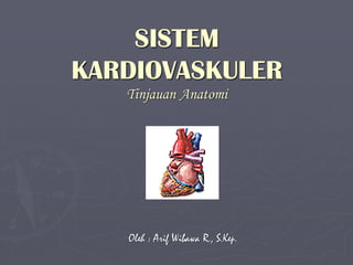
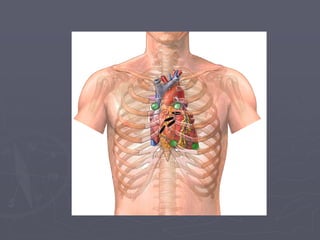
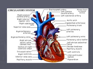
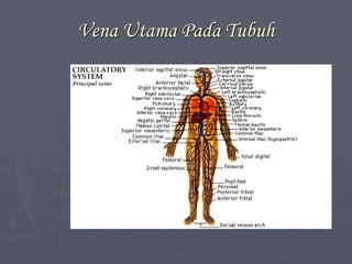
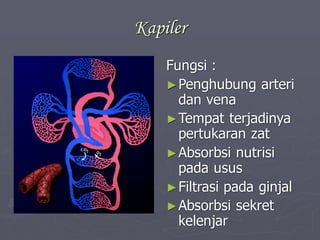
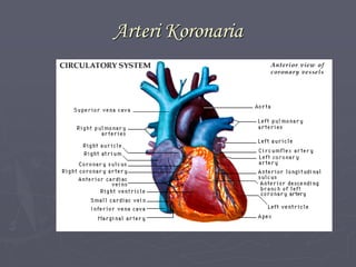
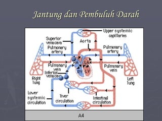
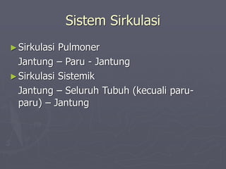
Sistem kardiovaskuler merupakan bagian dari
sistem sirkulasi
â–ş Sub sistem lain dari sistem sirkulasi adalah sistem
limfatik
â–ş Sistem berperan dalam mensuplai oksigen dan
nutrisi kepada sel dan jaringan
â–ş Suplai nutrisi dan oksigen diperlukan untuk
pembentukan energi melalui metabolisme
â–ş Energi diperlukan untuk mempertahankan fungsi
sel
Sistem Limfatik Sebagai Sub Sistem Dari Sistem
Sirkulasi
Bentuk Jantung
â–ş Seperti buah pir atau
seperti kerucut
â–ş Mempunyai basis &
apeks
► Beratnya 250 – 350 gr
pd orang dewasa
Letak Jantung
â–ş Terletak pada kantung perikardium pada rongga
mediastinum di antara ke 2 paru-paru
â–ş Terikat pada dinding thorak oleh ligamen
â–ş Bagian puncak (apex) terletak diatas diafragma
pada interkosta ke-5, 4 cm ke arah sinistra dari
garis medial. miring ke kiri.
â–ş Bagian dasar (basis) terletak pada garis yang
merupakan penghubung 2 titik. Titik pertama
terdapat pada kosta ke-3 dekstra, 2 cm dari
sternum. Titik ke-2 pada kosta ke-2 sinistra, 1cm
dari sternum.
Lapisan Jantung
â–şPerikardium
Perikardium Parietal
Cairan Serous
Perikardium Viseral
â–şMiokardium
â–şEndokardium
Miokardium
â–ş Berkontraksi secara ritmis
â–ş Bekerja secara
otomatis/miogenik
â–ş Tebal berbeda pada setiap
bagian
â–ş Ventrikel lebih tebal, LV
lebih tebal
â–ş Membentuk sinsitium
atrium & ventrikel,
terdapat diskus interkalaris
â–ş Berhubungan melalui
anoulus fibrosus
Ruangan Pada Jantung
â–şAtrium kanan (right atrium/RA)
â–şVentrikel kanan (right ventricle/RV)
â–şAtrium kiri (left atrium/LA)
â–şVentrikel kiri (left ventricle/LV)
Katup Jantung
Atrioventrikuler
Terikat pada dinding
ventrikel oleh korda
tendinae yang
berhubungan dengan
muskulus papilaris
â–ş Trikuspidalis
â–ş Bikuspidalis/Mitral
Semilunaris
â–ş Aortik
â–ş Pulmonal
Pembuluh Darah Yang Langsung Berhubungan Dengan
Jantung
â–şVena Kava Atrium Kanan
â–şArteri Pulmonalis Ventrikel Kanan
â–şVena PulmonalisAtrium Kiri
â–şAorta Ventrikel Kiri
Persarafan
â–şSaraf Simpatis
â–şSaraf Parasimpatis (Nervus Vagus)
Pembuluh Darah
â–ş Ada arteri, vena dan
kapiler
â–ş Terdiri dari 3 lapis
tunika intima
tunika media
tunika adventisia
Arteri Utama Pada Tubuh
Vena Utama Pada Tubuh
Kapiler
Fungsi :
â–şPenghubung arteri
dan vena
â–şTempat terjadinya
pertukaran zat
â–şAbsorbsi nutrisi
pada usus
â–şFiltrasi pada ginjal
â–şAbsorbsi sekret
kelenjar
Arteri Koronaria
Jantung dan Pembuluh Darah
Sistem Sirkulasi
â–şSirkulasi Pulmoner
Jantung – Paru - Jantung
â–şSirkulasi Sistemik
Jantung – Seluruh Tubuh (kecuali paru-
paru) – Jantung
Fetal Circulation
How does the fetal circulatory system work?
â–ş During pregnancy, the fetal circulatory system works
differently than after birth:
â–ş The fetus is connected by the umbilical cord to the
placenta, the organ that develops and implants in the
mother's uterus during pregnancy.
â–ş Through the blood vessels in the umbilical cord, the fetus
receives all the necessary nutrition, oxygen, and life
support from the mother through the placenta.
â–ş Waste products and carbon dioxide from the fetus are sent
back through the umbilical cord and placenta to the
mother's circulation to be eliminated.
Blood from the mother enters the fetus through the vein in the umbilical
cord. It goes to the liver and splits into three branches. The blood then
reaches the inferior vena cava, a major vein connected to the heart.
Inside the fetal heart
â–ş Blood enters the right atrium, the chamber on the upper right side of the
heart. Most of the blood flows to the left side through a special fetal opening
between the left and right atria, called the foramen ovale.
â–ş Blood then passes into the left ventricle (lower chamber of the heart) and
then to the aorta, (the large artery coming from the heart).
â–ş From the aorta, blood is sent to the head and upper extremities. After
circulating there, the blood returns to the right atrium of the heart through
the superior vena cava.
â–ş About one-third of the blood entering the right atrium does not flow through
the foramen ovale, but, instead, stays in the right side of the heart,
eventually flowing into the pulmonary artery.
â–ş Because the placenta does the work of exchanging oxygen (O2) and carbon
dioxide (CO2) through the mother's circulation, the fetal lungs are not used
for breathing. Instead of blood flowing to the lungs to pick up oxygen and
then flowing to the rest of the body, the fetal circulation shunts (bypasses)
most of the blood away from the lungs. In the fetus, blood is shunted from
the pulmonary artery to the aorta through a connecting blood vessel called
the ductus arteriosus.
Blood circulation after birth
â–ş With the first breaths of air the baby takes at
birth, the fetal circulation changes. A larger
amount of blood is sent to the lungs to pick up
oxygen.
â–ş Because the ductus arteriosus (the normal
connection between the aorta and the pulmonary
valve) is no longer needed, it begins to wither and
close off.
â–ş The circulation in the lungs increases and more
blood flows into the left atrium of the heart. This
increased pressure causes the foramen ovale to
close and blood circulates normally.
Darah
â–ş Plasma darah
â–ş Sel darah
eritrosit
leukosit
trombosit
Sistem Limfatik
Fungsi :
â–ş Mengembalikan cairan dan
protein ke sirkulasi darah
â–ş Mengangkut limfosit dari
kelenjar lemfe ke sirkulasi
darah
â–ş Membawa emulsi lemak
dari usus ke sirkulasi darah
â–ş Menyaring dan
menghancurkan
mikroorganisme
â–ş Menghasilkan antibodi
Terima Kasih

3 sistem kardiovaskuler

  • 1.
  • 2.
    Introduksi â–ş Sistem kardiovaskulermerupakan bagian dari sistem sirkulasi â–ş Sub sistem lain dari sistem sirkulasi adalah sistem limfatik â–ş Sistem berperan dalam mensuplai oksigen dan nutrisi kepada sel dan jaringan â–ş Suplai nutrisi dan oksigen diperlukan untuk pembentukan energi melalui metabolisme â–ş Energi diperlukan untuk mempertahankan fungsi sel
  • 3.
    Sistem Limfatik SebagaiSub Sistem Dari Sistem Sirkulasi
  • 4.
    Bentuk Jantung ► Sepertibuah pir atau seperti kerucut ► Mempunyai basis & apeks ► Beratnya 250 – 350 gr pd orang dewasa
  • 5.
    Letak Jantung â–ş Terletakpada kantung perikardium pada rongga mediastinum di antara ke 2 paru-paru â–ş Terikat pada dinding thorak oleh ligamen â–ş Bagian puncak (apex) terletak diatas diafragma pada interkosta ke-5, 4 cm ke arah sinistra dari garis medial. miring ke kiri. â–ş Bagian dasar (basis) terletak pada garis yang merupakan penghubung 2 titik. Titik pertama terdapat pada kosta ke-3 dekstra, 2 cm dari sternum. Titik ke-2 pada kosta ke-2 sinistra, 1cm dari sternum.
  • 7.
    Lapisan Jantung â–şPerikardium Perikardium Parietal CairanSerous Perikardium Viseral â–şMiokardium â–şEndokardium
  • 9.
    Miokardium â–ş Berkontraksi secararitmis â–ş Bekerja secara otomatis/miogenik â–ş Tebal berbeda pada setiap bagian â–ş Ventrikel lebih tebal, LV lebih tebal â–ş Membentuk sinsitium atrium & ventrikel, terdapat diskus interkalaris â–ş Berhubungan melalui anoulus fibrosus
  • 11.
    Ruangan Pada Jantung â–şAtriumkanan (right atrium/RA) â–şVentrikel kanan (right ventricle/RV) â–şAtrium kiri (left atrium/LA) â–şVentrikel kiri (left ventricle/LV)
  • 12.
    Katup Jantung Atrioventrikuler Terikat padadinding ventrikel oleh korda tendinae yang berhubungan dengan muskulus papilaris â–ş Trikuspidalis â–ş Bikuspidalis/Mitral Semilunaris â–ş Aortik â–ş Pulmonal
  • 13.
    Pembuluh Darah YangLangsung Berhubungan Dengan Jantung â–şVena Kava Atrium Kanan â–şArteri Pulmonalis Ventrikel Kanan â–şVena PulmonalisAtrium Kiri â–şAorta Ventrikel Kiri
  • 15.
  • 16.
    Pembuluh Darah â–ş Adaarteri, vena dan kapiler â–ş Terdiri dari 3 lapis tunika intima tunika media tunika adventisia
  • 17.
  • 18.
  • 19.
    Kapiler Fungsi : â–şPenghubung arteri danvena â–şTempat terjadinya pertukaran zat â–şAbsorbsi nutrisi pada usus â–şFiltrasi pada ginjal â–şAbsorbsi sekret kelenjar
  • 20.
  • 21.
  • 22.
    Sistem Sirkulasi ►Sirkulasi Pulmoner Jantung– Paru - Jantung ►Sirkulasi Sistemik Jantung – Seluruh Tubuh (kecuali paru- paru) – Jantung
  • 23.
    Fetal Circulation How doesthe fetal circulatory system work? â–ş During pregnancy, the fetal circulatory system works differently than after birth: â–ş The fetus is connected by the umbilical cord to the placenta, the organ that develops and implants in the mother's uterus during pregnancy. â–ş Through the blood vessels in the umbilical cord, the fetus receives all the necessary nutrition, oxygen, and life support from the mother through the placenta. â–ş Waste products and carbon dioxide from the fetus are sent back through the umbilical cord and placenta to the mother's circulation to be eliminated.
  • 24.
    Blood from themother enters the fetus through the vein in the umbilical cord. It goes to the liver and splits into three branches. The blood then reaches the inferior vena cava, a major vein connected to the heart.
  • 25.
    Inside the fetalheart â–ş Blood enters the right atrium, the chamber on the upper right side of the heart. Most of the blood flows to the left side through a special fetal opening between the left and right atria, called the foramen ovale. â–ş Blood then passes into the left ventricle (lower chamber of the heart) and then to the aorta, (the large artery coming from the heart). â–ş From the aorta, blood is sent to the head and upper extremities. After circulating there, the blood returns to the right atrium of the heart through the superior vena cava. â–ş About one-third of the blood entering the right atrium does not flow through the foramen ovale, but, instead, stays in the right side of the heart, eventually flowing into the pulmonary artery. â–ş Because the placenta does the work of exchanging oxygen (O2) and carbon dioxide (CO2) through the mother's circulation, the fetal lungs are not used for breathing. Instead of blood flowing to the lungs to pick up oxygen and then flowing to the rest of the body, the fetal circulation shunts (bypasses) most of the blood away from the lungs. In the fetus, blood is shunted from the pulmonary artery to the aorta through a connecting blood vessel called the ductus arteriosus.
  • 26.
    Blood circulation afterbirth â–ş With the first breaths of air the baby takes at birth, the fetal circulation changes. A larger amount of blood is sent to the lungs to pick up oxygen. â–ş Because the ductus arteriosus (the normal connection between the aorta and the pulmonary valve) is no longer needed, it begins to wither and close off. â–ş The circulation in the lungs increases and more blood flows into the left atrium of the heart. This increased pressure causes the foramen ovale to close and blood circulates normally.
  • 27.
    Darah â–ş Plasma darah â–şSel darah eritrosit leukosit trombosit
  • 28.
    Sistem Limfatik Fungsi : â–şMengembalikan cairan dan protein ke sirkulasi darah â–ş Mengangkut limfosit dari kelenjar lemfe ke sirkulasi darah â–ş Membawa emulsi lemak dari usus ke sirkulasi darah â–ş Menyaring dan menghancurkan mikroorganisme â–ş Menghasilkan antibodi
  • 30.