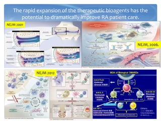
The rapid expansionof the therapeutic bioagents has the
potential to dramatically improve RA patient care.
NEJM 2001
NEJM, 2006.
NEJM 2012
Others:
小分子標靶藥物
Tofacitinib(Xeljanz
Or Anti-IL17…..
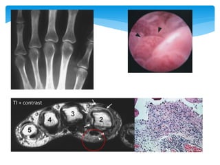
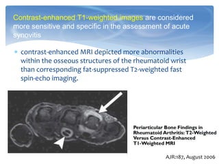
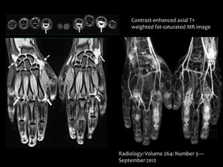
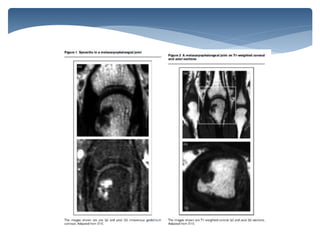
contrast-enhanced MRIdepicted more abnormalities
within the osseous structures of the rheumatoid wrist
than corresponding fat-suppressed T2-weighted fast
spin-echo imaging.
Contrast-enhanced T1-weighted images are considered
more sensitive and specific in the assessment of acute
synovitis
AJR:187, August 2006
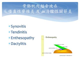
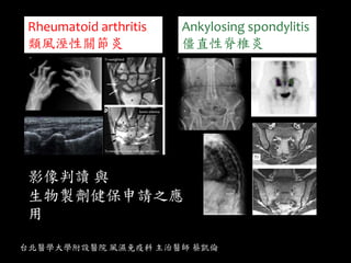
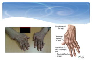
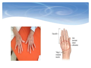
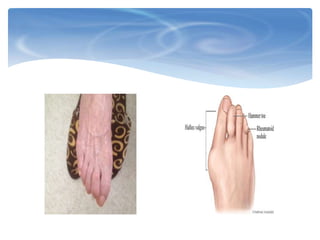
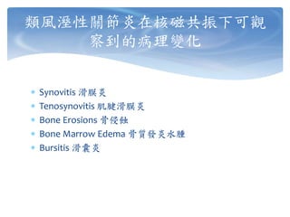
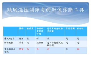
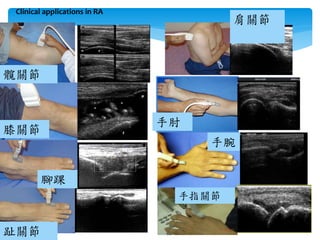
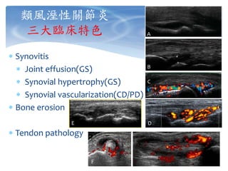
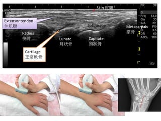
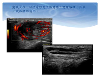
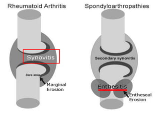
類風溼性關節炎
三大臨床特色
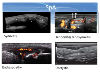
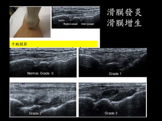
Synovitis
Jointeffusion(GS)
Synovial hypertrophy(GS)
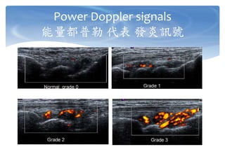
Synovial vascularization(CD/PD)
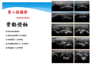
Bone erosion
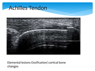
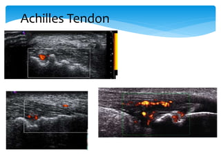
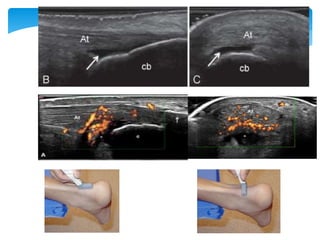
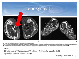
Tendon pathology
A
B
C
DE
F
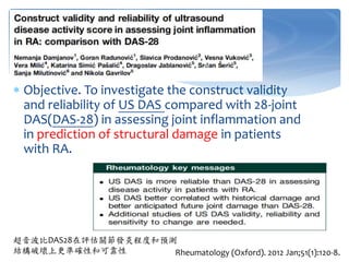
Objective. Toinvestigate the construct validity
and reliability of US DAS compared with 28-joint
DAS(DAS-28) in assessing joint inflammation and
in prediction of structural damage in patients
with RA.
Rheumatology (Oxford). 2012 Jan;51(1):120-8.
超音波比DAS28在評估關節發炎程度和預測
結構破壞上更準確性和可靠性
41.
是否可以當作退場機制申覆上的輔助工具?
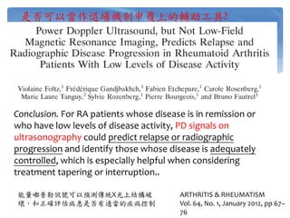
ARTHRITIS & RHEUMATISM
Vol.64, No. 1, January 2012, pp 67–
76
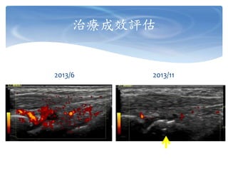
Conclusion. For RA patients whose disease is in remission or
who have low levels of disease activity, PD signals on
ultrasonography could predict relapse or radiographic
progression and identify those whose disease is adequately
controlled, which is especially helpful when considering
treatment tapering or interruption..
能量嘟普勒訊號可以預測傳統X光上結構破
壞,和正確評估病患是否有適當的疾病控制
[image tool: MRI]
modifiedby Medscape Rheumatology education, Expanding
the spectrum for TNF antagonist: Safty & Efficacy in the
Spondyloarthropathies: Ankylosing Spondylitis
Image tool: MRI,
Ultrasound
46.
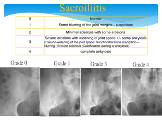
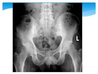
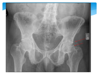
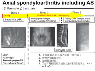
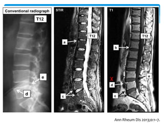
Sacroiliitis
0 Normal
1 Someblurring of the joint margins - suspicious
2 Minimal sclerosis with some erosions
3
Severe erosions with widening of joint space +/- some ankylosis
(Pseudo-widening of the joint space: Subchondral bone resorption—
blurring ; Erosion sclerosis ;Calcification leading to ankylosis)
4 complete ankylosis
Grade 0 Grade 1 Grade 3 Grade 4
T1 white, STIRdark
Bone marrow
edema, STIR white,
T1 dark
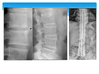
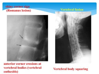
Romanus lesion
Active lesion
Chronic lesion
60.
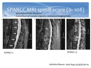
SPARCC MRI spinalscore (0–108)
Arthritis Rheum. 2005 Aug 15;53(4):502-9.
T2
SPARCC 2 SPARCC 13
Spondyloarthritis Research
Consortium of Canada magnetic resonance imaging index
![[image tool: MRI]
modified by Medscape Rheumatology education, Expanding
the spectrum for TNF antagonist: Safty & Efficacy in the
Spondyloarthropathies: Ankylosing Spondylitis
Image tool: MRI,
Ultrasound](https://image.slidesharecdn.com/20140802crctraingingimageforraas-140802102728-phpapp02/85/20140802-crc-trainging-image-for-ra-as-45-320.jpg)