20 Objective Structured Clinical Examination (OSCE) for trainees.doc
1.
QUESTIONS 1 -7: Name the tests/instruments. Name the purpose of each test.
QUESTION 1
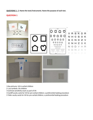
1-Kay pictures: VA in verbal children
2- Lea symbols: VA children
3 Contrast sensitivity exam as part of VA
4 Cardiff acuity cards for VA for pre-verbal children; a preferential looking procedure
5 Teller acuity cards for VA for pre-verbal children; a preferential looking procedure
2.
QUESTION 2
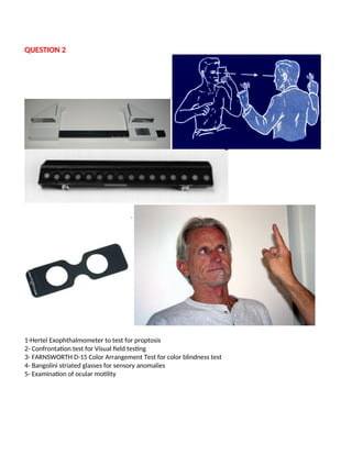
1-Hertel Exophthalmometerto test for proptosis
2- Confrontation test for Visual field testing
3- FARNSWORTH D-15 Color Arrangement Test for color blindness test
4- Bangolini striated glasses for sensory anomalies
5- Examination of ocular motility
3.
QUESTION 3
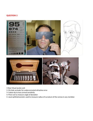
1-Near Visualacuity card
2- Pin hole occluder for undercorrected refractive error
3- Cotton tip to test corneal sensitivity
4- Prism set to measure angle of deviation
5- Javal ophthalmometer used to measure radius of curvature of the cornea in any meridian
4.
QUESTION 4
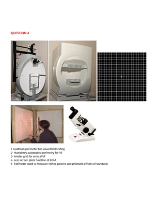
1-Goldman perimeterfor visual field testing
2- Humphrey automated perimetry for VF
3- Amsler grid for central VF
4- Lees screen plots function of EOM
5- Focimeter used to measure vertex powers and prismatic effects of spectacle
5.
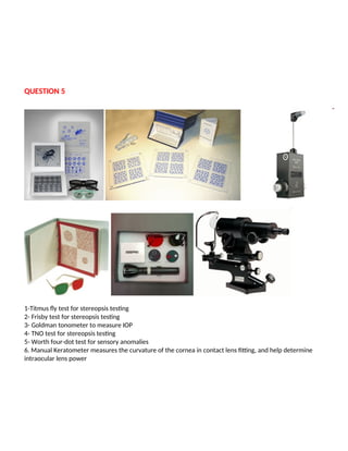
QUESTION 5
1-Titmus flytest for stereopsis testing
2- Frisby test for stereopsis testing
3- Goldman tonometer to measure IOP
4- TNO test for stereopsis testing
5- Worth four-dot test for sensory anomalies
6. Manual Keratometer measures the curvature of the cornea in contact lens fitting, and help determine
intraocular lens power
6.
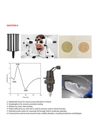
QUESTION 6
1- Optokineticdrum for visual acuity estimation in infants
2- Synoptophore for sensory anomalies testing
3- Ishihara for color vision testing
4- Pattern ERG print out. this test is used to measure central retinal function
5- Potential acuity meter for assessing VA through mild to moderate opacities
6. Fresnel prism used to treat several ocular motility disorders, including Strabismus and Diplopia
7.
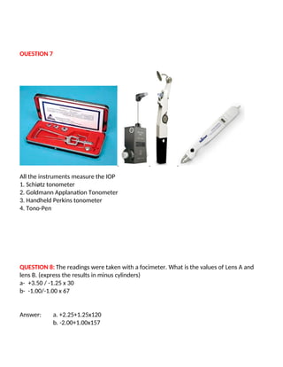
OUESTION 7
All theinstruments measure the IOP
1. Schiøtz tonometer
2. Goldmann Applanation Tonometer
3. Handheld Perkins tonometer
4. Tono-Pen
QUESTION 8: The readings were taken with a focimeter. What is the values of Lens A and
lens B. (express the results in minus cylinders)
a- +3.50 / -1.25 x 30
b- -1.00/-1.00 x 67
Answer: a. +2.25+1.25x120
b. -2.00+1.00x157
8.
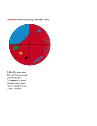
QUESTION 9: Retinaldrawing. Name the labels.
A (blue)Detached retina
B (green) Vitreous opacity
C (yellow) Exudate
D (black) Retinal pigment
E (hatched blue) Lattice
F (blue-red) retinal break
G (red) Flat retina
9.
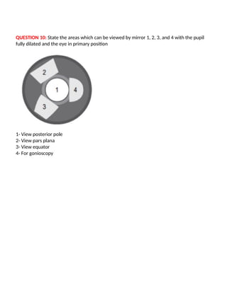
QUESTION 10: Statethe areas which can be viewed by mirror 1, 2, 3, and 4 with the pupil
fully dilated and the eye in primary position
1- View posterior pole
2- View pars plana
3- View equator
4- For gonioscopy
10.
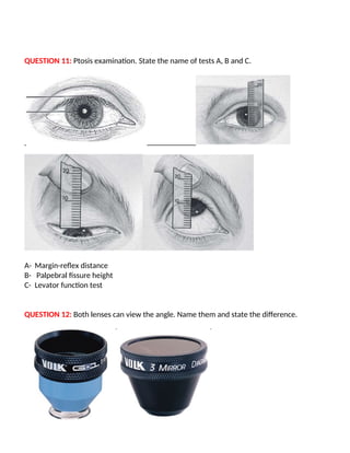
QUESTION 11: Ptosisexamination. State the name of tests A, B and C.
A- Margin-reflex distance
B- Palpebral fissure height
C- Levator function test
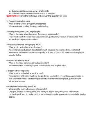
QUESTION 12: Both lenses can view the angle. Name them and state the difference.
11.
A- Sussman goniolens:can view 4 angles only
B- Goldman 3 mirror: can view from the macula to pars plana
QUESTION 13: Name the technique and answer the question for each.
1- Fluorescein angiography
What are the causes of hyperfluorescence?
Window defect, pooling, leakage and staining.
2- Indocyanine green (ICG) angiography
What is the main advantage over fluorescein angiography?
The detection of choroidal neovascularization, particularly if occult or associated with
hernorrhage, pigment or exudate.
3- Optical coherence tomography (OCT)
What are its main clinical applications?
Assessing various types of maculopathy such a cystoid macular oedema, epiretinal
membrane and central serous retinopathy. It is also of particular value in the staging of
macular holes.
4- A-scan ultrasonography
What is the most common clinical application?
Measurement of axial length prior to intraocular lens implantation.
5- B-scan ultrasonography
What are the main clinical applications?
The diagnosis of lesions involving the posterior segment in eyes with opaque media. In
eyes with clear media the technique is useful in differential diagnosis, particularly of
intraocular tumors.
6- Computerized tomography (CT)
What are the main advantages of over MRI?
Cheaper, shorter scanning time, and ability to depict bony structures and tumors
containing calcium. It can be used in patients with cardiac pacemakers or metallic foreign
bodies.
12.
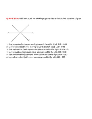
QUESTION 14: Whichmuscles are working together in the six Cardinal positions of gaze.
1- Dextroversion (both eyes moving towards the right side): RLR + LMR
2- Laevoversion (both eyes moving towards the left side): LLR + RMR
3- Dextroelevation (both eyes move upwards and to the right): RSR + LIO
4- Laevoelevation (both eyes move upwards and to the left): LSR + RIO
5- Dextrodepression (both eyes move down and to the right): RIR + LSO
6- Laevodepression (both eyes move down and to the left): LIR + RSO
13.
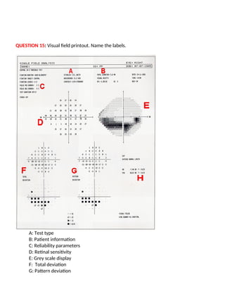
QUESTION 15: Visualfield printout. Name the labels.
A: Test type
B: Patient information
C: Reliability parameters
D: Retinal sensitivity
E: Grey scale display
F: Total deviation
G: Pattern deviation
14.
H: Global indices
QUESTION16: Cranial nerve examination. Name and write down the end-result of complete
damage to cranial nerves II till VII.
Second cranial Nerve: Optic Nerve.
Complete damage causes the following: Blindness
Third cranial Nerve: Oculomotor Nerve.
Complete damage causes the following: Ptosis and exotropia; Intorsion on downgaze; Limited elevation;
Limited depression. Dilated pupil and defective accommodation.
Fourth cranial nerve: Trochlear Nerve
Complete damage causes the following: Defective depression of the eye in adduction; In primary gaze - light
hypertropia; excycloduction; Compensatory head posture as follows: Head tilt, Face turn and Chin
depressed
Fifth cranial nerve: Trigeminal Nerve
Complete damage causes the following: sensory losses over the face or in the oral cavity; Damage to motor
fibers results in paralysis of the masticatory muscles
Sixth cranial nerve: Abducens Nerve
Complete damage causes the following: esotropia in the primary position; Defective abduction; Horizontal
diplopia; Compensatory face turn
Seventh cranial nerve: Facial Nerve
Complete damage causes the following: Bell's palsy
15.
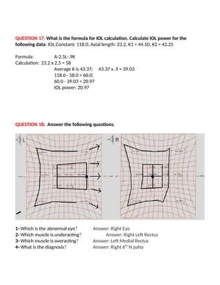
QUESTION 17: Whatis the formula for IOL calculation. Calculate IOL power for the
following data: IOL Constant: 118.0; Axial length: 23.2, K1 = 44.50, K2 = 42.25
Formula: A-2.5L-.9K
Calculation: 23.2 x 2.5 = 58
Average K is 43.37; 43.37 x .9 = 39.03
118.0 - 58.0 = 60.0;
60.0 - 39.03 = 20.97
IOL power: 20.97
QUESTION 18: Answer the following questions.
1- Which is the abnormal eye? Answer: Right Eye
2- Which muscle is underacting? Answer: Right Left Rectus
3- Which muscle is overacting? Answer: Left Medial Rectus
4- What is the diagnosis? Answer: Right 6th
N palsy
16.
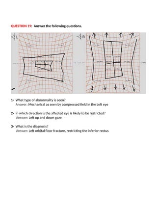
QUESTION 19: Answerthe following questions.
1- What type of abnormality is seen?
Answer: Mechanical as seen by compressed field in the Left eye
2- In which direction is the affected eye is likely to be restricted?
Answer: Left up and down gaze
3- What is the diagnosis?
Answer: Left orbital floor fracture, restricting the inferior rectus
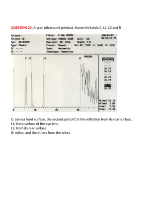
17.
QUESTION 20: A-scanultrasound printout. Name the labels C, L1, L2 and R
C: cornea front surface, the second part of C is the reflection from its rear surface.
L1: front surface of the eye lens
L2: from its rear surface.
R: retina, and the others from the sclera