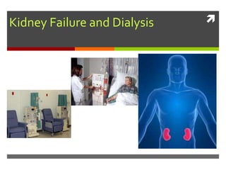
2. Function of Kidneys
• Production of urine and
elimination of waste
• Facilitates electrolyte
balance
• Facilitates acid-base
balance
• Manages water balance
and maintain blood
osmolality
• Influences blood
pressure and blood
volume
• Renal clearance
• Secretion of
prostaglandins
• Conversion of vitamin D
to it’s active form
• Assists with red blood
cell production
(erythropoietin)
(Day, Paul, Williams, Smeltzer, & Bare, 2010, p. 1405;
Tortora & Derrickson, 2009, p. 1020 )
6. Definition:
The kidneys failure to expel
wastes, maintain electrolyte
balance, concentrate urine,
and maintain chemicals in
the bloodstream that are
regulated by the kidneys
(ex. Renin) (Mosby’s
Dictionary of Medicine,
Nursing & Health
Professionals, 2006).
Can be acute or
chronic
Renal Failure
8. Acute Renal
Failure
“Acute renal failure (ARF) is a sudden
and almost complete loss of kidney
function over a period of hours to days”
(Day et al., 2010, p. 1435).
Oliguria: urine output of less then
400mL /day. is the most common
clinical manifestation (p.1435).
Anuria (less than 50 ml of urine a day)
Elevated BUN and creatinine
Reversible if treated promptly
9. Categories of ARF
1. Prerenal: Hypoperfusion of the kidneys.
2. Intrarenal: Acute damage to kidney tissue
3. Postrenal: obstruction to urine flow
10. Clinical Manifestations
Pt will appear critically ill
and lethargic, and confused
Skin and mucus membranes
will be dry from dehydration
drowsiness, headache,
muscle twitching, and
seizures.
dyspnea, crackles,
tachypnea,
(Day et al., 2010, p. 1436)
11. Nursing interventions
Monitor intake and output,
including all body fluids
May need to stimulate
production of urine with IV
fluids, diuretics.
Daily weights
Monitor lab results, CBC,
BUN, creatinine, urea, e’lyles
Watch hyperkalemia
symptoms: malaise,
anorexia, parenthesia, or
muscle weakness, EKG
changes
Maintain nutrition
Mouth care – dry mucus
membranes
Assess for signs of cardiac
involvement- dysthymias
Skin integrity problems.
Edema, itching –from toxins
Signs and symptoms of
infection
May need dialysis, or
continuous renal
replacement therapy.
12. Chronic Renal failure (CRF)
Definition: “ Chronic Renal failure is a progressive,
irreversible deterioration of renal function in which
the body ability to maintain metabolic, fluid and
electrolyte balance fails, resulting in uremia or
azotemia (retention of urea and other nitrogenous
waste in blood) (Day et al., 2010, p. 1440).
13. Stages of CRF
The normal glomerular filtration rate
(GFR) is 125ml/min/1.73m2 (Day et al., 2010,
p. 1440)
The stages of renal failure is determined
by the GFR (Day et al., 2010, p. 1440).
14. Signs & Symptoms of CRF
Ammonia-like taste in mouth or urinous breath
Edema of feet, hands, arms, face and around eyes
Hypertension
Extended neck veins
Anemia
Fatigue
Neurologic disturbances
Nausea, vomiting, and anorexia
Headaches and blurred vision
15. Signs & Symptoms of CRF
Pruritus
Shortness of breath
Bone and joint problems
Weakness, numbness, tremors, bone pain, and
paresthesia
Urine that is cloudy, tea-coloured, or bloody
Decreased urine output or trouble urinating
Foaming of urine
Proteinuria
16. Nursing interventions CRF
Assessing fluid status
Nutrition/Diet
Patient teaching
Assess emotional status and coping strategies
Assessing for complications
Administering Medications
19. Screening: UrineTesting
Creatinine clearance formula:
(Volume of urine [ml/min] X Urine creatinine [MMOL/L])
Serum Creatinine (mmol/L)
As renal function decreases, creatinine clearance decreases
Day et al., 2010, pp1410
20.
Treatment of Renal Failure
Medication
Proper Diet
Dialysis (2 types: peritoneal & hemodialysis)
Transplantation
Conservation Care
21. Treatment of Renal Failure
Medication: Medication may be used to help
maintain or improve kidney function, as well as,
treat complications of renal failure (eg.
Antihypertensives, kayexalate, etc.) (Day et al.,
2010, pp 1442).
22. Dialysis
When the kidneys are not removing fluid and uremic waste
from the body, dialysis can be used to do so
Dialysis can be acute or chronic
Acute dialysis is used for people with high levels of serum
potassium, fluid overload, or impending pulmonary edema,
increasing acidosis, pericarditis, and severe confusion
Acute dialysis may also be used to remove certain
medications or other toxins from the blood
23. Dialysis
Chronic dialysis is used for chronic renal failure
Dialysis can be used for years to help maintain people with
no renal function
Indications may include: uremic signs and symptoms
affecting all body systems, hyperkalemia, fluid overload,
pericardial friction rub, and lack of well being
24. Peritoneal Dialysis
Removes metabolic wastes and toxin’s so the body’s normal
fluid and electrolyte balance is re-established
The peritoneum that lines the abdominal cavity and covers
the abdominal organs acts as a semipermeable membrane
that allows metabolic end products to be removed from the
blood by means of diffusion and osmosis
25. Peritoneal Dialysis
An abdominal catheter allows sterile dialysate fluid to enter
the peritoneal cavity
The metabolic waste products in the blood move from an
area of high concentration (blood), across the peritoneal
membrane, to an area of low concentration (peritoneal
cavity with dialysate fluid)
26. Peritoneal Dialysis
The body’s excess fluid is removed by an osmotic gradient,
because the dialysate fluid in the peritoneal cavity has a
higher glucose concentration
the fluid is then removed from the peritoneal cavity and
discarded
This process is repeated 4-6 times ever 24hrs
The most common complication from peritoneal dialysis is
peritonitis
28. Peritoneal Dialysis Nursing management
Client and family education
Sterile technique (face mask, gloves, sterile field)
Signs and symptoms of peritonitis
Inspect site and dialysate solution for signs and
symptoms of infection
29. Hemodialysis
The most common type of dialysis
Purpose remains to remove toxins from the blood and excess
water from the body
Usually patients receive Hemodialysis 3 times per week
Treatment takes about 3-8 hours per treatment
30. Hemodialysis
The blood is delivered from the patient and to the dialysis
machine, where a dialyzer (artificial kidney) uses diffusion,
osmosis, and ultrafiltration to remove toxins from the blood,
which is then returned to the patient
The metabolic waste products in the blood move from an
area of high concentration (blood), to an area of low
concentration (dialysate)
31. Hemodialysis
Dialysate is a solution composed of electrolytes, which
concentration levels can be adjusted to accommodate the
desired electrolyte level in the patients blood
Osmosis and ultrafiltration is used to remove the body’s
excess water
32. Arteriovenous fistula- is made by
sewing a vein and artery together under
the skin. Fistulas may take 2 to 4
months to mature. A temporary access
device is usually needed until It matures
(Williams & Hopper 2007, p. 803).
Arteriovenous graft: uses a tube of
systhetic material to attach an artery
and a vein. Needles are inserted into
the graft to access the clients blood
(Williams & Hopper 2007, p. 803).
Hemodialysis:
Vascular
Access Device
33. Two tailed subclavian/ double lumen,
cuffed hemodialysis catheter used for
acute hemodialysis.
Red port: blood line
Blue port: return dialyzed blood to
client.
35. HemodialysisV.S peritoneal
Hemodialysis
Requires vascular access device.
Either temporary (ARF) or permanent
(CRF).
Requires a complex specialized
dialyzer
Requires a skilled hemodialysis nurse
Intermittent (q3-4days)
Principals of osmosis and diffusion
Preferred for end-stage renal failure
Peritoneal
Requires a insertion of a catheter
into the peritoneal cavity
Does not require specialized dialyzer
Can be done by client (sterile
technique)
Continuous (4-6 q 24hr)
Principals of osmosis and diffusion
Have few cardio side effects can be
used in unstable clients.
37. KidneyTransplantation
Nursing Management
Pre and postoperative teaching
Assessing patient coping and anxiety
Assessing for signs and symptoms of transplant rejection
Preventing infection
Monitoring urinary functioning
Psychological concerns
Monitoring and managing potential complications
Promoting home and community based care
Editor's Notes
Marie
Production of urine and elimination of waste :“By forming urine, the kidneys help excrete substances that have no function or useful purpose in the body. Some waste excreted in urine results from metabolic reactions in the body. These include ammonia and urea from the deamination of amino acids; bilirubin from the catabolism of hemoglobin; creatinine from the breakdown of creatine phosphate in muscle fibers; and uric acid from the catabolism of nucleic acid. Other wastes excreted in urine are forging substances from diet, such as drug and environmental toxins” (Tortora & Derrickson, 2009, p. 1020).
Facilitates electrolyte balance:The kidneys help regulate the blood levels of several ions, most importantly Na+, K+, calcium, chloride, and phosphate. (Tortora & Derrickson, 2009).
Facilitates acid-base balance: the kidneys excrete variable amount of hydrogen ions into the urine and conserve bicarbonate, which are an important buffer of hydrogen in the blood. Both of these activities help regulate blood pH (Tortora & Derrickson, 2009).
Manages water balance and maintain blood osmolality: By separately regulating loss of water and loss of solutes in the urine, the kidneys maintain a relatively constant blood osmolality- close to 300 milliosmoles per liter (Tortora & Derrickson, 2009).
Blood pressure: the kidneys help to regulate blood pressure by secreting the enzyme Renin, which activates the renin-angeotensin- aldosterone pathway-vasoconstriction. The increase in renin causes BP to increase (Tortora & Derrickson, 2009).
Blood volume- the kidneys adjust blood volumes by conserving or eliminating water in the urine. An increase in blood volume will blood pressure; a decrease in blood volume will decrease bp (Tortora & Derrickson, 2009).
Renal clearance refers to the ability of the kidneys to clear solute from plasma. A 24hr urine collection is the primary test for renal clearence. This test is used to evaluate how the kidneys are functioning as an excretory organ (Day et al., 2010, p. 1410).
The kidneys also produce prostaglandin E and prostacyclin , which have a vasodilatory effect and are important in maintaining renal blood flow Day et al., 2010, p. 1411).
The kidneys also produce calcitrol, the active form of vit D, which helps regulate calcium homeostasis (Tortora & Derrickson, 2009).
And
When the kidneys sense a decrease in the oxygen tension in renal blood flow, they release erythropoietin. Erythropoietin stimulated the bone marrow to produce RBC, thereby increasing the amount of hemoglobin available to carry O2 (Day et al., 2010, pp, 1410-1411)
MArie
The renal medulla- the inner portion of the of the kidneys
Renal pyramids- “the triangular division of the medulla of the kidney. Extension of the cortical tissue that dip down into the medulla between the renal pyramids are called renal columns” (Thibodeau & Patton, 2004, p.438).
Renal papilla- “narrow innermost end of the pyramid” (Thibodeau & Patton, 2004).
Renal pelvis- “an expanstion of the upper end of the ureter” (Thibodeau & Patton, 2004, p.438).
Calyx- “ a division of the renal pelvis (the papilla of a pyramid opens into each calyx)” (Thibodeau & Patton, 2004, p.438).
and the nephron, which I will discuss on the next slide
MArie
The nephron the functional unit of the kidneys.
According to Thibodeau & Patton “more then a million mirooscopic units called nephrons make up each kidney’s interior” (p. 438). The nephrons is composed of two major components; the renal corpuscle and the renal tubule. The renal corpuscle can be subdivided into two parts and the renal tubular into four regions
1) renal corpuscle
a. Bowman’s capsule- “the cup-shaped top of a nephron (the sac like Bowman’s capsule surrounds the glomerulus) (Thibodeau & Patton, 2004, p.438).
b. Glomerulus- “ a network of blood capillaries tucked into Bowman’s capsule
Blood plasma is filtered in the glomerulus capsule, and then the filtered fluids passes into the renal tubules in the order of:
Proximal convoluted tubule-
Loop of henle
Distal convoluted tubule
Collecting tubule
Go back to previous slide and talk about the movement of fluid through the kidneys
marie
Really nice diagram that sums it all up ..
G
Renal Failure can be either acute or chronic, and it is defined as the kidneys failure to expel wastes, maintain electrolyte balance, concentrate urine, and maintain chemicals in the bloodstream that are regulated by the kidneys (ex. Renin)
Mosby’s Dictionary of Medicine, Nursing & Health Professionals (8th Ed.). (2006). p. 1485 St.Louis, Missouri; Mosby Elsevier.
Gale
Renal Failure, can be acute or chronic.
Marie
“Acute renal failure (ARF) is a sudden and almost complete loss of kidney function over a period of hours to days” (Day et al., 2010, p. 1435).
(Day et al., 2010, p. 1436)
Subjective symptoms
Nausea
Loss of appetite
Headache
Lethargy
Objective symptoms
Oliguric phase –
vomiting
disorientation,
edema,
^K+
decrease Na
^ BUN and creatinine
Acidosis
uremic breath CHF and pulmonary edema
hypertension caused by hypovolemia, anorexia
sudden drop in UOP
convulsions, coma
changes in bowels
Diuretic phase
Increased UOP
Gradual decline in BUN and creatinine
Hypokalemia
Hyponaturmia
Tachycardia
Improved LOC
Day et al., 2010, pp 1473-1439
Azotemia: concentration of urea and other nitrogenous wastes in the blood
If caused by meds, must stop meds
If caused by obstruction, must remove obstruction
If caused by blockage of artery, must open artery
Dietary restrictions may include : low K+, adequate carbs, also may give TPN
Fluids : calculate closley I/O
Hyperkalemia is life threatening
Lower K+ with Kayexalate, glucose, insulin, NaBicarb, caalcium carbonate
“Each patient’s caloric needs are based on their height, weight,
age, sex and activity. The prescribed diet should ensure an intake
of 35 to 45 calories per kilogram of body weight.”
The vitamin and mineral intake of patients on a restricted protein, potassium and sodium diet often does not meet the recommended daily allowances for certain vitamins and minerals. This is especially true for the water-soluble vitamins such as Vitamin C and the B vitamins, which are abundant in high potassium foods such as fruits, vegetables, meat and milk. Also, there is a loss of water-soluble vitamins during dialysis treatments. When the protein restriction is less than 50 grams per day, the diet tends to be low in folic acid, niacin, riboflavin, thiamine
and vitamin B6.
“Patients on long-term low-protein, low-sodium, low-potassium diets should receive a multivitamin capsule and folic acid daily to ensure adequate intake and to make up for losses that
occur during the course of dialysis” (Somers, 2008)
m
G
As Marie had mentioned,
“Glomerular filtration rate (GFR) is the volume of plasma filtered at the glomerulus into the kidney tubules each minute” (Day et al., 2010, pp 1405).
The normal glomerular filtration rate (GFR) is 125ml/min/1.73m2
The different stages of renal failure is determined by the GFR
Stages of CRF (Day et al., 2010, p. 1440)
Gale
Assessing for signs and symptoms of CRF is important.
Lab values and urine screening may indicate CRF, but additional signs and symptoms may be present, such as:
Ammonia – like taste or breath because of uremia in the blood
(“Uremia – excess amounts of urea and other nitrogenous waste products in the blood, as occurs in renal failure”)
(Mosby’s Dictionary of Medicine, Nursing & Health Professionals (8th Ed.). (2006). p. 1778 St.Louis, Missouri; Mosby Elsevier.)
Edema of the feet, hands, arms, face and around eyes, caused by fluid overload
HTN develops from elevated serum sodium levels, extracellular fluid volume expansion, and renin hypersecretion
Extended veins in the neck from fluid overload
Anemia from decreased RBC production and shortened RBC lifespan because or uremic toxins
Fatigue related to anemia and toxins in the blood
Neurologic disturbances including lethargy, impaired mental status, and sleep pattern due to the uremic toxins
Retention of urea and metabolic waste products can cause nausea, vomiting, and anorexia
Headaches and blurred vision may be caused by uremic toxins and extracellular volume expansion
(Cannon, 2004)
More S+S of CRF include:
Increased levels of phosphorus, calcium, and aluminum in the body can cause pruritus (itchy)
Shortness of breath is caused by the fluid overload and can lead to pulmonary edema if left untreated
“Bone and Joint problems from bone resorption associated with vitamin D deficiency, demineralization, and calcium and phosphate imbalances” (Cannon, 2004, pp 52)
Uremic toxins can also cause weakness, numbness, tremors, bone pain, and paresthesia
Urine that is cloudy, tea-coloured, or bloody may indicate CRF
And Decreased urine output or trouble urinating
Foaming of urine and Proteinuria is not normal and may also be a sign of CRF
In addition to watching for these signs and symptoms of renal failure, you should also be mind-full of risk factors that increase a persons likely-hood of developing renal problems (ex. History of diabetes), as well as monitoring lab values (creatinine and urine protein levels are early indicators of renal disease) (Cannon, 2004)
(Cannon, 2004; Kidney Foundation of Canada, 2012)
Nursing Interventions are basically the same as ARF
Assessing fluid status by monitoring intake and output, daily weight, respiratory rate and effort, edema, and distension of neck veins (Day et al., 2010, pp 1445 )
Nutrition status should be assessed by monitoring for changes in weight, nutritional dietary patterns, and factors that may contribute to altered nutrition (eg. Depression, stomatitis, anorexia, nausea, and vomiting)
Pt teaching is important!!! Teaching is required about treatment options, and things like the vascular access device and monitoring it for patency and using precautions, such as, not having the blood pressure taken in the same arm as the device (fistula).
-The client with CRF also needs teaching about when to contact a doctor about increased signs and symptoms of renal failure (nausea, vomiting, changes in urine output, ammonia odour on breath),
-Client’s need teaching about how to assess for access problems such as clotted fistula, or infection, and client needs to know to watch for signs of hyperkalemia (muscle weakness, diarrhea, abdominal cramps), (Day et al., 2010, pp 1443 – 1444)
Assessing clients emotional status and coping strategies is important for those who are diagnosed with CRF or End Stage Renal Disease, This may involve providing support for patients and families, as well as, accessing additional resources for supports (eg. Kidney support groups, social work, etc.)
It is also important for the nurse to be assessing for complications related to dialysis, medications, dietary restrictions, lifestyle, and so on. (Day et al., 2010. p. 1444)
(Williams & Hopper, 2007).
Renal ultrasound
KUB- Kidneys, ureters and bladder x-ray : also known as a flat plate of the abdomen- help to identify if there is a blockage ie stone
CT scan- note any abnormalities size shape. Can be used to identify non-functioning kidney , renal stones, obstruction, infections nursing mamagement for CT include NPO for 4 hr before the procedure, contrasting dye may be given, if given with compromised renal function assess signs and symptoms of acute failure, also assess for allergies,
MRI: claustrophobia, remove all metal objects, may be given contrasting dye
IVP intravenous Pyelogram: this is an invasive procedure- dye is injected into a large vein, the dye is cleared from the blood by the kidneys. Because the x-ray cannot penetrate the dye, the dye outlines the renal structure. The test will provide a rough estimate of renal function. nursing considerations include: NPO for 8hr prior to the procedure, enema given the evening before to empty the colon; the client should be warned about a warm flushing sensation when the dye is injected. A strange test may occur as well. After the procedure client should be encouraged to drink lots of fluids to flush the dye from the body. On rare occasion the client may develop acute renal failure because the dye is highly concentrated and can obstruct the kidney tubules. Urine output should be monitored after test.
Nephrotomogram: same prep as IVP. This procedure takes various x-rays from different 3D angles. Test is useful in identifying of renal cysts, tumours, areas of non perfusion an renal fractures or laceration post trauma.
Renal angiogram:: to visualize renal blood vessels. Catheter is inserted in femoral artery and dye is injected. Same pre procedure nursing management as the IVP. But because catheter Is inserted into the femoral artery the nurse will need to monitor for signs and symptoms of bleeding post the procedure. After the procedure the client may be on bed rest for up to 24 hr to prevent bleeding from injection site, check distal pulse q 30 to 60 min. instructed client not to bend leg and HOB is not raised more tthan 45 degrees. The nurse will need to monitor V/S, the dressing, frequently.
Renal scan: this is a nuclear scan. Radioactive isotope injected into blood and can be detected by gamma camera similar to x-ray. These isotopes are picked up by this gamma camera. This test measures: kidney function; measure renal blood flow; GFR, kidney shape; diagnosis of renal HTN. The major advantage of this diagnostic test is that is does not use contrasting dye. Therefor, it is better to use with clients with acute or chronic failure. No special prep is needed for this procedure, but may be asked to drink two glasses of water beforehand. Nurse should assess if client is taking NSAID’s or antihypertensive drugs because the may interfere with test. The level of radiation is very low , but is not recommended for pregnant.
Renal biopsy: Renal tissue is obtained for lab analysis. Nursing management: NPO 8hr; do not take anticoagulants; CBC and coagulation labs done prior; pt is prone and biopsy is taken through flank area; keep in prone position; assess fro bleeding-kidney highly vascular; bedrest 24hr; assess urin for blood.
This slide identifies the normal ranges for of various substances in the blood that are related to kidney function.
When the kidneys are not functioning properly, these ranges may be outside of the normal limits.
Urea and Creatinine are some of the waste products that are removed from the body via the kidneys.
When kidneys are not functioning properly, the amount of these waste products in the blood increase and can become toxic.
Urea is one of the waste product of protein break down, and Creatinine is a waste product of muscle.
In addition to removing waste, the kidneys also produce hormones that help regulate some body functions, such as, blood pressure, red blood cell production, and calcium absorption. (Kidney Foundation of Canada, 2012)
Again Note that “Renal functions test results may be within normal limits until the GFR is reduced to less than 50% of normal” (Day et al., 2010, pp1418)
Don’t read this again:
“Glomerular filtration rate (GFR) is the volume of plasma filtered at the glomerulus into the kidney tubules each minute” (Day et al., 2010, pp 1405)
Creatine clearance is a good means of determining the Glomerular filtration rate (GFR), and therefore, determining renal function.
Creatinine clearance, can be measured by taking a 24hr urine sample and collecting a serum creatinine level midway through the urine collection process, to determine the glomerular filtration rate (GFR).
The formula for Creatinine Clearance is:
(Volume of urine [ml/min] X Urine creatinine [MMOL/L])
Serum Creatinine (mmol/L)
As renal function decreases, creatine clearance decreases
(Day et al., 2010, pp1410)
Additional urine testing can include specific gravity, which indicates the kidneys ability to concentrate solutes in urine ( Day et al., 2010 pp 1419).
Gale
Tx include: Medication
Proper Diet
Dialysis (2 types: peritoneal & hemodialysis)
Transplantation
Conservation Care
Medication may be used to help maintain or improve kidney function, as well as, treat some of the complications of renal failure (eg. Antihypertensives, kayexalate, etc.) (Day et al., 2010, pp 1442).
Day, 2010, pp 1444.
Day, 2010, pp 1444.
Don’t Read:
Uremic signs and symptoms include nausea and vomiting, severe anorexia, increasing lethargy, mental confusion
Fluid overload that does not respond to diuretics and fluid restriction
(Day, 2010, pp 1455; Kidney Foundation of Canada, 2012)
Note:
With peritoneal dialysis, the blood is cleaned without leaving the body, where as with hemodialysis the blood is cleaned while it runs through a machine and then is returned to the body
(Day, 2010, pp1456; Kidney Foundation of Canada, 2012)
“With peritoneal dialysis you always have dialysis fluid in you peritoneal cavity, so your blood is constantly being cleaned. The fluid is changed at regular intervals throughout the day”, (Kidney Foundation of Canada, 2012).
Peritonitis is caused by bacteria and causes inflammation of the peritoneum.
Signs and symptoms include “abdominal distension, rigidity and pain, rebound tenderness, decreased or absent bowel sounds, nausia, vomiting, and tachycardia. The patient has chills and fever; breathes rapidly and shallowly; is anxious, dehydrated, and unable to defecate. Leukocytosis, an electrolyte imbalance, and hypovolemia are usually present, and shock and heart failure may ensue.” (Mosby’s Dictionary, 2006, pp 1321)
Mosby’s Dictionary of Medicine, Nursing & Health Professionals (8th Ed.). (2006).
St.Louis, Missouri; Mosby Elsevier.
the peritoneal catheter is implanted through the abdomen wall. Dacron cuffs and a subQ tunnel provides protection against bacterial infection. The dialysate (1500-2000 m/L) flows by gravity though the peritoneal cavity. After a prescribed period of time, the fluid is drained by gravity and disregarded. New solution is then infused the peritoneal cavity until the next drainage.
Continues peritoneal dialysis continues on a 24hr basis, during which the client is allowed to move around and engage in usual activities (Day et al., 2010, p. 1458) acute
What do you need.
A bag of peritoneal dialysate hung above high.
Marie 803
1457 day
Client and family education is extremely important for peritoneal dialysis to be successful. The client must be taught and be able to demonstrate that he oe she is able to do a successful exchange. Sterile technique while performing the exchange is imperative and the exchange should be done in a clean environment. As mentioned earlier peritonitis (infection if the peritoneum), witch can be life threatening. The major cause of peritonitis is poor technique when connecting the bag of dialyzing solution to the peritoneal catheter. The first sign of peritonitis is pain. Client should notify physician immediately. (Williams & Hopper, 2007, p. 803)
(Day, 2010, pp 1449)
Patients usually receive dialysis three times per week for about 4 hours per treatment, although some patients may require treatment more frequently or for longer durations
(Day, 2010, pp 1449)
(Day, 2010, pp 1449)
Osmosis – “the water move from an area of higher solute concentration (the blood) to an area of lower solute concentration (dialysate bath).”
“Ultrafiltration is defined as water moving under high pressure to an area of lower pressure.”
Hemodialysis requires a permanent way to access the blood stream for blood removal and return to the body during dialysis (Williams & Hopper 2007).
Blood leaves the artery and goes through a BP monitor then the Blood from is pumped into the dialyzer where is flows through the cellophane tubes, which acts a semipermeable membrane. The dialysate, which has the same chemical composition as the blood except for urea and waste products, flows in and around the tubules. The waste products in the blood diffuse through the semipermeable membrane onto the dialysate.
After the blood leaves the dialyzer it is pumped through an air filter trap and clot trap before it is returned to the client via the vein.
Ask Cathy if they always add heparin to blood ? Conflict in the lit
In a research study done that compared survival advantages of hemodialysis relative to peritoneal dialysis in clients with end stage renal disease and congestive heart failure found that mortality risk was higher with peritoneal dialysis clients. (Sens, Schott-Pethelaz, , Labeeuw, Colin, Villar, & Rein Registry, 2011). In saying that, although hemodialysis is peered to peritoneal dialysis for clients with end stage renal failure, hemodialisis is associated with an increase risk of stroke (Power, Chan, Singh, Taube & Duncan, 2012).
Kidney Transplantation is when they surgically transplant a functioning kidney into a patient with end-stage renal disease.
Also know as renal replacement therapy (RRT) for those with end-stage renal disease.
The donated kidney may be from either a living donor or a deceased donor.
Prior to transplant surgery, patient will have to have a complete physical examination, for things like tissue typing, blood typing, antibody screen, and etc.;
A thorough past medical history is collected (eg. Hx of MI).
In addition to , a psychosocial evaluation to assess coping, supports systems, financial resources, and so on.
The patient must be free from infection prior to surgery to help prevent complications, and attempts to improve patients current health status may be made as a way of preparing the patient for surgery (Day et al., 2010, pp 1471-1474).
Postoperatively, treatment will be directed toward maintaining homeostasis (monitoring blood levels, some ppl have dialysis until new kidney begins to work, not all transplant pts though) , usual abdominal postoperative interventions (Intake + Output….restrictions, early ambulation, deep breathing/incentive spirometer, wound, etc) and preventing complications (Day et al., 2010, pp 1471-1474).
Transplant pt’s are required to use immunosuppressive agents such as cyclosporine, to block the bodies immune response to the transplanted kidney.
These medications are used to try and avoid transplant rejection.
Patients must remain on immunosuppressive therapy for as long as they have the transplanted organ (often for the rest of their life) (Day et al., 2010, pp 1471-1474).
Careful assessment of cyclosporine levels are important because of the adverse effects that toxic levels can have.
These include tremors, acute confusion, status epilepticus (continuous seizures lasting longer than 30 min), speech abnormalities, and even coma.
Because transplant patients are on immunosuppressant's, monitoring of blood work is essential because of immunosuppressive effects such as depressing leukocyte formation and platelets.
In addition, patients need to be assessed carefully for signs and symptoms of infection do to these immunosuppressant's (Day et al., 2010, pp 1471-1474).
Patient teaching involves teaching the patient about what they can expect before and after surgery.
This includes things like pulmonary hygiene, pain management, diet, invasive lines such as IV, and catheters, early ambulation, infection prevention, medication regime, and so on (Day et al., 2010, pp 1471-1474).
It important to acknowledge patient’s worries and fears.
Many patients are anxious for a variety of reasons, about things like the surgical procedure and transplant rejection.
You may be able to help reduce anxiety by supporting the client, further teaching, or consulting further support systems (Day et al., 2010, pp 1471-1474).
“Signs and symptoms of transplant rejection include oliguria, edema, fever, increasing blood pressure, weight gain, and swelling or tenderness over the transplanted kidney or graft.
Patients receiving cyclosporine may not exhibit the usual signs and symptoms of acute rejection” , They may only show signs of an elevation in serum creatinine level (Day et al., 2010, pp 1473).
Preventing infection and assessing for infection is important because of the patients increased risk due to immunosuppressant therapy.
Some signs and symptoms of infection may include, fever, tachycardia, chills, shaking, tachypnea, and an increase or decrease in WBC’s (Day et al., 2010, pp 1471-1474).
Monitoring urinary functioning involves assessing fluid and electrolyte status (eg. intake and output, IV fluid administration, lab values) (Day et al., 2010, pp 1471-1474).
Psychological concerns of the patient are important because of the immense impact that a kidney transplant can have on patients and their family (fears of transplant rejection, financial concerns, complications, etc.) (Day et al., 2010, pp 1471-1474).
Try to recognize, minimize, and avoid complications by using “strategies that promote surgical recovery (breathing exercises, early ambulation, care of the surgical incision)” and assess for complication associated with corticosteroid use ( GI ulceration, fungal colonization) (Day et al., 2010, pp 1474).
Promoting home and community based care by doing ongoing teaching and assessing the patient’s understand, and contacting appropriate resources for patient (eg. Support groups, etc.), and ensuring that patient is aware the follow up care is a life long process (Day et al., 2010, pp 1471-1474).