Bangalore Call Girls Marathahalli 📞 9907093804 High Profile Service 100% Safe
Ear Anatomy and Physiology
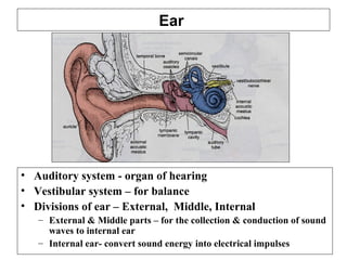
1. Ear
• Auditory system - organ of hearing
• Vestibular system – for balance
• Divisions of ear – External, Middle, Internal
– External & Middle parts – for the collection & conduction of sound
waves to internal ear
– Internal ear- convert sound energy into electrical impulses
4. External Ear
• Vestigial in humans
• Role in sound localization &
amplification
• parts:
– Auricle: or pinna, made of skin,
hair follicles, sweat &
sebaceous glands, Elastic
cartilage
– External Acoustic Meatus
(EAM):
• lateral ⅓ - elastic cartilage,
lining is same as pinna except
ceruminous glands in place of
sweat glands
• Medial ⅔ - within Temporal
bone, with lining of thinner skin,
fewer hair & glands
Clinical : Cerumen or earwax ( secretions of ceruminous and sebaceous glands)
if accumulates in excess ear ache and deafness
5. Middle Ear
• Located in air filled space of temporal bone
Tympanic cavity
• Boundaries
– laterally : TM
– Medially: Internal Ear
– Anteriorly : ET
– Posteriorly : Mastoid with air cells
• Function: convert sound vibrations to mechanical
vibrations and send to internal ear
• Openings
– Oval window – vestibular
– Round window – cochlear
• Structures
– Tympanic membrane
– Ossicular chain
– Muscles (tensor tympani & Stapedius)
7. Middle Ear
• Ossicular chain
– Malleus –attaches to TM
– Incus -links Malleus with
stapes
– Stapes with its foot
process attaches oval
window
Clinical : calcification at foot plate of Stapes Ankylosis (otosclerosis) causes
deafness
8. Internal Ear
• Location
– Petrous part of temporal bone
• Divisions
– Bony labyrinth
– Membranous labyrinth
• Spaces
– Endolymphatic space: within membranous labyrinth,
contains endolymph ( similar to intracellular fluid)
– Perilymphatic space: between bony and membranous
labryinth, contains perilymph ( similar to ECF)
– Cortilymphatic space: with organ of corti, contains
cortilymph ( like ECF)
9. Internal Ear
• Bony labryinth
– Vestibule with utricle
and saccule
– Semicircular canals
– cochlea
10. Internal Ear
• membranous
labryinth
– Cochlear labryinth with
cochlear duct
connected to saccule
– Vestibular labryinth
having semicircular
canals(3) & utricle &
Saccule
11. Membranous labryinth
• Hair cells :
– specialized cells of
membranous labryinth
– Form a hair bundle with
stereocilia and tallest
kinocilium
– Mechanoelctric transduction
in stereocilia lead to influx of
K+ gated ion channels
opening of voltage gated
Ca++ channels release of
neurotransmitter
generation of action potential
in afferent nerve endings
19. The ‘HAIRS’ of HAIR CELLS
The electron microscope revealed that each
‘hair’ consists of one kinocilium at the side of
an array of many non-motile sensory
stereocilia. (These stereocilia are not the
absorptive kind found in the male repoductive tract.)
A cell - View from on top
Cilium
Stereocilia
Viewed from the side, the
stereocilia differ regularly
in height, becoming
shorter going away from
the tall kinocilium
They are more numerous
than shown (70 per cell),
and are attached by links
near their tips
20. Viewed from the
side, the stereocilia
vary regularly in
height, becoming
shorter going away
from the tall
kinocilium
HAIR-CELL DIRECTIONAL SENSITIVITY
Vesicles &
tubules
Sensitive to bending:
Kinocilium
Stereocilia
Plate for
attachment of
actin cores of
stereocilia
Tip links
between
stereocilia
Synapse
Towards kinocilium
causes cell
depolarization, and
increased afferent
fiber firing
Bending away from
kinocilium causes cell
hyperpolarization, and
decreased afferent
fiber firing
Afferent axon
21. CUPULA
CANAL FLUID
HAIR CELLS
The bending of the hairs sometimes is co-
ordinated and amplified by imbedding the
hairs in a gelatinous body called the cupula
CUPULA
22. HAIR-CELL SIGNAL TRANSDUCTION
How does bending towards kinocilium
cause cell depolarization, and increased
afferent fiber firing?
Kinocilium
Vesicles &
tubules
Stereocilia
Tip links
between
stereocilia
Synapse
Transduction channels
for cations, e.g., Ca2 +
, K+
are opened by the bending
The entering cations
depolarize the cell
which increase s transmitter
release at the base,
raising the firing rate in
the axon
23. Supporting, basal, mantle, etc cells
Electron microscopy also revealed that the
‘supporting’ cells were of various different
kinds
Nerve fibers and synapses were both afferent,
and coming from the CNS as effferent
(controlling)
Afferent synapses were of more than one kind,
as are the hair cells
Although there was a fundamental pattern,
species differences were widepsread in ‘hairs,
sensory cells, ‘supporting’ cells, and almost all
aspects of the receptor structures
Certain of the supporting cells secrete the
gelatinous (glycoprotein) cupula
24. VESTIBULAR APPARATUS I
The fluid in the bags - endolymph -
has a special ionic composition to
allow for efficient depolarization,
when the hair-cell stereocilia are
deflected.
Spaces form in the skull’s temporal
bone on each side for three differently
oriented CANALS communicating with
a larger space - VESTIBULE - to hold a
system of fluid-filled bags & tubes
Each canal, and the hair cells
positioned within it, provide nervous
signals responsive to movement of
the head in a particular way.
The three mutually perpendicular
canals on each side can thus inform
on any angularly accelerated (rotary)
head movement
25. VESTIBULAR APPARATUS II Semicircular canal & duct
BONE SEMICIRCULAR CANAL containing
SEMICIRCULAR DUCT containing
Perilymph
Endolymph
Always an initial source of confusion - the semicircular space
in the bone is the CANAL
Inside, and attached to the wall, is the smaller membranous tube -
the DUCT
The rest of the space in the canal is taken by a loose arachnoid-
like tissue, occupied by CSF-like perilymph
The duct is filled with endolymph, high in K+
, & made elsewhere
When the head moves in the plane of the canal, the endolymph
lags a little in relation to the canal’s & duct’s movement
26. VESTIBULAR APPARATUS III Duct’s Ampulla & Christa
At one end of the canal, where it opens into the bony vestibule,
the duct swells out, then constricts, creating the ampulla
BONE
SEMICIRCULAR
CANAL
SEMICIRCULAR DUCT
Perilymph
AMPULLA
Raised ridge -
CRISTA - with hair
cells & gelatinous
cupula
Opening into utricle
Endolymph
CUPULA
ENDOLYMPH
27. VESTIBULAR APPARATUS IV Duct & Christa Activity
As th head moves so , the endolymph in this duct lags
BONE
SEMICIRCULAR
CANAL
SEMICIRCULAR DUCT
Perilymph
Endolymph
CUPULA
along with the cupula
ENDOLYMPH
But moving with the
head are the tissues,
including the hair
cells
So the hair cells are
bent by the dragging
cupula
causing opening or
closing of the cation
channels, with
change in hair-cell
polarization &
synaptic drive
to the christa axons
28. Ampulla of superior
semicircular duct
start of superior
semicircular duct
UTRICLE
SACCULE
MACULA
of Utricle
MACULA
of Saccule
Saccular
Duct
Utricular Duct
VESTIBULAR APPARATUS V Saccule & Utricle
SACCULE
29. MACULA
of Saccule
UTRICLE
SACCULE
MACULA
of Utricle
Saccular
Duct
Utricular Duct
VESTIBULAR APPARATUS VI Saccule versus Utricle
Both contain endolymph & are connected via the U & S ducts
Both utricle & saccule contain a macula with hair cells
Both maculae are covered with a gelatinous otolithic membrane
The utricle is much larger
The maculae are oriented differently
The utricle has the six openings
for the 3 semicircular ducts
but
Saccule’s near vertical;
Utricle’s near horizontal
30. VESTIBULAR APPARATUS VII Macula Structure
Crystalline OTOCONIA
on gelatinous
OTOLITHIC
MEMBRANE
HAIR CELLS
Basement membrane
AXONS of vestibular
ganglion neurons
Supporting cells
Being in pairs, and in different orientations, the maculae
can sense the head’s position and its linear movement
The OTOCONIA of calcium salts and protein contribute to the effect
of gravity on the hair cells, providing a vestibular drive to eventually
keep ‘postural’ skeletal muscles active in maintaining one’s posture
OTOLITHIC
MEMBRANE
Connective
tissue
Endolymph
31. VESTIBULAR GANGLION
Bipolar
neurons VESTIBULAR
NERVE
start of superior
semicircular duct
Ampulla of
superior
semicircular
duct
SACCULE
MACULA
of Utricle
MACULA
of Saccule
VESTIBULAR APPARATUS VIII Vestibular nerve & Ganglion
UTRICLE
CRISTA The vestibular
ganglion & nerve
lie in the bony
internal acoustic
meatus
32. Also, within the bone, spaces must be found for the air
vibrations to be conveyed to the cochlea; while air
pressure has to be equilibrated across the ear drum
The cochlea has to have its own coiled space in the bone
We have seen that: the semicircular ducts require three
canals in each temporal bone; the utricle and ampullae,
& the saccule, need a vestibule in the bone; and the
vestibular ganglion & nerve need a passageway
(meatus) to reach the brainstem.
TEMPORAL BONY SPACES
Finally, passages (aqueducts) are needed to keep the
two fluids - perilymph and endolymph - in balance
The intricate result is best depicted initially as a crude
diagram for learning parts and relations
39. The signals are turned into nerve-cell electrical activity
by mechanoreception for sensing fluid movement
EAR, HEARING & BALANCE
In the inner ear are the organs for the senses of hearing
and balance - the cochlea and the vestibular apparatus
The outer and middle ear get airborne sound to the
inner ear.
W Beresford
40. COCHLEAR APPARATUS I
The cochlear duct inside contains
endolymph , with a special ionic
composition to allow for efficient
depolarization, when the hair-cell
stereocilia are deflected.
Spaces form in the skull’s temporal
bone on each side for three differently
oriented CANALS communicating with
a larger space - VESTIBULE - to hold a
system of fluid-filled bags & tubes
The deflection arises from membrane
deflections, ultimately derived from air
vibrations outside the head
Also, coming off the vestibule is the
snail-like bony cochlea with 21/2 turns
41. Also, within the bone, spaces must be found for the air
vibrations to be conveyed to the cochlea; while air
pressure has to be equilibrated across the ear drum
The cochlea has to have its own coiled space in the bone
We have seen that: the semicircular ducts require three
canals in each temporal bone; the utricle and ampullae,
& the saccule, need a vestibule in the bone; and the
vestibular ganglion & nerve need a passageway
(meatus) to reach the brainstem. (Other nerves pass by.)
TEMPORAL BONY SPACES
Finally, passages (aqueducts) are needed to keep the
three fluids - perilymph, endolymph, & CSF - in balance
The intricate result is best depicted initially as a
diagram for learning parts and relations, but first a more
anatomical overview of the whole system
44. COCHLEA IV Bony Modiolus
HELICOTREMA
where Scalae
vestibuli & tympani
connect
Scala vestibuli
Scala
tympani
COCHLEAR
DUCT or
Scala media
M
O
D
I
O
L
U
S
The cochlea
spirals around a
bony core - the
Modiolus
Note that
although, in
a section, we
see five
profiles, the
structures
spiral
continously
e.g.,
OSSEOUS
SPIRAL
LAMINA
45. COCHLEA IV Spiral ganglion & Modiolus
The modiolus is very
spongy bone , filled with
nerve fibers becoming the
cochlear nerve
ORGAN of
CORTI
SPIRAL
GANGLION
Also, the VIIIth nerve
has incoming efferent
fibers to influence the
outer hair cells in the
Organ of Corti
’efferent’ - from
brain-stem neurons
Axons to Inner
hair cells derive
from spiral-
ganglion cell
bodies
47. BONE
Basilar membrane
ORGAN of
CORTI
Scala vestibuli
Scala tympani
BONE
PERILYMPH
PERILYMPH
COCHLEA VII Basilar membrane II
It vibrates well to low
frequency sounds at its apex
Vibrations from oval
window of vestibule
The basilar membrane is vibrated by fluid pressures in the Scala typani
The spiralling hides that the basilar membrane is LONG
Its WIDTH & STIFFNESS alter regularly along its length, so that
The high-frequency
response is at the base
COCHLEAR
DUCT or
Scala media
The particular component
frequencies of a ‘sound’
produce a pattern of
vibrations along the basilar
membrane,
detectable by the inner hair
cells attached to the active
regions of the
48. Scala tympani
Basilar
membrane
INNER HAIR CELL
TECTORIAL
MEMBRANE
with attached
ENDOLYMPH
innervated by axon
from spiral-
ganglion neuron
Tectorial membrane & Inner Hair Cell
SPIRAL LIMBUS
Support for Reissner’s
membrane & Tectorial
membrane
TECTORIAL MEMBRANE is gelatinous, like the cupula, but is
attached at one side, aside from its hair-cell connections
49. Organ of Corti - cell types
Crista & Macula -- “Electron microscopy also revealed that the
‘supporting’ cells were of various different kinds”. Far more true for
the Organ of Corti, and detectable already in the 19th century, hence
some eponyms
Basilar membrane
OUTER HAIR CELLSTECTORIAL MEMBRANE
INNER & OUTER
PILLAR CELLS
SPIRAL LIMBUS
INNER HAIR CELL
INNER & OUTER PHALANGEAL CELLS
DEITER’S
TECTORIAL CELLS
HENSEN &
CLAUDIUS
CELLS
50. Stria vascularis & K+
recycling I
Basilar membrane
OUTER HAIR CELLS
INNER & OUTER
PILLAR CELLS
INNER HAIR CELL
OUTER PHALANGEAL CELLS
DEITER’S
HENSEN &
CLAUDIUS
CELLS
FIBROBLASTS
STRIA CELLS
K+
The Kcc4 channel gets the K+ into the Deiter’s cells, whence it
goes via gap junctions to theStria for pumping into the
endolymph
51. Stria vascularis II
The Stria vascularis was so named because, quite unusually, capillaries
are found amongst the three kind of epithelial cells
Basilar membrane
HENSEN &
CLAUDIUS
CELLS
STRIA CELLS
52. Also, within the bone, spaces must be found for the air
vibrations to be conveyed to the cochlea; while air
pressure has to be equilibrated across the ear drum
The cochlea has to have its own coiled space in the bone
We’ll return to the schematic of the whole auditory
system for:
SOUND CONDUCTION TO THE INNER EAR
The membrane-sealed openings - oval & round windows
- from vestibule to middle ear, allowing transmission of
pressures, but keeping in the perilymph
The tympanic membrane (ear drum) separating outer
auditory meatus from the middle ear
57. AUDITORY OSSICLES II
MALLEUS
INCUS
STAPES
EXTERNAL
CANAL EAR DRUM
OVAL WINDOW
ROUND WINDOW
MIDDLE EAR
MALLEUS
INCUS
STAPES
The malleus (hammer) is vibrated by air impinging
on the tympanic membrane (ear-drum). Malleus
movements drive the incus (anvil), which in its
turn moves the stapes (stirrup) in and out of the
oval window, so pulsating the fluid (perilymph) in
the vestibule. The bony chain & geometry amplify
the air’s initial force.
VESTIBULE
To relieve fluid pressures
in the vestibule
58. AUDITORY OSSICLES II
MALLEUS
INCUS
STAPES
EXTERNAL
CANAL EAR DRUM
OVAL WINDOW
ROUND WINDOW
MIDDLE EAR
MALLEUS
INCUS
STAPES
The malleus (hammer) is vibrated by air impinging
on the tympanic membrane (ear-drum). Malleus
movements drive the incus (anvil), which in its
turn moves the stapes (stirrup) in and out of the
oval window, so pulsating the fluid (perilymph) in
the vestibule. The bony chain & geometry amplify
the air’s initial force (& match impedance)
VESTIBULE
To relieve fluid pressures
in the vestibule
59. AUDITORY OSSICLES III
The malleus (hammer) is vibrated by air impinging on the
tympanic membrane (ear-drum). Malleus movements
drive the incus (anvil), which in its turn moves the stapes
(stirrup) in and out of the oval window, so pulsating the
fluid (perilymph) in the vestibule. The bony chain &
geometry amplify the air’s initial force.
OVAL WINDOW
MALLEUS
INCUS
STAPES
EAR DRUM
60. Stapedius muscle & Facial nerve
INCUS
STAPES
Tympanic cavity/
Middle ear
VESTIBULE
FACIAL
NERVE
Stapedius muscle
Other long spaces in
the bone house the
Facial nerve &
the Stapedius muscle
whose contraction
hinders the
movement of the
so
protecting the ear
from loud sounds
along with Tensor
tympani‘s action
(Next slide)
The two responses
constitute Sound
attenuation reflex
Oval
window
61. COCHLEA
VESTIBULE
AURICLE
EAR CANAL
MIDDLE EAR
TENSOR TYMPANI
Auditory TUBE
Tensor tympani muscle TT tendon
Malleus
Tensor tympani muscle has its bony tunnel parallel to Eustachian tube’s
TT contraction limits Malleus movement for protection from loud sounds
V th
NERVE
62. EAR PATHOLOGY
FACIAL
NERVE
VIIIth
NERVE
COCHLEA
V
AURICLE
EAR CANAL
MIDDLE EAR
Auditory/
Eustachian TUBE
Nasopharynx
CARTILAGE
EAR DRUM
Angle tumor
-Neuroma of
VIIIth
N - bad
balance
/hearing
Lost Hair
cells - loud
noises, age,
streptomycin,
neomycin,
cisplatin
Blocked tube Perforated ear-drum
-infection, blast injury
Excess endolymph - hydrops
Otitis media - middle ear infection; Cholesteatoma - kerat strat squam ep
Ankylosed ossicles
Wax, foreign
bodies in canal
Meningitis,
abscess
Overgrowth of bone - Otosclerosis
63. EAR PATHOLOGY II
Lost/damaged Hair cells from - loud noises, age;
ototoxic agents - streptomycin, neomycin (aminoglycoside
antibiotics), cisplatin (anticancer agent)
Congenital deafness - One of a number of defects in genes can
impair the development of the inner ear, or the differentiation and
functioning of hair cells
Hypothyroidism and iodine deficiency in pregnancy can result in
defective development of the fetus’ Organ of Corti
64. EAR PATHOLOGY III
Angle tumor -Neuroma
of VIIIth N - bad
balance /hearing
Lost Hair cells - loud
noises, age, streptomycin,
Blocked Eustachian tube
Perforated ear-drum
-infection, blast
Excess endolymph -
hydrops
Otitis media - middle ear
infection
Ankylosed
ossicles
Wax, foreign
bodies in canal
Meningitis,
abscess
Overgrowth of bone -
Otosclerosis
Congenital deafness - defects in genes