The document provides information on the anatomy of the eye and its surrounding structures. It discusses the bones that make up the orbit, the layers of the eyelid, muscles that control eyelid movement, blood supply to different parts of the eye, and the structures that make up the iris, ciliary body, choroid, retina, and extraocular muscles. The document also describes the circulation of aqueous humor and the vascular supply to different parts of the eye.
1-IT IS A MIDDLE VASCULAR COAT OF EYEBALL.
2-IT MAINLY CONSIST OF THREE PARTS IRIS, CHOROID, CILIARY BODY.
3- CILIARY BODY CAN HOLD THE LENS AND PLAY IMPORTANT ROLE IN ACCOMODATION.
1-IT IS A MIDDLE VASCULAR COAT OF EYEBALL.
2-IT MAINLY CONSIST OF THREE PARTS IRIS, CHOROID, CILIARY BODY.
3- CILIARY BODY CAN HOLD THE LENS AND PLAY IMPORTANT ROLE IN ACCOMODATION.
Servers: Servers are the backbone of e-commerce websites. They store the website data, including product information, customer details, and transactional data. They are responsible for processing customer requests, generating dynamic content, and serving web pages to customers.
Storage devices: Storage devices such as hard disk drives (HDDs) or solid-state drives (SSDs) are used to store the website data and application files. They provide the necessary storage capacity to accommodate large amounts of data, such as product images, videos, and customer information.
Routers and switches: Routers and switches are used to connect the e-commerce website to the internet and facilitate data transfer between different devices. They help to ensure that data is transmitted quickly and reliably, and that the website is accessible to customers from anywhere in the world.
Firewalls: Firewalls are used to protect the e-commerce website from unauthorized access and malicious attacks. They monitor incoming and outgoing network traffic
There are several challenges associated with the trade cycle in e-commerce, which can affect the overall efficiency and effectiveness of the process. Some of these challenges include:
Security: One of the main challenges in e-commerce is ensuring the security of the transaction. This includes protecting sensitive data such as credit card information and personal details from theft, fraud, and other cyber threats.
Logistics: Shipping and delivery can be a significant challenge in e-commerce, particularly for products that require special handling or transportation. This includes ensuring timely delivery, tracking shipments, and dealing with returns and exchanges.
Payment processing: Payment processing can be complex, particularly for cross-border transactions involving different currencies and payment systems. It is essential to ensure that payment methods are secure, reliable, and convenient for customers.
The trade cycle in e-commerce refers to the various stages involved in a typical online transaction between a buyer and a seller. The trade cycle typically includes the following stages:
Product search and selection: The buyer searches for a product or service online and selects the desired item from the e-commerce website. This may involve browsing product categories, using search filters, and reading product descriptions and reviews.
Shopping cart and checkout: Once the buyer has selected the desired item, they add it to their shopping cart and proceed to checkout. At this stage, they may be required to enter their personal and payment information, such as name, address, and credit card details.
Order processing: After the buyer has completed the checkout process, the seller receives the order and processes it. This may involve verifying the availability of the product, preparing it for shipment, and generating a shipping label.
Payment processing: Once the order has been processed, the payment is processed by the payment gateway. This involves verifying the payment information and authorizing the transaction.
Shipping and delivery: The seller ships the product to the buyer's address using a third-party logistics provider or their own delivery service. The buyer is provided with tracking information to monitor the status of the shipment.
Returns and refunds: If the buyer is not satisfied with the product, they may initiate a return or exchange. The seller handles the return or exchange process and ensures that the buyer is satisfied with their purchase.
Customer service: The seller provides customer service to address any issues or concerns that the buyer may have regarding the product or service.
Title: Sense of Smell
Presenter: Dr. Faiza, Assistant Professor of Physiology
Qualifications:
MBBS (Best Graduate, AIMC Lahore)
FCPS Physiology
ICMT, CHPE, DHPE (STMU)
MPH (GC University, Faisalabad)
MBA (Virtual University of Pakistan)
Learning Objectives:
Describe the primary categories of smells and the concept of odor blindness.
Explain the structure and location of the olfactory membrane and mucosa, including the types and roles of cells involved in olfaction.
Describe the pathway and mechanisms of olfactory signal transmission from the olfactory receptors to the brain.
Illustrate the biochemical cascade triggered by odorant binding to olfactory receptors, including the role of G-proteins and second messengers in generating an action potential.
Identify different types of olfactory disorders such as anosmia, hyposmia, hyperosmia, and dysosmia, including their potential causes.
Key Topics:
Olfactory Genes:
3% of the human genome accounts for olfactory genes.
400 genes for odorant receptors.
Olfactory Membrane:
Located in the superior part of the nasal cavity.
Medially: Folds downward along the superior septum.
Laterally: Folds over the superior turbinate and upper surface of the middle turbinate.
Total surface area: 5-10 square centimeters.
Olfactory Mucosa:
Olfactory Cells: Bipolar nerve cells derived from the CNS (100 million), with 4-25 olfactory cilia per cell.
Sustentacular Cells: Produce mucus and maintain ionic and molecular environment.
Basal Cells: Replace worn-out olfactory cells with an average lifespan of 1-2 months.
Bowman’s Gland: Secretes mucus.
Stimulation of Olfactory Cells:
Odorant dissolves in mucus and attaches to receptors on olfactory cilia.
Involves a cascade effect through G-proteins and second messengers, leading to depolarization and action potential generation in the olfactory nerve.
Quality of a Good Odorant:
Small (3-20 Carbon atoms), volatile, water-soluble, and lipid-soluble.
Facilitated by odorant-binding proteins in mucus.
Membrane Potential and Action Potential:
Resting membrane potential: -55mV.
Action potential frequency in the olfactory nerve increases with odorant strength.
Adaptation Towards the Sense of Smell:
Rapid adaptation within the first second, with further slow adaptation.
Psychological adaptation greater than receptor adaptation, involving feedback inhibition from the central nervous system.
Primary Sensations of Smell:
Camphoraceous, Musky, Floral, Pepperminty, Ethereal, Pungent, Putrid.
Odor Detection Threshold:
Examples: Hydrogen sulfide (0.0005 ppm), Methyl-mercaptan (0.002 ppm).
Some toxic substances are odorless at lethal concentrations.
Characteristics of Smell:
Odor blindness for single substances due to lack of appropriate receptor protein.
Behavioral and emotional influences of smell.
Transmission of Olfactory Signals:
From olfactory cells to glomeruli in the olfactory bulb, involving lateral inhibition.
Primitive, less old, and new olfactory systems with different path
The prostate is an exocrine gland of the male mammalian reproductive system
It is a walnut-sized gland that forms part of the male reproductive system and is located in front of the rectum and just below the urinary bladder
Function is to store and secrete a clear, slightly alkaline fluid that constitutes 10-30% of the volume of the seminal fluid that along with the spermatozoa, constitutes semen
A healthy human prostate measures (4cm-vertical, by 3cm-horizontal, 2cm ant-post ).
It surrounds the urethra just below the urinary bladder. It has anterior, median, posterior and two lateral lobes
It’s work is regulated by androgens which are responsible for male sex characteristics
Generalised disease of the prostate due to hormonal derangement which leads to non malignant enlargement of the gland (increase in the number of epithelial cells and stromal tissue)to cause compression of the urethra leading to symptoms (LUTS
Anti ulcer drugs and their Advance pharmacology ||
Anti-ulcer drugs are medications used to prevent and treat ulcers in the stomach and upper part of the small intestine (duodenal ulcers). These ulcers are often caused by an imbalance between stomach acid and the mucosal lining, which protects the stomach lining.
||Scope: Overview of various classes of anti-ulcer drugs, their mechanisms of action, indications, side effects, and clinical considerations.
Couples presenting to the infertility clinic- Do they really have infertility...Sujoy Dasgupta
Dr Sujoy Dasgupta presented the study on "Couples presenting to the infertility clinic- Do they really have infertility? – The unexplored stories of non-consummation" in the 13th Congress of the Asia Pacific Initiative on Reproduction (ASPIRE 2024) at Manila on 24 May, 2024.
Ethanol (CH3CH2OH), or beverage alcohol, is a two-carbon alcohol
that is rapidly distributed in the body and brain. Ethanol alters many
neurochemical systems and has rewarding and addictive properties. It
is the oldest recreational drug and likely contributes to more morbidity,
mortality, and public health costs than all illicit drugs combined. The
5th edition of the Diagnostic and Statistical Manual of Mental Disorders
(DSM-5) integrates alcohol abuse and alcohol dependence into a single
disorder called alcohol use disorder (AUD), with mild, moderate,
and severe subclassifications (American Psychiatric Association, 2013).
In the DSM-5, all types of substance abuse and dependence have been
combined into a single substance use disorder (SUD) on a continuum
from mild to severe. A diagnosis of AUD requires that at least two of
the 11 DSM-5 behaviors be present within a 12-month period (mild
AUD: 2–3 criteria; moderate AUD: 4–5 criteria; severe AUD: 6–11 criteria).
The four main behavioral effects of AUD are impaired control over
drinking, negative social consequences, risky use, and altered physiological
effects (tolerance, withdrawal). This chapter presents an overview
of the prevalence and harmful consequences of AUD in the U.S.,
the systemic nature of the disease, neurocircuitry and stages of AUD,
comorbidities, fetal alcohol spectrum disorders, genetic risk factors, and
pharmacotherapies for AUD.
Title: Sense of Taste
Presenter: Dr. Faiza, Assistant Professor of Physiology
Qualifications:
MBBS (Best Graduate, AIMC Lahore)
FCPS Physiology
ICMT, CHPE, DHPE (STMU)
MPH (GC University, Faisalabad)
MBA (Virtual University of Pakistan)
Learning Objectives:
Describe the structure and function of taste buds.
Describe the relationship between the taste threshold and taste index of common substances.
Explain the chemical basis and signal transduction of taste perception for each type of primary taste sensation.
Recognize different abnormalities of taste perception and their causes.
Key Topics:
Significance of Taste Sensation:
Differentiation between pleasant and harmful food
Influence on behavior
Selection of food based on metabolic needs
Receptors of Taste:
Taste buds on the tongue
Influence of sense of smell, texture of food, and pain stimulation (e.g., by pepper)
Primary and Secondary Taste Sensations:
Primary taste sensations: Sweet, Sour, Salty, Bitter, Umami
Chemical basis and signal transduction mechanisms for each taste
Taste Threshold and Index:
Taste threshold values for Sweet (sucrose), Salty (NaCl), Sour (HCl), and Bitter (Quinine)
Taste index relationship: Inversely proportional to taste threshold
Taste Blindness:
Inability to taste certain substances, particularly thiourea compounds
Example: Phenylthiocarbamide
Structure and Function of Taste Buds:
Composition: Epithelial cells, Sustentacular/Supporting cells, Taste cells, Basal cells
Features: Taste pores, Taste hairs/microvilli, and Taste nerve fibers
Location of Taste Buds:
Found in papillae of the tongue (Fungiform, Circumvallate, Foliate)
Also present on the palate, tonsillar pillars, epiglottis, and proximal esophagus
Mechanism of Taste Stimulation:
Interaction of taste substances with receptors on microvilli
Signal transduction pathways for Umami, Sweet, Bitter, Sour, and Salty tastes
Taste Sensitivity and Adaptation:
Decrease in sensitivity with age
Rapid adaptation of taste sensation
Role of Saliva in Taste:
Dissolution of tastants to reach receptors
Washing away the stimulus
Taste Preferences and Aversions:
Mechanisms behind taste preference and aversion
Influence of receptors and neural pathways
Impact of Sensory Nerve Damage:
Degeneration of taste buds if the sensory nerve fiber is cut
Abnormalities of Taste Detection:
Conditions: Ageusia, Hypogeusia, Dysgeusia (parageusia)
Causes: Nerve damage, neurological disorders, infections, poor oral hygiene, adverse drug effects, deficiencies, aging, tobacco use, altered neurotransmitter levels
Neurotransmitters and Taste Threshold:
Effects of serotonin (5-HT) and norepinephrine (NE) on taste sensitivity
Supertasters:
25% of the population with heightened sensitivity to taste, especially bitterness
Increased number of fungiform papillae
Report Back from SGO 2024: What’s the Latest in Cervical Cancer?bkling
Are you curious about what’s new in cervical cancer research or unsure what the findings mean? Join Dr. Emily Ko, a gynecologic oncologist at Penn Medicine, to learn about the latest updates from the Society of Gynecologic Oncology (SGO) 2024 Annual Meeting on Women’s Cancer. Dr. Ko will discuss what the research presented at the conference means for you and answer your questions about the new developments.
ARTIFICIAL INTELLIGENCE IN HEALTHCARE.pdfAnujkumaranit
Artificial intelligence (AI) refers to the simulation of human intelligence processes by machines, especially computer systems. It encompasses tasks such as learning, reasoning, problem-solving, perception, and language understanding. AI technologies are revolutionizing various fields, from healthcare to finance, by enabling machines to perform tasks that typically require human intelligence.
Pulmonary Thromboembolism - etilogy, types, medical- Surgical and nursing man...VarunMahajani
Disruption of blood supply to lung alveoli due to blockage of one or more pulmonary blood vessels is called as Pulmonary thromboembolism. In this presentation we will discuss its causes, types and its management in depth.
New Directions in Targeted Therapeutic Approaches for Older Adults With Mantl...i3 Health
i3 Health is pleased to make the speaker slides from this activity available for use as a non-accredited self-study or teaching resource.
This slide deck presented by Dr. Kami Maddocks, Professor-Clinical in the Division of Hematology and
Associate Division Director for Ambulatory Operations
The Ohio State University Comprehensive Cancer Center, will provide insight into new directions in targeted therapeutic approaches for older adults with mantle cell lymphoma.
STATEMENT OF NEED
Mantle cell lymphoma (MCL) is a rare, aggressive B-cell non-Hodgkin lymphoma (NHL) accounting for 5% to 7% of all lymphomas. Its prognosis ranges from indolent disease that does not require treatment for years to very aggressive disease, which is associated with poor survival (Silkenstedt et al, 2021). Typically, MCL is diagnosed at advanced stage and in older patients who cannot tolerate intensive therapy (NCCN, 2022). Although recent advances have slightly increased remission rates, recurrence and relapse remain very common, leading to a median overall survival between 3 and 6 years (LLS, 2021). Though there are several effective options, progress is still needed towards establishing an accepted frontline approach for MCL (Castellino et al, 2022). Treatment selection and management of MCL are complicated by the heterogeneity of prognosis, advanced age and comorbidities of patients, and lack of an established standard approach for treatment, making it vital that clinicians be familiar with the latest research and advances in this area. In this activity chaired by Michael Wang, MD, Professor in the Department of Lymphoma & Myeloma at MD Anderson Cancer Center, expert faculty will discuss prognostic factors informing treatment, the promising results of recent trials in new therapeutic approaches, and the implications of treatment resistance in therapeutic selection for MCL.
Target Audience
Hematology/oncology fellows, attending faculty, and other health care professionals involved in the treatment of patients with mantle cell lymphoma (MCL).
Learning Objectives
1.) Identify clinical and biological prognostic factors that can guide treatment decision making for older adults with MCL
2.) Evaluate emerging data on targeted therapeutic approaches for treatment-naive and relapsed/refractory MCL and their applicability to older adults
3.) Assess mechanisms of resistance to targeted therapies for MCL and their implications for treatment selection
- Video recording of this lecture in English language: https://youtu.be/lK81BzxMqdo
- Video recording of this lecture in Arabic language: https://youtu.be/Ve4P0COk9OI
- Link to download the book free: https://nephrotube.blogspot.com/p/nephrotube-nephrology-books.html
- Link to NephroTube website: www.NephroTube.com
- Link to NephroTube social media accounts: https://nephrotube.blogspot.com/p/join-nephrotube-on-social-media.html
HOT NEW PRODUCT! BIG SALES FAST SHIPPING NOW FROM CHINA!! EU KU DB BK substit...GL Anaacs
Contact us if you are interested:
Email / Skype : kefaya1771@gmail.com
Threema: PXHY5PDH
New BATCH Ku !!! MUCH IN DEMAND FAST SALE EVERY BATCH HAPPY GOOD EFFECT BIG BATCH !
Contact me on Threema or skype to start big business!!
Hot-sale products:
NEW HOT EUTYLONE WHITE CRYSTAL!!
5cl-adba precursor (semi finished )
5cl-adba raw materials
ADBB precursor (semi finished )
ADBB raw materials
APVP powder
5fadb/4f-adb
Jwh018 / Jwh210
Eutylone crystal
Protonitazene (hydrochloride) CAS: 119276-01-6
Flubrotizolam CAS: 57801-95-3
Metonitazene CAS: 14680-51-4
Payment terms: Western Union,MoneyGram,Bitcoin or USDT.
Deliver Time: Usually 7-15days
Shipping method: FedEx, TNT, DHL,UPS etc.Our deliveries are 100% safe, fast, reliable and discreet.
Samples will be sent for your evaluation!If you are interested in, please contact me, let's talk details.
We specializes in exporting high quality Research chemical, medical intermediate, Pharmaceutical chemicals and so on. Products are exported to USA, Canada, France, Korea, Japan,Russia, Southeast Asia and other countries.
5. The roof: the lesser wing of the sphenoid, the orbital plate of the frontal.
The lateral wall: the greater wing of the sphenoid, Zygomatic.
The floor: Zygomatic, Maxillary, Palatine.
The Medial wall: Maxillary, Lacrimal, Ethmoid, Sphenoid.
10. Eyelid Structure
1. Orbicularis oculi (sphincter muscle); closes eyelids.
2. Levator muscle of the upper eyelid (striate muscle); opens eyelids.
3. Tarsal plate (connective tissue); contributes to eyelid form and support.
4. Locus of tarsal plate; set as a requirement for eyelid manipulation
11.
12. Orbicularis oculi
Two main parts:
• Orbital part: plays a role when the eyelids need to be tightly shut.
• Palpebral portion that plays a role in winking and blinking.
Nerve supply: facial nerve
13. Levator Palpebrae Superioris
Origin: Roof of the orbit, above the optic foramen
Insertion: Anterior surface of superior tarsus and upper eyelid
Innervation: Oculomotor nerve (CN III)
Action: Retraction and elevation of eyelid.
22. Lacrimal gland
• Located within the lacrimal fossa in the superior and
outer edge of the orbital roof.
• The tendon of the levator palpebrae muscle divides the
lacrimal gland into a larger orbital part (2/3) and a smaller
palpebral part (1/3).
• Parasympathetic secretomotor nerve supply comes from
the nervus intermedius.
• Blood supply: lacrimal artery (a branch of the ophthalmic
artery
23.
24.
25.
26. Tear drainage
The superior and inferior puncta lacrimales collect the
tears, which then drain through the superior and inferior
lacrimal canaliculi into the lacrimal sac. From there they
pass through the nasolacrimal duct into the inferior
concha
28. The cornea is the transparent tissue that covers the front
of the eye, forms anterior 1/6th of the outer coat of
eyeball.
• Diameter 11.5 mm. (11.7 mm * 11mm).
It forms 3/4th of the total refractive power of eye.
• Refractive power: 45D.
• Refractive index: 1.376.
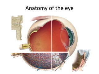
32. Iris
• Iris is the anterior-most part of the uveal tract.
• It is a thin, circular structure, forms a diaphragm like structure in
front of the crystalline lens.
• It has a central aperture known as the pupil.
• The pupil determines the amount of light entering the eye.
33. Muscles in iris
Sphincter pupillae: circular muscle, 1 mm wide, encircles the pupil.
Innervated by the parasympathetic system.
Contraction of the sphincter causes the pupil to constrict (miosis).
Dilator pupillae: extends radially from the iris root to the sphincter.
innervated by the sympathetic system
Contraction of the dilator muscle causes the pupil to dilate
(mydriasis).
34. Ciliary body
The ciliary body is the middle part of the uveal tract forms a ring-
shaped structure that projects posteriorly from the scleral spur.
Anteriorly: it is confluent with the periphery of the iris (iris root).
Posteriorly: it has a scalloped periphery, known as ora serrata, where
it is continuous with the choroid and retina.
35.
36. Ciliary muscle
Nonstriated muscle primarily situated in the anterior 2/3 of
the ciliary body stroma.
The muscle has three parts: Outer longitudinal portion,
Middle oblique portion, Inner circular portion.
Action: Accommodation
Nerve supply: The parasympathetic stimulation activates
the muscle for contraction, whereas sympathetic innervation
likely has an inhibitory effect.
37. Pars plicata: the portion of the ciliary body that contains finger-like
projections (ciliary processes), extend into the posterior chamber.
Pars plana: the smooth part of the ciliary body, terminates at the
ora serrata, which is the transitional zone between the ciliary body
and choroid.
40. Choroid
• The choroid is a thin but highly vascular layer, lining the inner
surface of the sclera.
• It extends from ora serrata anteriorly to the optic nerve posteriorly.
• The outer surface is attached to the sclera at the optic nerve and at
the exit of the vortex veins, The inner surface is attached to the
retinal pigmented epithelium (RPE).
42. Blood supply of uveal tract:
The blood supply of the uveal tract is mainly from three
arteries namely
• Short posterior ciliary arteries
• Long posterior ciliary arteries
• Anterior ciliary arteries.
The posterior ciliary arteries are branches of the
ophthalmic artery
48. The optic disc lies a 3 mm medial to the center of the macula
(fovea). There are no normal retinal layers in this zone (blind spot)
as ganglion cell axons from the retina pierce the sclera to enter the
optic nerve.
49. Vascular supply to the retina
The inner layers of the retina (the internal limiting membrane
through the inner nuclear layer) are supplied by the central
artery of the retina. This originates at the ophthalmic artery,
enters the eye with the optic nerve, and branches on the inner
surface of the retina. It is a terminal artery without
anastomoses and divides into four main branches
Because the central artery is a terminal artery, occlusion will
lead to retinal infarction.
The outer layers (outer plexiform layer through the pigment
epithelium) contain no capillaries. They are nourished by
diffusion primarily from the richly supplied capillary layer of the
choroid.
50. diagram of the human visual pathways and their neuronal components.