1. CLINICAL
systems of life
NT 4 April 2006 Vol 102 No 14 www.nursingtimes.net26
Homeostasis
Authors Brendan Doherty, MSc, PGCE, RN, is
nurse patient access manager, Prince of Wales
Hospital, Sydney, Australia; Colette Foudy, RN,
GradDip, is clinical care coordinator, intensive care
unit, St George Private Hospital, Sydney, Australia.
This article, the first in a series of four, looks at the
anatomy and physiology of homeostasis.
Homeostasis comprises the dynamic processes that
enable optimum conditions to be maintained for
cells, in spite of continual changes taking place
internally and externally (Clancy and McVicar, 1995).
All the systems of the human body are involved,
with particular contributions by the endocrine,
nervous, respiratory and renal systems. Whenever an
imbalance occurs, regulatory systems become active
to restore optimum conditions, usually by a process
known as negative feedback in which a deviation
from the normal level is detected and initiates
changes that bring the level back to where it should
be (Clancy and McVicar, 1995). These systems have to
endure for survival and adapt to modifications of the
environment so must therefore evolve.
Anatomy and physiology
Many factors affect the suitability of body fluids to
sustain life. These will be explored later in this series.
They include:
l Oxygen (O2
) and carbon dioxide (CO2
)
concentrations;
l The pH of the internal environment;
l Concentrations of nutrients and waste products;
l Concentration of salt and other electrolytes
(osmoregulation);
l Volume and pressure of extracellular fluid;
l Body temperature;
l Blood glucose level.
As these properties affect the chemical reactions that
sustain life, there are built-in physiological mechanisms
to maintain them at desirable levels. The body needs
homeostasis to maintain stability and survive by
ensuring that the internal environment remains
relatively constant (Tortora and Anagnostakos, 2003).
To enable cells to survive, the composition of the
intracellular and extracellular fluids must be
accurately maintained at all times. Intracellular fluid
accounts for two-thirds of total water content (Tortora
and Anagnostakos, 2003). Extracellular fluid includes
gases, nutrients, plasma (in blood vessels) and ions,
all of which are necessary for maintaining life
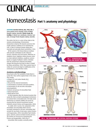
(Tortora and Anagnostakos, 2003) (Fig 1).
Extracellular fluid circulates constantly within
Part 1: anatomy and physiology
Brain Ear
Spinal cord
Eyes
Taste and smell
sensors
Skin
sensors
Nerves
Pineal gland
Thyroid
Andrenals
Ovaries
Testes
Hypothalamus
Pituitary
Pancreas
Fig 1. extracellular
fluid and blood vessels
Artery
Arteriole
Capillary
Venule
Vein
Capillary
Plasma Extracellular
fluid
Nervous system endocrine system
Fig 2. The endocrine and central nervous systems
Thymus
Cell
JohnnyZygo
2. This article has been double-blind
peer-reviewed.
For related articles on this subject
and links to relevant websites see
www.nursingtimes.net
NT 4 April 2006 Vol 102 No 14 www.nursingtimes.net
keywords n Anatomy and physiology n Homeostatic control
27
the blood and lymphatic system and is known as
‘the body’s internal environment’ (Tortora and
Anagnostakos, 2003).
The body is said to be in homeostasis when its
internal environment contains:
l Optimum levels of gases, ions, water and nutrients;
l Is at optimal temperature;
l Has optimal pressure for the health of cells.
A disturbance in these optimum conditions causes
failure of the organs and may lead to death (Tortora
and Anagnostakos, 2003).
Homeostatic control
The endocrine and central nervous systems are
the major control systems for regulating homeostasis
(Tortora and Anagnostakos, 2003) (Fig 2). The
endocrine system consists of a series of glands
that secrete chemical regulators (hormones). The
nervous system can detect deviation from the
body’s normal equilibrium (state of homeostasis)
and sends messages to the affected organ to
counteract this disturbance. Over a relatively
short time it restores the required balance. Both
systems act mostly automatically but there is some
voluntary control over the nervous system
(Sherwood, 1997).
The control and maintenance of blood sugar levels
is an example of homeostatic regulation by the
endocrine system. Blood sugar is maintained by two
hormones secreted by the pancreas: insulin and
glucagon. Blood sugar rises after digestion of food. In
response, pancreas cells are stimulated to secrete
insulin, which enables sugar uptake by cells and the
storage of sugar in the liver and muscles. In effect,
insulin decreases blood sugar levels to normal
(Tortora and Anagnostakos, 2003).
The respiratory system provides an example
of homeostatic regulation by the nervous system.
In normal breathing there is a state of homeostasis.
During exercise the respiratory system must work
faster to keep the O2
in the extracellular fluid and
in the cells within normal limits, preventing excessive
build-up of CO2
and disturbance to the blood pH
through the accumulation of acid (Tortora and
Anagnostakos, 2003). Because muscles require
more O2
during exercise, more CO2
is released and
therefore also needs to be excreted (Tortora and
Anagnostakos, 2003).
These chemical changes are detected in certain
nerve cells, which send this message to the cardio-
respiratory centre in the medulla oblongata in
the base of the brain (Docherty, 2002). The brain
sends a message to the heart to increase its pumping
action (heart rate) to take on more O2
and enable
the blood to give up excess CO2
. Respiratory muscles
also receive instructions from the brain to contract
faster, enabling a rise in both O2
delivery and
CO2
exhalation.
Negative feedback
Homeostasis is constantly disturbed by external
factors, which can be described as a form of stress
on the internal environment (Tortora and
Anagnostakos, 2003). These stresses can be:
l Internal: occurring within the body, for example in
pain, or as a result of high or low blood pressure;
l External: occurring outside the body, for example as
a result of heat, cold or loud noises.
Since these stresses affect the chemical reactions
sustaining life, there are built-in physiological
mechanisms to maintain or return them to
desirable levels.
Each organ or structure has its own intrinsic way
of keeping the internal environment within normal
limits. When homeostasis is altered there are two
possible responses:
l In negative feedback the system responds
to reverse the direction of change. As this tends
to keep things constant, it allows the maintenance
of homeostasis. For example, if there is a fall
in calcium in the blood, the parathyroid glands
sense the decrease and secrete more parathyroid
hormone, thereby increasing calcium release from
the bones;
l Positive feedback increases the variable in the
same direction, a destabilising effect that does
not result in homeostasis. Positive feedback is used
when rapid change is needed. For example in
childbirth the hormone oxytocin is produced to
stimulate and enhance labour contractions
(Bodyguide, 2005).
Components of homeostasis
A system requires three components for homeostasis:
l A receptor;
l A control centre;
l An effector.
These components do specific jobs that allow
regulation of the internal environment.
A receptor detects external changes that could
influence the internal environment. For example,
the following are involved in the regulation of
blood pressure:
l Receptors are in the baroreceptor system;
l The control centre is the medulla oblongata;
l The effector is the cardiovascular system.
When there is an increase in heart rate more blood
is pumped into the arteries resulting in an increase in
blood pressure (Docherty, 2005). This is detected by
the baroreceptors, which are located in the walls of
certain arteries.
These receptors send impulses to the control
centre (the medulla oblongata), which interprets
the message and sends impulses to the effectors
(the cardiovascular system). These slow the
pulse, decreasing blood pressure (Tortora and
Anagnostakos, 2003). n
References
Bodyguide (2005) Endocrine
System. www.besthealth.com/
besthealth/bodyguide/reftext/html/
endo_sys_fin.html#homeostatic.
Clancy, J., McVicar, A. (1995)
Physiology and Anatomy:
A Homeostatic Approach.
London: Edward Arnold Publishers.
Docherty, B. (2005) The
arteriovenous system: part one, the
anatomy. Nursing Times; 101: 34,
28–29.
Docherty, B. (2002)
Cardiorespiratory physical
assessment for the acutely ill: part
one. British Journal of Nursing;
11: 11, 750–758.
Sherwood, L. (1997) Human
Physiology: From Cells to Systems.
Pacific Grove, CA: West Publishing.
Tortora, G.J., Anagnostakos, N.P.
(2003) Principles of Anatomy and
Physiology. New York, NY: John
Wiley and Sons.