Calcinosis in Scleroderma Treatment Options
•Download as PPTX, PDF•
0 likes•48 views

This document discusses calcinosis, a complication of scleroderma where calcium deposits form in the soft tissues. It outlines that calcinosis most commonly affects the hands, joints, knees and muscles. While the calcium levels in the blood are normal, inflammation and low blood flow in tissues are thought to contribute to calcinosis formation. The deposits contain bone-like proteins and crystals. 18F-NaF PET imaging may help detect early or active calcinosis. Treatments discussed include protecting joints, surgical removal of deposits, topical or injected sodium thiosulfate, medications like tofacitinib, NSAIDs, colchicine and minimizing scleroderma complications to reduce calcinosis risk over

Recommended
More Related Content
Similar to Calcinosis in Scleroderma Treatment Options
Similar to Calcinosis in Scleroderma Treatment Options (20)
More from Scleroderma Foundation of Greater Chicago
More from Scleroderma Foundation of Greater Chicago (20)
Recently uploaded
Recently uploaded (20)
Calcinosis in Scleroderma Treatment Options
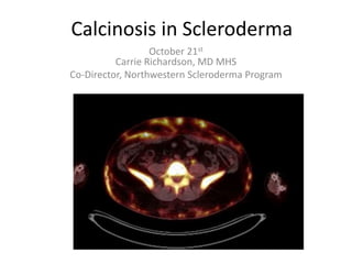
- 1. Calcinosis in Scleroderma October 21st Carrie Richardson, MD MHS Co-Director, Northwestern Scleroderma Program
- 2. Disclosures • Consultant for Cabaletta Pharmaceuticals
- 3. Presentation Outline • Part 1 – Calcinosis Overview • Part 2 – The Science of Calcinosis • Part 3 – Treatments We Use for Calcinosis
- 4. Key Questions • What is scleroderma-related calcinosis? • What do we know about what causes calcinosis? • What do we know about how to treat calcinosis?
- 5. Part 1 – Calcinosis Overview
- 6. What Is Scleroderma-Related Calcinosis? • Calcium-phosphate crystal deposition in the soft tissue • Not exactly bone but shares some similarities • Calcium and phosphorous levels in the bloodstream are NORMAL Pai et al. Mod Rheumatol 2018 Bartoli et al. Rheumatol 2016
- 7. Calcinosis Most Commonly Affects the Hands Thumb of the dominant hand is the most likely to be affected – A VERY BAD SPOT!!! Calcinosis
- 8. Calcinosis Commonly Occurs At Joints This can make it hard to bend!!! Or it can pinch nerves at the elbow
- 9. The Knee is Another Area Where We Commonly See Calcinosis These can take a VERY long time to heal if they open up
- 10. Calcinosis May Also Encase the Muscles These deposits can be VERY extensive and can feel like a plate
- 11. Why Are These Specific Areas So Affected? • Tendon insertions (which are located in or near joints) and muscles are prone to calcification, especially with pressure or injury • If the joints, tendons, and/or muscles get inflamed, the inflammation can eventually result in calcinosis as the body tries to heal • People with scleroderma are especially prone to this calcification process
- 12. Who Is Affected by Calcinosis? • Affects to 40% of patients with scleroderma • People with LONGER disease duration and more scleroderma-related damage have more calcinosis • Myositis and PM/Scl antibodies increase the risk of calcinosis
- 13. More Scleroderma Complications for a Longer Time Period => More Calcinosis Scleroderma Complications People with the Most Complications have the most Calcinosis Richardson et al. Rheumatology (Oxford) 2022
- 14. What Do We Think Causes Calcinosis? • Scleroderma-related inflammation… • PLUS low blood flow in the tissue and scarring… • Causes tissue-resident stem cells to behave like bone cells as the body tries to repair damage
- 15. A Simple Schematic of Calcinosis Inflammation Tissue Hypoxia (Low Oxygen State) Fibrosis Tissue-level factors (cytokines/ chemokines/ adipokines, resident stem cells, loss of calcification inhibitors) Calcinosis
- 16. Why Does Calcinosis Matter? • No known effective medication to prevent or treat this • Disabling and disfiguring • Understanding calcinosis in scleroderma may help us understand common problems like ”hardening of the arteries” or rotator cuff problems
- 17. Calcinosis Can Get Infected and Require Surgery
- 18. Calcinosis May Make it Hard to Feel Comfortable
- 19. Part 2 – The Science of Calcinosis
- 20. Calcinosis Does Not Look Quite Like Bone … Vs.
- 21. …But Calcinosis Shares Some Similarities With Bone Formation • Bone-associated proteins are present in calcinosis deposits • Mechanical loading important Lian et al. Biochem Biophys Res Comm 1976 Hughes et al. Semin Arthritis Rheum 2020 Urganus et al. Arthritis Rheumatol 2009
- 22. Calcinosis Lesions And the Blood Vessels Near Them Contain Bone- Associated Proteins OPN ON OC RUNX2 MGP ALP BSP DPP DMP1 OC ON MGP DPP BSP ALP OPN DMP1 Vasculature OPN DPP DMP1 BSP ALP RUNX2 Deposits TRAP Urganus et al. Arthritis Rheum 2009
- 23. The Calcium Crystal in Calcinosis is the Same as in Bone Hydroxyapatite – Ca10(PO4)6(OH)2 Cochrane and Davies Ann Rheum Dis 1965
- 24. What Does Calcinosis Look Like Under the Microscope? • “Morphea with dystrophic calcification” • Some look disorganized, some liquid, others grossly resembling bone • Some have tissue macrophages (“big eater” inflammatory cells), some have few cells
- 25. Calcinosis Under the Microscope Images from Olsen et al. AJR Am J Roentgenol 2004 Macrophages Irregular calcification Fibrosis
- 26. How Can We Measure Calcinosis? Xray – fine for identifying calcinosis but gives a 2D image CT – Provides a 3D view but does not tell us how “active” calcinosis is NaF PET CT- Could this give us an indicator of calcinosis activity?
- 27. 18F-NaF PET Deposits in Areas of Growing Calcinosis CT of knee shows calcinosis (white arrow) but no calcinosis in the back of the knee Radiotracer lights up the calcinosis but also an area in the back of the knee – is this a very early calcinosis deposit?
- 28. 18F-NaF PET Can Show Active Calcinosis in Areas Where We Cannot See Calcinosis Yet on CT Calcinosis (white markings) looks pretty minimal The whole pelvic area lights up with the radiotracer
- 29. 18F NaF PET Might Be a Way to Identify Calcinosis Before We Can Even Feel It Are calcinosis deposits THIS big…. …going to turn into calcinosis deposits THIS big?
- 30. Part 3 – Calcinosis Treatments
- 31. Treatments We Use for Calcinosis* • Protection from mechanical injury • Treatment of any ongoing inflammatory activity in the joints or muscles • Surgical resection in select cases • Sodium thiosulfate (topical or injected) • Tofacitinib (Xeljanz) • Anakinra (Kineret) • Non-steroidal anti-inflammatories like celecoxib, ibuprofen • Minocycline • Colchicine • Low-dose prednisone • Methotrexate • Sulfasalazine *Note: There are NO FDA-approved medications for calcinosis
- 32. Would Treating Scleroderma Better Eliminate Calcinosis?!!!!! Could we reduce or eliminate calcinosis by decreasing scleroderma complications???? People with the most scleroderma complications have the most calcinosis People with fewer scleroderma complications have less calcinosis
- 33. How Do We Treat Scleroderma Better? • Check for myositis with a CK level and strength exam and treat (consider IVIG or rituximab) if myositis is present • Treat for arthritis if exam shows joint swelling or if an ultrasound shows joint inflammation • Maximize Raynaud’s medications to improve blood flow
- 34. Protection from Mechanical Injury • Finger cots • Adhesive bandages • Arm rest cushions • Chair cushions
- 35. Surgical Removal • Helpful for SMALL deposits in inconvenient spots, like the hand • Also helpful for infected deposits or areas that are causing pinched nerves • Deposits are often connected to muscle, tendons and can be difficult to remove • Removal can leave chronic open wounds
- 36. Sodium Thiosulfate • Prevents calcium crystals from clumping together • ~75% of patients notice some benefit based on a Mayo Clinic study • Can be used as a cream on the skin or injected into calcinosis • Often makes deposits liquify and come to the surface or pop out
- 37. How Do You Get Sodium Thiosulfate? • Compounded preparation, requires prescription • We use Petranek’s Pharmacy in Illinois or Mayo Clinic Pharmacy • 25% sodium thiosulfate, compounded in zinc oxide or Vanicream
- 38. Tofacitinib (Xeljanz) • Inhibitor of Jak1 and 3 (inflammatory proteins) • Oral medication, typically 5 mg twice daily but higher doses can be used
- 39. Xeljanz Considerations • Infection risk, particularly shingles (see picture) • Elevated liver enzymes, high cholesterol, low white blood cell count
- 40. Minocycline • Oral medication • Typical dose is 50-100 mg daily • May have some benefits for inflammatory arthritis
- 41. Minocycline Considerations • Causes permanent blue/gray skin and nail discoloration • Can also turn calcinosis deposits blue/gray • Risk of development of drug-induced autoimmune disease
- 42. Colchicine • Common treatment for gout and pseudogout (other disorders caused by crystals) • Anti-inflammatory, blocks cells called neutrophils from migrating to cause inflammation • Typical dose is 0.6-1.2 mg daily
- 43. Anakinra • Also used for gout, pseudogout • Daily injection, typically 100 mg daily • Causes injection site reactions • Expensive, difficult to get insurance to cover
- 44. Non-Steroidal Anti- Inflammatories • Examples: Ibuprofen/Motrin, naproxen/Aleve, celecoxib/Celebrex • Can cause issues with GI tract, kidney, and heart especially with regular use • Diclofenac gel/Voltaren gel is a topical NSAID that is gentler on the body
- 45. A Note On Calcinosis and Osteoporosis • People with calcinosis are at increased risk of osteoporosis (low bone density) • If you have calcinosis, consider getting screened for osteoporosis!!! • We generally try to avoid using teriparatide/Forteo (bone-building osteoporosis medication) in people with calcinosis
- 47. Special Thank You to Stephanie Gresh for Organizing!