International Journal of Clinical Cardiology & Research
•
0 likes•11 views

A Primary Percutaneous Coronary (PCI) intervention and pharmaco-invasive Primary Percutaneous Coronary (PCI) continues to be the optimal reperfusion therapy in patients with anterior ST Elevation Myocardial Infarction (STEMI) however, the optimal treatment in patients with anterior STEM equivalents (wellness and DeWinter syndromes) is unknown.
![SCIRES Literature - Volume 3 Issue 2 - www.scireslit.com Page -032
International Journal of Clinical Cardiology & Research
INTRODUCTION
American College of Cardiology/American Heart Association
guidelines recommend primary Percutaneous Coronary Intervention
(PCI) for patients with ST-segment-elevation myocardial infarction
presenting ≤ 12 hours of symptom onset with a first medical contact-
to-device time goal of ≤ 120 minutes. Pharmacoinvasive strategy
where fibrinolysis is followed by immediate transfer to a PCI-capable
hospital for either rescue PCI in case of failed fibrinolysis or routine
coronary angiography and PCI in case of successful fibrinolysis
may be a valid alternative to primary PCI and part of the primary
reperfusion in patients with ST-segment-elevation myocardial
infarction in whom long PCI-related delay is anticipated [1].
Wallen’s’ syndrome or “Widow Maker”, is referred to an
Electrocardiogram (ECG) pattern (There are two ECG patterns with
difference incidence: Pattern A with biphasic T wave in V2-V3n and
pattern B with deep T wave in V2-V3) for proximal Left Anterior
Descending (LAD) artery lesion). ECG abnormalities present at the
Emergency Department (ED) in patients admitted with angina during
pain free periods. Cardiac biomarkers are usually normal or slightly
elevated, this syndrome requires urgent coronary angiography and
PCI to avoid the development of extensive anterior Myocardial
Infarction (MI) [2].
The De Winter syndrome was reported as an indicator of acute
Left Anterior Descending (LAD) coronary artery occlusion. The
ECG pattern typically include ST-depression and peaked T-waves in
precordial leads, and it can be seen in around 2% of patients with
anterior myocardial infarction [3].
Wellness and De Winter syndrome are considered an anterior
ST-Elevation Myocardial Infarction (STEMI) equivalents [4,5].
The aim of our trial to evaluate the efficacy of the patients
presenting within 2 hours of symptom onset of acute anterior
ST elevation myocardial infarction to primary PCI < 2 hours or
pharmaco-invasivePCI2-24hoursafterstreptokinase,comparedwith
acute anterior ST elevation myocardial infarction equivalents to early
PCI < 24 hours The primary (efficacy) endpoint a composite of death,
shock and congestive heart failure at 30 days after hospitalization.
METHODS
Study design
This prospective, cohort study was conducted on 173 patients
who had symptoms onset of myocardial ischemia within 12 hours
divided according to ECG into two groups:
Group 1: (Anterior STEMI): ST elevation in anterior precordial
leads.
Group 2: (Anterior STEMI Equivalents): Biphasic T wave in V2-
V3 or deep T wave in V2-V3 (Wallen’s syndrome) and upsloping
ST depression with peaked T-waves in precordial leads (De winter
syndromes) (Figure 1).
The aim of the our study is to comparison of the efficacy of
Primary PCI (< 2 hours) and/or pharmaco-invasive PCI (< 24 hours)
in patient with anterior STEMI group compared to early PCI (< 24
hours) in anterior STEMI equivalents group in patients presents with
onset of myocardial ischemia within 12 hours and less than 2 hours of
admission to emergency room in Gaza.
ABSTRACT
Background: A Primary Percutaneous Coronary (PCI) intervention and pharmaco-invasive Primary Percutaneous Coronary (PCI)
continues to be the optimal reperfusion therapy in patients with anterior ST Elevation Myocardial Infarction (STEMI) however, the optimal
treatment in patients with anterior STEMI equivalents (wellness and De Winter syndromes) is unknown.
Methods: Patients presenting within 2 hours of symptom onset, divided according to Electrocardiography (ECG) into two groups, the
first group: acute anterior STEMI treated by primary PCI < 2 hours or pharmaco-invasive PCI 2-24 hours after streptokinase, and second
group: acute anterior STEMI equivalents (wellness and De Winter syndromes) treated with early PCI < 24 hours. The primary (efficacy)
endpoint a composite of death, shock and congestive heart failure at 30 days after hospitalization.
Results: Total 173 patients (134 patients: anterior STEMI, 39 patients: Anterior STEMI Equivalents) with mean age 56.2 ± 9.4 years,
the mean age was 55.1 ± 9.2 years for the Anterior STEMI group, and 59.7 ± 9.1 years in Anterior STEMI equivalents group. (p = 0.007).
The female gender was higher in Anterior STEMI Equivalents compared with anterior STEMI (p = 0.036). Efficacy (primary) endpoint:
total death, shock or heart failure at 30 days after hospitalization was seen in myocardial infarction was significantly higher in patient with
anterior STEMI group compared with anterior STEMI equivalent group (p = 0.011). The total death, shock and heart failure were significant
in anterior STEMI group (p = 0.047, 0.024 and 0.028) respectively.
Conclusion: A strategy of early PCI less than 24 hours in patients with Anterior STEMI Equivalents has safe and less adverse events
than anterior STEMI with primary PCI < 2 hours and/or pharmaco-invasive PCI 2-24 hours after streptokinase administration.
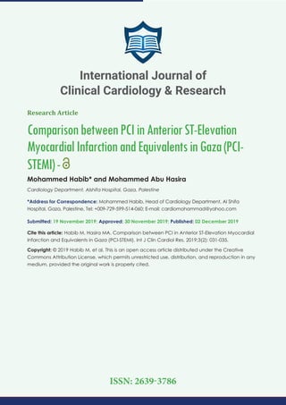
Initial treatment in Emergency Room for all patients (N: 173)
Aspirin 100-300mg tab, clopidogrel 600 mg tab, heparin 5000 u IV
Primary PCI (60 patients)
Or pharmaco -invasive PCI (74 patients )
Early PCI within < 24 hours Wallen's (36
patients) or De winter (3 patients) Syndromes
Patients with onset of ischemic symptoms < 12 hours and < 120 min of admission to the
emergency room (N: 181 patient). 8 patients were excluded (5 from anterior equivalents
group and 3 from anterior STEMI group)
Anterior SETMI Equivalents group (N: 39
patients)
Anterior ST EMI group
(N: 134 patients)
Study Endpoint
Primary Endpoint: Death, shock or heart failure
Figure 1: The study flow diagram of patients.](data:image/gif;base64,R0lGODlhAQABAIAAAAAAAP///yH5BAEAAAAALAAAAAABAAEAAAIBRAA7)
Recommended
More Related Content
What's hot
What's hot (20)
Similar to International Journal of Clinical Cardiology & Research
Similar to International Journal of Clinical Cardiology & Research (20)
More from SciRes Literature LLC. | Open Access Journals
More from SciRes Literature LLC. | Open Access Journals (20)
Recently uploaded
Recently uploaded (20)
International Journal of Clinical Cardiology & Research
- 1. Research Article ComparisonbetweenPCIinAnteriorST-Elevation MyocardialInfarctionandEquivalentsinGaza(PCI- STEMI)- Mohammed Habib* and Mohammed Abu Hasira Cardiology Department, Alshifa Hospital, Gaza, Palestine *Address for Correspondence: Mohammed Habib, Head of Cardiology Department, Al Shifa Hospital, Gaza, Palestine, Tel: +009-729-599-514-060; E-mail: cardiomohammad@yahoo.com Submitted: 19 November 2019; Approved: 30 November 2019; Published: 02 December 2019 Cite this article: Habib M, Hasira MA. Comparison between PCI in Anterior ST-Elevation Myocardial Infarction and Equivalents in Gaza (PCI-STEMI). Int J Clin Cardiol Res. 2019;3(2): 031-035. Copyright: © 2019 Habib M, et al. This is an open access article distributed under the Creative Commons Attribution License, which permits unrestricted use, distribution, and reproduction in any medium, provided the original work is properly cited. International Journal of Clinical Cardiology & Research ISSN: 2639-3786
- 2. SCIRES Literature - Volume 3 Issue 2 - www.scireslit.com Page -032 International Journal of Clinical Cardiology & Research INTRODUCTION American College of Cardiology/American Heart Association guidelines recommend primary Percutaneous Coronary Intervention (PCI) for patients with ST-segment-elevation myocardial infarction presenting ≤ 12 hours of symptom onset with a first medical contact- to-device time goal of ≤ 120 minutes. Pharmacoinvasive strategy where fibrinolysis is followed by immediate transfer to a PCI-capable hospital for either rescue PCI in case of failed fibrinolysis or routine coronary angiography and PCI in case of successful fibrinolysis may be a valid alternative to primary PCI and part of the primary reperfusion in patients with ST-segment-elevation myocardial infarction in whom long PCI-related delay is anticipated [1]. Wallen’s’ syndrome or “Widow Maker”, is referred to an Electrocardiogram (ECG) pattern (There are two ECG patterns with difference incidence: Pattern A with biphasic T wave in V2-V3n and pattern B with deep T wave in V2-V3) for proximal Left Anterior Descending (LAD) artery lesion). ECG abnormalities present at the Emergency Department (ED) in patients admitted with angina during pain free periods. Cardiac biomarkers are usually normal or slightly elevated, this syndrome requires urgent coronary angiography and PCI to avoid the development of extensive anterior Myocardial Infarction (MI) [2]. The De Winter syndrome was reported as an indicator of acute Left Anterior Descending (LAD) coronary artery occlusion. The ECG pattern typically include ST-depression and peaked T-waves in precordial leads, and it can be seen in around 2% of patients with anterior myocardial infarction [3]. Wellness and De Winter syndrome are considered an anterior ST-Elevation Myocardial Infarction (STEMI) equivalents [4,5]. The aim of our trial to evaluate the efficacy of the patients presenting within 2 hours of symptom onset of acute anterior ST elevation myocardial infarction to primary PCI < 2 hours or pharmaco-invasivePCI2-24hoursafterstreptokinase,comparedwith acute anterior ST elevation myocardial infarction equivalents to early PCI < 24 hours The primary (efficacy) endpoint a composite of death, shock and congestive heart failure at 30 days after hospitalization. METHODS Study design This prospective, cohort study was conducted on 173 patients who had symptoms onset of myocardial ischemia within 12 hours divided according to ECG into two groups: Group 1: (Anterior STEMI): ST elevation in anterior precordial leads. Group 2: (Anterior STEMI Equivalents): Biphasic T wave in V2- V3 or deep T wave in V2-V3 (Wallen’s syndrome) and upsloping ST depression with peaked T-waves in precordial leads (De winter syndromes) (Figure 1). The aim of the our study is to comparison of the efficacy of Primary PCI (< 2 hours) and/or pharmaco-invasive PCI (< 24 hours) in patient with anterior STEMI group compared to early PCI (< 24 hours) in anterior STEMI equivalents group in patients presents with onset of myocardial ischemia within 12 hours and less than 2 hours of admission to emergency room in Gaza. ABSTRACT Background: A Primary Percutaneous Coronary (PCI) intervention and pharmaco-invasive Primary Percutaneous Coronary (PCI) continues to be the optimal reperfusion therapy in patients with anterior ST Elevation Myocardial Infarction (STEMI) however, the optimal treatment in patients with anterior STEMI equivalents (wellness and De Winter syndromes) is unknown. Methods: Patients presenting within 2 hours of symptom onset, divided according to Electrocardiography (ECG) into two groups, the first group: acute anterior STEMI treated by primary PCI < 2 hours or pharmaco-invasive PCI 2-24 hours after streptokinase, and second group: acute anterior STEMI equivalents (wellness and De Winter syndromes) treated with early PCI < 24 hours. The primary (efficacy) endpoint a composite of death, shock and congestive heart failure at 30 days after hospitalization. Results: Total 173 patients (134 patients: anterior STEMI, 39 patients: Anterior STEMI Equivalents) with mean age 56.2 ± 9.4 years, the mean age was 55.1 ± 9.2 years for the Anterior STEMI group, and 59.7 ± 9.1 years in Anterior STEMI equivalents group. (p = 0.007). The female gender was higher in Anterior STEMI Equivalents compared with anterior STEMI (p = 0.036). Efficacy (primary) endpoint: total death, shock or heart failure at 30 days after hospitalization was seen in myocardial infarction was significantly higher in patient with anterior STEMI group compared with anterior STEMI equivalent group (p = 0.011). The total death, shock and heart failure were significant in anterior STEMI group (p = 0.047, 0.024 and 0.028) respectively. Conclusion: A strategy of early PCI less than 24 hours in patients with Anterior STEMI Equivalents has safe and less adverse events than anterior STEMI with primary PCI < 2 hours and/or pharmaco-invasive PCI 2-24 hours after streptokinase administration. Initial treatment in Emergency Room for all patients (N: 173) Aspirin 100-300mg tab, clopidogrel 600 mg tab, heparin 5000 u IV Primary PCI (60 patients) Or pharmaco -invasive PCI (74 patients ) Early PCI within < 24 hours Wallen's (36 patients) or De winter (3 patients) Syndromes Patients with onset of ischemic symptoms < 12 hours and < 120 min of admission to the emergency room (N: 181 patient). 8 patients were excluded (5 from anterior equivalents group and 3 from anterior STEMI group) Anterior SETMI Equivalents group (N: 39 patients) Anterior ST EMI group (N: 134 patients) Study Endpoint Primary Endpoint: Death, shock or heart failure Figure 1: The study flow diagram of patients.
- 3. SCIRES Literature - Volume 3 Issue 2 - www.scireslit.com Page -033 International Journal of Clinical Cardiology & Research Study population The study population was single Center trial derived from al shifa hospital between March 2018 and October 2019. We identified 173 patients (≥ 18 years) with anterior STEMI (< 12 hours) and < 2 hours of presentation eligible for either pharmaco-invasive strategy or Primary PCI or early PCI < 24 hours in patients with anterior STEMI Equivalents. All subjects received acetylsalicylic acid, clopidogrel, unfractionated heparin and high dose statin (Atorvastatin 80 mg) according to our guidelines. Patients were excluded from the study if their age was < 18 years or if they have contraindications of fibrinolytic therapy or coronary artery disease not suitable for revascularization by PCI or patients who didn’t underwent coronary angiogram and PCI within 24 hours. Clinical definitions: Anterior STEMI was defined as chest pain suggestive of myocardial ischemia for ≈30 minutes, ST-segment elevation > 0.1 mV in ≥ 2 contiguous leads in VI-6 and elevated cardiac markers (creatine kinase-MB or troponin I/T). PPCI was defined as PCI within 12 hours of symptom onset in a patient not receiving streptokinase. Time to start of reperfusion therapy was defined as time to intravenous injection of fibrinolytics and time to balloon inflation in patients treated with fibrinolysis and PPCI, respectively. Pharmacoinvasive strategy was defined as fibrinolysis followed by rescue or by routine elective PCI (beyond 2 hours of fibrinolytic administration). The Wallen’s’ syndrome is presentation of anterior ischaemia and it is characterised by deeply inverted or biphasic T waves in multiple precordial lead The De Winter ECG pattern typically displays tall T-waves with Upsloping ST depression in multiple precordial lead. End points Efficacy (primary) endpoint: total death, shock or heart failure at 30 days after myocardial hospitalization. Statistical analysis Data was analyzed by SPSS version 19. Continuous variables were presented as Mean ± Standard Deviation (SD) and categorical variables as absolute numbers and percentages. Comparison of demographic and clinical data among the groups was performed using independent t-test for continuous variables and chi-square (χ2) for categorical variables. Pearson’s correlation coefficients were calculated to illustrate certain relationships. P = < 0.05 were considered significant. RESULTS Baseline characteristics Total 173 patients (134 patients: anterior STEMI, 39 patients: Anterior STEMI Equivalents) with mean age 56.2 ± 9.4 years, the mean age was 55.1 ± 9.2 years for the Anterior STEMI group, and 59.7 ± 9.1 years in Anterior STEMI equivalents group. (p = 0.007). The female gender was higher in Anterior STEMI Equivalents compared with anterior STEMI (p = 0.036), the history of PCI was higher in anterior STEMI group compared with STEMI equivalents group (p = 0.025), no significant difference between two groups in risk factors diabetes mellitus, hypertension, smoking, history of CABG, history of stroke, presence of COPD and family history of coronary artery disease (Table 1). Efficacy (primary) endpoint Total death, shock or heart failure at 30 days after hospitalization was seen in myocardial infarction was significantly higher in patient with anterior STEMI group compared with anterior STEMI equivalent group (p = 0.011). The total death, shock and heart failure were significant in anterior STEMI group (p = 0.047, 0.024 and 0.028) respectively (Table 2). No difference between two groups in the number of significant coronary artery stenosis (p = 0.72), but the effected LCX was higher in anterior STEMI group (p = 0.024) (Table 3). Type of intervention In acute anterior STEMI, 60 (44.8%) patients underwent standard primary PCI and 75 (55.2%) patient underwent pharmacoinvasive strategy but in this group 28 patients (37.8%) had failed streptokinase and need emergency angiography and PCI. In acute anterior STEMI equivalents: 3 patients with De Winter syndrome underwent standard primary PCI within 2 hours and 36 patients with wellens syndrome underwent standard primary PCI within 24 hours. Door to needle and door to balloon time Table 1: Total (n = 173) Anterior STEMI (n = 134) Anterior STEMI equivalents (n = 39) p value Age, years 56.2 ± 9.4 55.1 ± 9.2 years 59.7 ± 9.1years 0.007 Female gender 26 (15%) 16 (11.9%) 10 (25.6%) 0.036 Diabetes mellitus 62 (34.8%) 49 (36.6%) 13 (33%) 0.71 Hypertension 103 (57.9%) 78 (58.2%) 25 (64.1%) 0.51 Smoking 75 (42.1%) 62 (46.3%) 13 (56.4%) 0.15 Previous PCI 23 (12.9%) 22 (16.4%) 1 (2.6%) 0.025 Previous CABG 2 (1.1%) 0 (0%) 2 (5.1%) 0.44 Previous Stroke/ TIA 2 (1.1%) 1 (0.07%) 1 (2.6%) 0.93 COPD 14 (7.9%) 12 (8.9%) 2 (5.1%) 0.44 Family history of CAD 29 (16.3%) 22 (16.4%) 7 (18%) 0.82 PCI: Percutaneous Coronary Intervention; CABG: Coronary Artery By-Pass Grafting; TIA: Transient Ischemic Attack; COPD: Chronic Obstructive Pulmonary Disease; CAD: Coronary Artery Disease Table 2: Primary outcome between two groups. Anterior STEMI (n = 134) Anterior STEMI equivalents (n = 39) p value Primary endpoint 31 (23%) 2 (5.1%) 0.011 Death 8 (5%) 0 (0%) 0.047 Shock 16 (12%) 0 (0%) 0.024 Heart failure 27 (20.1%) 2 (5.1%) 0.028
- 4. SCIRES Literature - Volume 3 Issue 2 - www.scireslit.com Page -034 International Journal of Clinical Cardiology & Research In anterior STEMI: Average time from medical contact to streptokinase administration was 33.17 ± 15 min. in patient with successful streptokinase all Coronary angiography was performed with 24 hours with average time 16 ± 3.4 hrs., but in patient with failed streptokinase emergency angiography was required in 28 patients (37.8%) of the patients the median time 6 ± 1.3 hours after randomization. In primary PCI: The Door to balloon time was 75.25 ± 20 min. and the Door to balloon was performed in 81% in the time < 90 min. In anterior STEMI equivalents: The Door to balloon time was (8 ± 1.2) hours. SUBGROUP ANALYSIS In the anterior STEMI equivalent group The LAD totally occlusion was present in all 3 patients with De winter Syndrome and severe stenosis in all patients with Wallen’s syndrome except one patient with wellens syndrome have aneurysmal dilatation and slow flow in LAD without significant stenosis was treated medically. Only one patient underwent PCI after 48 hours because anterior STEMI due to subacute stent thrombosis, primary PCI was done and the patient discharged from hospital with mild impaired left ventricle dysfunction and symptoms of heart failure, and 4 patients were died: One patient died within 24 h of presentation before coronary angiography. One patient had cardiac arrest just before reaching to the catheterization laboratory and cardiopulmonary resuscitation was done but patient died. One patient underwent diagnostic coronary angiography but PCI not done at that time because of the size of stent was not available in our center and the patient was transferred elective to another center to make PCI but the patient was died within 48 hours before PCI. One patient was missed diagnosed and discharged with medical treatment, 2 weeks after discharge, acute massive anterior MI and cardiogenic shock was developed the patient was admitted to coronary care unit, norepinephrine infusion was started, the patient intubated and primary PCI was done to LAD but patient died within 12 hours of admission. Totally five patients were excluded from our analysis in this group because of 4 patients died before coronary angiography and PCI and one patient who had aneurysmal dilatation and slow flow in LAD without significant stenosis. Our analysis including only patients who underwent coronary angiogram and PCI within 24 hours. All three patients with De winter syndrome underwent coronary angiogram and PCI less than 2 hours. In the anterior STEMI group Three patients died within 2 hours of presentation, one patient during streptokinase infusion, and 2 patients just after reaching to emergency room. These patients were excluded from our analysis because of our analysis including only patients who underwent coronary angiogram and PCI within 24 hours. DISCUSSION In our trial, we report on the efficacy PCI (primary or pharmacoinvasive) strategy in patients with anterior STEMI compared with early PCI < 24 hours in patients with anterior STEMI equivalents (Wallen’s and De winter syndromes) strategy in Gaza, we found that: • First, the efficacy end point : composite of total death, shock or heart failure at 30 days after hospitalization was significantly higher in patient with anterior STEMI group compared with anterior STEMI equivalent group (p = 0.011). • Second, actually, it has to be considered that patients enrolled in our trial were at low risk with a 30-day mortality of 5% in patient with anterior STEMI group compared with no death in anterior STEMI equivalent group who underwent early coronary angiography and PCI (two patients were died before coronary angiography). • Third, the rate of heart failure and shock was higher in anterior STEMI group • Forth, a 37.8% of patients in the acute anterior myocardial infarction with thrombolysis group required urgent coronary angiography. Despite advancements in percutaneous coronary interventions, acute ST elevation myocardial infarction is still one of the global leading causes of death [6]. And its incidence is increasing [7]. One of the most important factors in treating STEMI patients is to achieve early reperfusion. Primary Percutaneous Coronary Intervention (PCI) is superior to fibrinolytic therapy but difference in mortality between primary PCI or pharmaco-invasive strategy [8]. In our trial we treated only 3 patients with early PCI with an ECG pattern “up-sloping ST depression and symmetrical long and distinct T waves starting from J-point in precordial leads” and in coronary angiography we found totally occluded in proximal LAD and successfully treated by PCI without any complications de Winter, et al. [9]. Reported this ECG pattern that is found in about 2% of patients with proximal left anterior descending artery occlusion Although de Winter T-wave ECG pattern was suggested to be managed as STEMI equivalent [10]. Wellens’ syndrome was first described by De Zwann, et al. [2] in a study group of 145 patients with unstable angina, and 26 patients had the ECG pattern of Wellens’ syndrome. Twelve patients out of this group did not undergo coronary revascularization and subsequently developed anterior myocardial infarction within a few weeks’ time. Later on, a larger sample was used (1,260 patients, 180 patients with Wellens ECG findings) and all of the patients who showed these ECG changes were found to have stenosis in the proximal LAD ranging from 50% to 100% [11]. Rhinehardt, et al. [12] described the following diagnostic criteria for Wellens’ syndrome: • Deeply inverted or biphasic T waves in V2-3 (may extend to V1-6); Table 2: Primary outcome between two groups. Anterior STEMI (n = 134) Anterior STEMI equivalents (n = 39) p value Primary endpoint 31 (23%) 2 (5.1%) 0.011 Death 8 (5%) 0 (0%) 0.047 Shock 16 (12%) 0 (0%) 0.024 Heart failure 27 (20.1%) 2 (5.1%) 0.028
- 5. SCIRES Literature - Volume 3 Issue 2 - www.scireslit.com Page -035 International Journal of Clinical Cardiology & Research • Isoelectric or minimally elevated ST-segment (< 1 mm); • No precordial Q waves; • Preserved precordial R wave progression; • A recent history of angina; • ECG pattern present in the pain-free state; • Normal or slightly elevated serum cardiac markers. In our trial 36 patient with wellens syndrome were treated by early PCI (in Wellens’ syndrome: one patient was treated medically due to aneurysmal dilatation and slow flow in LAD was excepted from analysis, and only one patient have anterior STEMI after 48 hours of PCI related to subacute stent thrombosis). The STREAM (Strategic Reperfusion Early after Myocardial Infarction) trial compared fibrinolysis followed by PCI within 6 to 24 h to primary PCI. The investigators found that there was no difference in the composite endpoint of death, shock, congestive heart failure, or reinfarction at 30 days between the 2 treatments. In this study, tenecteplase was given as the lytic agent followed by catheterization and failure rate of tenecteplase was 36%, in our trial we use the streptokinase in emergency Room and the failure rate was 37.8% [8]. So that we recommended ideally, the presence of Wellens or de Winter as urgent as STEMI with catheter laboratory activation for coronary angiography and PCI. However, despite these findings, Wallen’s syndrome and de Winter patterns are yet to be included in the ESC guidelines for management of acute coronary syndromes in both persistent (2017) or without persistent ST-elevation (2015) and ACC/AHA guidelines regarding STEMI (2013) and NSTEMI (2014) [13,14]. CONCLUSIONS The LAD occlusion was present in all 3 patients with De winter Syndrome and severe stenosis in 36 patients with Wallen’s syndrome and only one patient have aneurysmal dilatation and slow flow in LAD without significant stenosis. The purpose of this trial was to emphasize the importance of this ECG pattern because it is as important as STEMI and these patients need to undergo immediate or early percutaneous coronary intervention in the optimal time to reduce morbidity and mortality. Wallen’s and de Winter are not mentioned in any guidelines regarding acute coronary syndromes and until now there are no clear recommendations. We recommended that in patients with De winter syndrome with onset of myocardial ischemic symptoms less than 12 hours, PCI < 2 hours or fibrinolysis may be considered and in patients with wellens syndrome the early PCI < 24 or immediately PCI < 2 hours after presentation is recommended. REFERENCES 1. O’Gara PT, Kushner FG, Ascheim DD, Casey DE Jr, Chung MK, de Lemos JA, et al. 2013 ACCF/AHA guideline for the management of STelevation myocardial infarction: A report of the American College of Cardiology Foundation/American Heart association task force on practice guidelines. J Am Coll Cardiol. 2013; 61: 485-510. PubMed: https://www.ncbi.nlm.nih.gov/ pubmed/23256913 2. de Zwaan C, Bar FW, Wellens HJ. Characteristic electrocardiographic pattern indicating a critical stenosis high in left anterior descending coronary artery in patients admitted because of impending myocardial infarction. Am Heart J. 1982; 103: 730-736. PubMed: https://www.ncbi.nlm.nih.gov/ pubmed/6121481 3. de Winter RJ, Verouden NJ, Wellens HJ, Wilde AA. A new ECG sign of proximal LAD occlusion. N Engl J Med. 2008; 359: 2071-2073. PubMed: https://www.ncbi.nlm.nih.gov/pubmed/18987380 4. Verouden NJ, Koch KT, Peters RJ, Henriques JP, Baan J, van der Schaaf RJ, et al. Persistent precordial “hyperacute” T-waves signify proximal left anterior descending artery occlusion. Heart. 2009; 95: 1701-1706. PubMed: https:// www.ncbi.nlm.nih.gov/pubmed/19620137 5. M Shams, A Sullivan, S Abudureyimu, B Hassouna, V Jayanthi, R Amdur, et al. Optimizing electrocardiogram interpretation and catheterization laboratory activation in ST-segment elevation myocardial infarction: A teaching module for medical students. J Am Coll Cardiol. 2016; 67: 643. http://www.onlinejacc. org/content/67/13_Supplement/643 6. Reddy K, Khaliq A, Henning RJ. Recent advances in the diagnosis and treatment of acute myocardial infarction. World J Cardiol. 2015; 7: 243-276. PubMed: https://www.ncbi.nlm.nih.gov/pubmed/26015857 7. Yusuf S, Reddy S, Ounpuu S, Anand S. Global burden of cardiovascular diseases: Part II: Variations in cardiovascular disease by specific ethnic groups and geographic regions and prevention strategies. Circulation. 2001; 104: 2855-2864. PubMed: https://www.ncbi.nlm.nih.gov/pubmed/11733407 8. Armstrong PW, Gershlick AH, Goldstein P, Wilcox R, Danays T, Lambert Y, et al. Fibrinolysis or primary PCI in ST-segment elevation myocardial infarction. N Engl J Med. 2013; 368: 1379-1387. PubMed: https://www.ncbi.nlm.nih.gov/pubmed/23473396 9. de Winter RJ, Verouden NJ, Wellens HJ, Wilde AA. A new ECG sign of proximal LAD occlusion. N Engl J Med. 2008; 359: 2071-2073. PubMed: https://www.ncbi.nlm.nih.gov/pubmed/18987380 10. Rokos IC, French WJ, Mattu A, Nichol G, Farkouh ME, Reiffel J, et al. Appropriate cardiac cath lab activation: Optimizing electrocardiogram interpretation and clinical decision-making for acute ST-elevation myocardial infarction. Am Heart J. 2010; 160: 995-1003. PubMed: https://www.ncbi.nlm. nih.gov/pubmed/21146650 11. de Zwaan C, Bar FW, Wellens HJ. Characteristic electrocardiographic pattern indicating a critical stenosis high in left anterior descending coronary artery in patients admitted because of impending myocardial infarction. Am Heart J. 1982; 103: 730-736. PubMed: https://www.ncbi.nlm.nih.gov/ pubmed/6121481 12. Rhinehardt J, Brady WJ, Perron AD, Mattu A. Electrocardiographic manifestations of Wellens’ syndrome. Am J Emerg Med. 2002; 20: 638-643. PubMed: https://www.ncbi.nlm.nih.gov/pubmed/12442245 13 Ibanez B, James S, Agewall S, Antunes MJ, Bucciarelli-Ducci C, Bueno H, et al. 2017 ESC Guidelines for the management of acute myocardial infarction in patients presenting with ST-segment elevation: The Task Force for the management of acute myocardial infarction in patients presenting with ST-segment elevation of the European Society of Cardiology (ESC). Eur Heart J. 2018; 39: 119-177. PubMed: https://www.ncbi.nlm.nih.gov/ pubmed/28886621 14. PTO’Gara, Frederick GK, Deborah DA, Donald EC, Mina KC, JA de Lemos, et al. 2013 ACCF/AHA guideline for the management of ST-elevationmyocardial infarction: A report of the American College of Cardiology Foundation/ American Heart Association task force on practice guidelines. Circulation. 2013; 127: e362-e425. PubMed: https://www.ahajournals.org/doi/ full/10.1161/cir.0b013e3182742cf6