
Recommended
More Related Content
What's hot
What's hot (20)
Similar to Fluid and electrolyte balance regulation
Similar to Fluid and electrolyte balance regulation (20)
More from sai kiran Neeli
Recently uploaded
Recently uploaded (20)
Fluid and electrolyte balance regulation
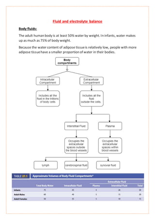
- 1. Fluid and electrolyte balance Body fluids: The adult human body is at least 50% water by weight. In infants, water makes up as much as 75% of body weight. Because the water content of adipose tissueis relatively low, people with more adipose tissuehave a smaller proportion of water in their bodies.
- 2. Regulationof water content: Before regulation, let’s discuss aboutwater input and output, The body’s water content is regulated so that its total volumeremains constant. Thus, the volume of water taken into the body is equal to the volume lost each day. The total volume of water entering the body each day is 1500–3000 mL.
- 3. i.water input: Although fluid consumption is heavily influenced by habit and by socialsettings, water ingestion does depend, at least in part, on thirst regulatory mechanisms. The sensation of thirst results primarily from an increase in the osmolality of the extracellular fluids and from a reduction in plasma volume, which lowers blood pressure.
- 4. The thirstsensation is temporarily reduced after a person drinks a smallamount of liquid. At least two factors are responsiblefor this temporary interruption of the thirstsensation. First, when the oral mucosa becomes wet after it has been dry, sensory neurons conduct action potentials to the thirst center of the hypothalamus and temporarily decrease the sensation of thirst. Second, consumed fluid increases the digestive tract volume, and stretch of the digestive tract wall initiates sensory action potentials in stretch receptors. The sensory neurons conduct action potentials to the thirst center of the hypothalamus, where they temporarily suppress the sensation of thirst Longer-termsuppression of thirstresults when extracellular fluid osmolality and blood pressurearewithin their normal ranges. ii.water output: 1.Kidneys: The kidneys are the primary organs thatregulate the composition and volume of body fluids by controlling the volume and concentration of water excreted in the formof urine. 2.Evaporation:Respiratory passages- The volume of water lost through the respiratory passages depends on the temperature and humidity of the air, body temperature, and the volume of air expired. Skin- Insensible perspiration- Water lostthrough thediffusionand evaporation of water from theskin is called insensible perspiration and it regulates heat loss. Sensible perspiration- Sweat, or sensible perspiration, is secreted by the sweat glands in contrast to insensible perspiration, it contains solutes. During exercise, elevated environmentaltemperature, or fever, the volume of sweatincreases substantially and plays an important role in heat loss.
- 5. NOTE: During severedehydration, the change can be great enough to cause blood viscosity to increase substantially, which increases the heart’s workload enough that heart failure can result. 3.Feces- Relatively little water is lost by way of feces from the digestive tract. Exceptions areseverevomiting and diarrhea,whichcan resultin a largevolumeof fluid loss. 1.Regulation of Extracellular Fluid osmolality: Increased blood osmolality affects hypothalamic neurons, and decreased blood pressure (BP) affects baroreceptors in the aortic arch, carotid sinuses, and atrium. as a result of these stimuli, the rate of antidiuretic hormone (ADH) secretion from the posterior pituitary increases, which increases water reabsorption by the kidneys.
- 7. (1) Blood osmolality is in the normal range. (2) blood osmolality increases outside the normal range, which causes homeostasis to be disturbed. (3) The control center responds to the change in blood osmolality. (4) The control center causes ADH to be secreted, which increases water reabsorption at the distal convoluted tubule and the collecting duct. (5) These changes cause blood osmolality to decrease. (6) blood osmolality returns to the normal range and homeostasis is restored.
- 9. (1) Blood volume is in the normal range. (2) Blood volume increases outside the normal range, which causes homeostasis to be disturbed. (3) The control centers respond to the change in blood volume. (4) The control centers cause ADH and aldosterone secretion to decrease, which reduces water reabsorption. The control centers also cause dilation of renal arteries, which increases urine production. The heart secretes ANH, which also increases urine production. (5) These changes cause blood volume to decrease. (6) Blood volume returns to the normal range and homeostasis is restored 3.Regulationof specific electrolytes in the extracellular fluid: Electrolytes are molecules or atoms with an electrical charge. The major extracellular ions are Na+, Cl-, K+, Ca2+, Mg2+, and phosphate ions (Po43-). Electrolytes are in the food and water we ingest. Organs—such as the kidneys and, to a lesser degree, the liver, skin, and lungs—remove them from the body. The concentrations of electrolytes in the extracellular fluid are regulated, so that they do not change unless the individual is growing, gaining weight, or losing weight. The regulation of each electrolyte involves the coordinated participation of several organ systems. i.Regulationof sodium- Sodium ions are the dominantextracellular cations. Because of their abundance in the extracellular fluid, they exert substantial osmotic pressure Although less than 0.5 g is required to maintain homeostasis, the average individual ingests approximately 10–15 g of sodium chloride daily. Therefore, regulation of the body’s Na+ content depends primarily on the excretion of excess quantities of Na+. On the other hand, when Na+ intake is very low, the mechanisms for conserving Na+ in the body take effect.
- 11. A reduced plasma Na+ concentration leads to hyponatremia; an elevated plasma Na+ concentration results in hypernatremia ii.Regulationof Chloride- Theelectrical attraction of anionsand cations makesit difficultto separatethese charged particles. Consequently, the regulatory mechanisms that influence the concentration of cations in the extracellular fluid also influence the concentration of anions. The mechanisms that regulate Na+, K+, and Ca2+ levels in the body are important in influencing Cl- levels. Because Na+ predominates, the mechanisms that regulate extracellular Na+ concentration are the most important in regulating the extracellular Cl- concentration.
- 12. iii.Regulation of potassium ion- Because the concentration gradient of potassium ions across the plasma membrane has a major influence on the resting membrane potential of electrically excitable cells, K+ concentrations are tightly regulated. They are actively reabsorbed in the proximal convoluted tubules and actively secreted in the distal convoluted tubules and collecting ducts.
- 13. (1) Blood K+ is in the normal range. (2) Blood K+ increases outside the normal range, which causes homeostasis to be disturbed. (3) The control center responds to the change in blood K+. (4) The control center causes aldosterone to be secreted, which increases K+ secretion at the distal convoluted tubule and the collecting duct. (5) These changes cause blood K+ to decrease. (6) Blood K+ returns to the normal range and homeostasis is restored. An abnormally low level of K+ in the extracellular fluid is called hypokalemia; an abnormally high level of K+ in the extracellular fluid is called hyperkalemia.
- 14. iv.Regulation of calcium: Almost 99% of total body calcium is contained in bone The kidneys, digestive tract, and bones are important in maintaining extracellular Ca2+ levels They are actively reabsorbed in the proximal convoluted tubules, loop of henle and Distal convoluted tubule a) Increase of Calcium levels: Parathyroid hormone (PTH), secreted by the parathyroid glands, increases extracellular Ca2+ levels and reduces extracellular phosphate levels. The rate of parathyroid hormone secretion is regulated by extracellular Ca2+ levels. Parathyroidcell receptorsactas extracellular Ca2+level sensors.Elevated Ca2+levels inhibit parathyroidhormonesecretion, and reduced levels stimulate it. Parathyroid hormone causes increased osteoclast activity, which results in the degradation of bone and the release of Ca2+ and phosphate ions into body fluids. Parathyroid hormone increases the rate of Ca2+ reabsorption from nephrons in the kidneys and increases the concentration of phosphate ions in the urine. It also increases the rate at which vitamin D is converted to 1,25- dihydroxycholecalciferol, or active vitamin D. Active vitamin D acts to increase Ca2+ absorption across the intestinal mucosa. Increased in Calcium levels leads to decreasesecretion of parathyroid hormone by feedback mechanism. b) Decrease of calcium levels: Calcitonin,whichissecreted by theparafollicularcells ofthe thyroidgland, helps reduce extracellular Ca2+ levels. The major effect of calcitonin is in bone, where it inhibits osteoclasts. Thus, calcitonin prevents bone degeneration, which keeps blood Ca2+ levels from rising. Hypocalcemia is a below-normal level of Ca2+ in the extracellular fluid, and hypercalcemia is an above-normal level of Ca2+ in the extracellular fluid
- 15. Changesin the extracellular concentration of Ca2+markedly affectthe electrical properties of excitable tissues. Hypocalcemia increases theplasma membrane’s permeability to Na+. As a result, nerveand muscletissuesundergo spontaneous action potential generation. Hypercalcemia decreases the plasma membrane’s permeability to Na+, preventing normal depolarization of nerve and muscle cells. High extracellular Ca2+ levels cause the deposition of calcium carbonate salts in soft tissues, resulting in irritation and inflammation of those tissues
- 16. V.Regulation of Magnesium: Most of the magnesium in the body is stored in the bones or intracellular fluid. Less than 1% of the total are ions in the extracellular fluid. Magnesium ions are cofactors for intracellular enzymes, such as the sodium- potassium pump involved in actively transporting Na+ out of and K+ into cells
- 17. (1) Blood Mg2+ is in the normal range. (2) Blood Mg2+ increases outside the normal range, which causes homeostasis to be disturbed. (3) The control center responds to the change in blood Mg2+. (4) The control center prevents Mg2+ reabsorption in the kidney. (5) These changes cause blood Mg2+ to decrease. (6) Blood Mg2+ returns to the normal range and homeostasis is restored Hypomagnesemiais a below-normalblood level of magnesium, and hypermagnesemia is an above-normalblood level of magnesium
- 18. vi.Regulation of phosphates: About 85% of the phosphate in the body is in the form of calcium phosphate salts in bone (hydroxyapatite) and teeth. Most of the remaining phosphate is inside cells. Many of the phosphate ions are covalently bound to other organic molecules. Phosphate ions are bound to lipids (to form phospholipids), proteins, and carbohydrates, and they are important components of DNA, RNA, and ATP. Phosphates also play important roles in regulating enzyme activity, and phosphate ions dissolved in the intracellular fluid act as buffers
- 19. (1) Blood PO43- is in the normal range. (2) Blood PO43- increases outside the normal range, which causes homeostasis to be disturbed. (3) The control center responds to the change in blood PO43-. (4) The control center prevents PO43- reabsorption in the kidney. (5) These changes cause blood PO43- to decrease. (6) Blood PO43- returns to the normal range and homeostasis is restored. A below-normalblood level of phosphateis called hypophosphatemia, and an above-normalblood level of phosphateis called hyperphosphatemia