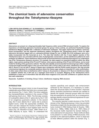
2. FIGURE 1. Primary sequence and secondary structure of the L-21 G414 version of the Tetrahymena group I intron (Cech
et al+, 1994)+ This ribozyme binds the oligonucleotide CCCUCdTAAAAA and transfers the AAAAA onto the 39 end of the
intron in a reaction analogous to the reverse of the second step of splicing+ Numbering of the nucleotides discussed in the
text is shown, as are the names of the helical (P1–P9) and single-stranded regions (J6/6a, J8/7, etc+) of the RNA+ The three
A-platforms in P4-P6 are shown as adjacent A’s with a heavy underline+ The long thin lines indicate regions known to make
tertiary interactions within the three-dimensional structure+ Thick lines designate connectivity of the RNA strand+
Adenosine conservation in the Tetrahymena ribozyme 499
3. Cate et al+, 1996b)+ This motif involves a side by side
alignment of two consecutive A’s to form a pseudo-
base pair that serves as a platform for tertiary stacking
interactions+ In one of the three occurrences, the
A-platform makes tertiary interactions with another ex-
ample of an A-rich motif, the GAAA tetraloop frequently
found at the end of RNA hairpin loops (Woese et al+,
1990)+ The A’s in this and related GNRA tetraloops
FIGURE 2. A: Scheme for the reaction of the L-21 G414 ribozyme with oligonucleotide substrate+ This reaction is analo-
gous to the reverse of the second step of splicing (Beaudry & Joyce, 1992; Mei & Herschlag, 1996)+ The ribozyme binds
the substrate to form the P1 helix, which docks into the active site+ The terminal guanosine (G414) nucleophilically attacks
the substrate and transfers the 39-terminus onto the 39 end of the intron+ The equilibrium constant for the chemical step of
this reversible reaction is approximately 1 (Mei & Herschlag, 1996)+ B: Scheme for the identification of the chemical groups
important for RNA activity by NAIM (Strobel & Shetty, 1997)+ The phosphorothioate-tagged nucleotide analogue (indicated
as daS) is randomly incorporated into the transcript in place of A+ If the analogue does not interfere with function at a
particular position (left side), then ribozymes with the analogue at that site perform the ligation reaction and become
radiolabeled+ If the analogue disrupts activity (right side), then the subset of ribozymes that have the analogue incorporated
at the susceptible site do not perform the ligation reaction and are not radiolabeled+ Cleavage of the phosphorothioate
linkages by treatment with iodine and resolution of the cleavage products by PAGE produces a sequencing ladder with gaps
that correspond to sites intolerant of analogue substitution+ AaS serves as a control to insure that loss of activity is not due
to the phosphorothioate group+ Unreacted RNA is also 59 end-labeled to ensure that the gap in the sequencing ladder is not
due to lack of analogue incorporation at a given site (not shown)+
500 L. Ortoleva-Donnelly et al.
4. appear to be widely utilized in tertiary interactions (Mur-
phy & Cech, 1994; Pley et al+, 1994; Costa & Michel,
1995; Costa et al+, 1997)+ A third example of an A-rich
motif in the P4-P6 structure is the asymmetric A-rich
bulge (AAUAA; positions 183–187; Fig+ 1), which makes
extensive tertiary interactions with the minor groove of
the P4 helix (Cate et al+, 1996a)+ The A-rich bulge also
has a corkscrew turn in the phosphate backbone that
forms the binding site for two divalent metal ions (Cate
et al+, 1997)+
The importance of A nucleotides among this wide
variety of structural motifs explains an observation made
several years ago about the conservation and se-
quence distribution of A’s within ribosomal RNA phy-
logeny+ A simple analysis of the 16S rRNA secondary
structure revealed that there is a bias for unpaired A’s
(Gutell et al+, 1985)+ More than 60% of the A’s are un-
paired, whereas only 30% of the G, C, and U nucleo-
tides were unpaired+ Among the unpaired A’s, there is
also a preference for the A’s to occur in adjacent posi-
tions+ Furthermore, of the universally conserved nucle-
otides, more than 80% of theA’s were unpaired, whereas
only 50% of the U, G, and C’s were unpaired+ At the
time of this analysis (1985), there were no structural or
functional explanations for this strong bias in favor of
unpaired A’s+ We now know that there are a number of
structural motifs that utilize A’s and require this pattern
of sequence conservation+
Several chemical reagents are available that cova-
lently modify functional groups on A and other nucleo-
tides (Stern et al+, 1989)+ For example, dimethylsulfate
(DMS) is used to explore the role of the N1 of A (von
Ahsen & Noller, 1993) and diethylpyrocarbonate (DEPC)
modifies A at the N7 position (Peattie, 1979)+ These
reagents have proven valuable in biochemical struc-
tural and functional studies of RNA (Inoue & Cech,
1985; Moazed et al+, 1986; von Ahsen & Noller, 1993;
Butcher & Burke, 1994; Beattie et al+, 1995)+ However,
these chemical reagents have some severe limitations+
They leave several functional groups unmodified, and
therefore untested+ They also probe the RNA by adding
steric bulk to the nucleotide, which may not correlate
with involvement of the unmodified functional group in
the RNA structure+
Given the importance of A-rich motifs within the struc-
ture of the Tetrahymena intron and the prevalence of A
residues in the unpaired regions of several different
RNA molecules, we focused on developing improved
biochemical methods to analyze these sequences+
Nucleotide Analog Interference Mapping (NAIM) is an
efficient biochemical approach to identify individual func-
tional groups and tertiary hydrogen bonds essential for
RNA activity (Fig+ 2B; Strobel & Shetty, 1997)+ In this
approach, a nucleotide analogue that contains an al-
teration of one functional group is covalently tagged
with a 59-phosphorothioate linkage and randomly in-
corporated into an RNA transcript by T7 RNA polymer-
ase at a level of approximately 5%+ The RNA transcripts
are then separated into active and inactive fractions
based upon their ability to perform a defined function+
For the group I intron experiments described here, this
function is the ability to perform the 39-exon ligation
reaction, which is analogous to the reverse of the sec-
ond step of splicing (Fig+ 2A; Beaudry & Joyce, 1992;
Mei & Herschlag, 1996)+ Cleavage of the phosphoro-
thioate linkages with iodine (Gish & Eckstein, 1988)
and resolution of the cleavage products by PAGE yields
a sequencing ladder with gaps that correspond to sites
where analogue substitution is detrimental to activity+
This interference approach makes it possible to simul-
taneously, yet individually, test the importance of spe-
cific functional groups at every position within an RNA+
Our initial efforts with this chemogenetic approach
tested the role of the N2 amine of G using the
phosphorothioate-tagged derivative of inosine as the
nucleotide analogue (Strobel & Shetty, 1997)+ Other
groups have tested the importance of the 29-OH using
29-deoxy and 29-O-methoxy derivatives of A, C, G, and
U (Gaur & Krupp, 1993; Conrad et al+, 1995; Hardt
et al+, 1996)+ In principle, NAIM can be extended to any
nucleotide analogue that can be incorporated into an
RNA by in vitro transcription+ In this paper, we utilize
eight derivatives of adenosine that each make an in-
cremental change in either a nucleobase or ribosyl
functional group (Fig+ 3)+ We report the synthesis,
incorporation, and sites of interference for each ana-
logue within the Tetrahymena ribozyme+ These inter-
ference data provide several biochemical constraints
for modeling the intron active site+ They also confirm
the biochemical relevance of some of the A-rich motifs
within the P4-P6 domain (Cate et al+, 1996a, 1996b)+
The data indicate that some of the tertiary interactions
proposed within the phylogenetic model of the group I
intron require revision (Michel & Westhof, 1990; Leh-
nert et al+, 1996)+ Given the importance and diversity of
A-rich folding motifs, these A analogues are likely to be
valuable reagents for investigating the relationship of
structure and function within a variety of RNAs+
RESULTS AND DISCUSSION
Eight nucleotide analogues of adenosine were se-
lected for use in NAIM (Fig+ 3)+ In this collection, three
analogues (NMe
AaS, PuraS, and 2APaS; abbreviations
defined in Fig+ 3) were used to examine the role of the
N6 amino group+ These nucleotides make it possible to
explore the requirement for the N6 position by deleting
the amine (2APaS and PuraS) or by replacing one
proton of the amine with a methyl group (NMe
AaS)+ In-
terpretation of NMe
AaS interference is somewhat com-
plicated because the methyl group can adopt either the
s-cis or s-trans rotamer+ In the context of a nucleoside
free in solution, the N6 methyl group prefers the s-cis
rotamer 20:1 (Engel & von Hippel, 1974); however, in
Adenosine conservation in the Tetrahymena ribozyme 501
5. the context of a DNA duplex, the methyl group is s-trans
and occupies the major groove face of the helix (Faza-
kerley et al+, 1985; Lingbeck et al+, 1996)+ Despite the
s-cis rotameric preference, NMe
A substitution destabi-
lizes a duplex by about 1 kcal{molϪ1
(Engel & von
Hippel, 1978; Guo et al+, 1995), which makes it an an-
alogue suitable for probing the helical major groove for
tertiary interactions+
Two of the analogues (DAPaS and 2APaS) were
used to determine the compatibility of an additional N2
amino group on the minor groove face of the nucleo-
tide+ This is a functional group present only on G, and
therefore interference with these two nucleotides indi-
cates sites that should be incompatible with G muta-
tion+ A fifth analogue (7dAaS) was designed to test the
role of the N7 imino group of A in the major groove by
changing the N7 from a hydrogen bond acceptor to
C-H+ We also attempted to prepare 3-deaza-adenosine,
but several attempts to synthesize the triphosphate from
commercially available unprotected nucleoside were
unsuccessful+
Three analogues (OMe
AaS, F
AaS, and dAaS) were
used to differentiate the contribution made by the 29-OH+
dAaS and F
AaS both delete the 29-OH, but replace it
with atoms vastly different in electronegativity+ dAaS
replaces the 29-OH with a proton, whereas F
AaS has a
fluorine at the 29-position+ dAaS can neither donate nor
accept a hydrogen bond, but the 29-fluoro group of
F
AaS can still act as a hydrogen bond acceptor (With-
ers et al+, 1988; Herschlag et al+, 1993a)+ OMe
AaS can
also act as a hydrogen bond acceptor, but only if there
is sufficient space to accommodate the additional ste-
ric bulk of the methyl group+ In addition to the effects on
the hydrogen bonding character of the 29-position, these
analogues have an indirect effect on the sugar pucker
of the ribose ring (Guschlbauer & Jankowski, 1980)+
The more electronegative the substituent, the more the
C39-endo sugar conformation is preferred (Uesugi
et al+, 1979)+ The propensity toward C39-endo sugar
pucker is F
AaS . AaS . dAaS+
Analogue incorporation: Efficiency
and accuracy
Each of the analogues was synthesized as the 59-O-
(1-thio)nucleoside triphosphate derivative using the pro-
cedure described by Arabshahi and Frey (1994)+ The
triphosphate facilitates incorporation of the analogue
into a growing RNA transcript, and the phosphorothio-
FIGURE 3. Eight nucleotide analogues used for NAIM in this study+ Each analogue was synthesized as the 59-O-
(1-thio)nucleoside triphosphate (Eckstein, 1985; Arabshahi & Frey, 1994)+ Each analogue is shown as the monophosphate
derivative, the form in which it is incorporated during transcription+ Functional group(s) modified relative to the parental A
nucleotide (box) are identified by a shadowed box+ Numbering of the adenosine rings is indicated on the parental nucleotide,
and the abbreviation used in the text for each analogue is listed in parentheses+
502 L. Ortoleva-Donnelly et al.
6. ate serves as a chemical tag to identify the sites of
analogue incorporation+
Prior to their use in NAIM, we determined the effi-
ciency and accuracy of analogue incorporation through-
out the RNA transcript+ Each of the triphosphates was
randomly incorporated into the L-21 G414 form of the
group I intron (Table 1; Strobel & Shetty, 1997)+ The
resulting RNAs were 59 end-labeled and the phospho-
rothioate linkages were cut with iodine (Fig+ 4A)+ All the
analogues were incorporated exclusively as an A, how-
ever, the relative incorporation efficiency at each posi-
tion varied somewhat between the analogues+ Efficient
analogue incorporation was observed for the nucleo-
tides DAPaS, 7dAaS, and NMe
AaS+ The intensity of
analogue incorporation at each site throughout the mol-
ecule was approximately equivalent to AaS (Fig+ 4A,
lanes 1–4)+ These three analogues do not alter any of
the functional groups necessary for Watson–Crick base
pairing+ The low concentration of DAPTPaS needed to
achieve 5% incorporation (25 mM DAPTPaS and 1 mM
ATP, Table 1) suggests that the additional N2 amino
group makes a positive contribution by hydrogen bond-
ing to the O2 of T during transcription (Strobel et al+,
1994)+ The fact that NMe
AaS is incorporated efficiently
throughout the RNA transcript provides additional evi-
dence that the N6 methyl group can occupy the major
groove face of the nucleotide+
Somewhat surprisingly, PuraS was also accurately
incorporated into the RNA, although the efficiency of
incorporation was quite uneven at several positions in
the transcript (Fig+ 4A, lane 5)+ PuraS lacks the N6
amino group and can form only one hydrogen bond to
the opposing T during transcription+ As a result of the
poor hydrogen bonding of PuraS with T, high ratios of
PurTPaS to ATP (2:0+5 mM) were required to achieve
efficient incorporation and, even then, PuraS was not
incorporated at all positions within consecutive runs of
A’s+ At four positions within the transcript (A103–A105,
A171–A173, A218–A219, A268–A270), PuraS was in-
corporated efficiently at the first (or 59-most) A in the
series, but it was not incorporated at the subsequent
A’s (Fig+ 4A, lane 5)+ However, this was not the case at
all consecutive A’s because normal incorporation lev-
els were observed at each position within several other
runs of A (e+g+, A64–A66, A87–A90, and A151–A153)+ It
is unclear how sequence context affects incorporation
at these sites+ Independent of the cause, seven posi-
tions within the L-21 G414 sequence (A104,A105,A172,
A173, A219, A269, and A270) were uninformative for
PuraS due to lack of incorporation+
2APaS, like PuraS, omits the N6 amine found on A;
however, 2APaS has an additional N2 amino group
that might hydrogen bond to the O2 of T during tran-
scription+ We found that 2APaS incorporated slightly
better than PuraS+ Weak, but detectable incorporation
was found at all seven of the sites that were uninfor-
mative for PuraS (Fig+ 4A, lane 12)+ Thus, it was pos-
sible to obtain information about the N6 amine at all the
resolvable sites within the molecule+ Interpretation of
the 2APaS interference data is complicated by the si-
multaneous changes at both the N2 and N6 positions
within the nucleotide+ In order to make conclusions about
the importance of the N6 amine, effects from 2APaS
must be compared to both AaS (phosphorothioate ef-
fects) and DAPaS (N2 amine effects) controls+ Exper-
iments with 2APaS were further complicated because
RNAs containing this analogue were unstable and had
a half-life of only a few days in the freezer, which made
it necessary to transcribe the RNA containing 2APaS
prior to each experiment+
Use of the T7 RNA polymerase mutant Y639F
for efficient analogue incorporation
Previous work with 29-deoxy and 29-methoxy deriva-
tives reported reasonably efficient analogue incorpora-
tion into tRNA using the wild-type version of T7 RNA
polymerase with high analogue to NTP ratios, and tran-
scription buffers containing Mn2ϩ
(Conrad et al+, 1995)+
Unfortunately, we were unable to incorporate efficiently
(,1%) dAaS, OMe
AaS, or F
AaS into the L-21 G414
RNA using these transcription conditions (data not
shown)+ Given that the 29-hydroxyl is likely to be used
widely in RNA folding and recognition, it is imperative
that a procedure be developed for the efficient incor-
poration of 29-substituted analogues+ Sousa and Pa-
dilla (1995) have reported that a Y639F point mutant of
the T7 RNA polymerase efficiently incorporates 29-
deoxynucleotides into what would otherwise be an RNA
polymer+ Subsequent work demonstrated that this poly-
merase can incorporate 29-methoxy analogues in the
presence of Mn2ϩ
, and that it can also incorporate 29-
deoxy-29-fluoro-NTPs, and 29-deoxy-29-thio-CTP (Huang
TABLE 1+ Concentration of ATP and analogue used to achieve
approximately 5% analogue incorporation during RNA transcription+a
Analogue
dTPaS
[dTPaS]
(mM)
[ATP]
(mM)
Polymerase
(wt or Y639F)
AaS (SP isomer) 0+05 1+0 wt
7dAaS 1+0 1+0 wt
NMe
AaS 0+4 1+0 wt
DAPaS 0+025 1+0 wt
PuraS 2+0 0+5 wt
2APaS 2+0 0+5 wt
dAaS 1+5 1+0 Y639F
OMe
AaS 2+0 0+1 Y639F
F
AaS 1+0 1+0 Y639F
a
In all cases, 1 mM CTP, UTP, and GTP, 40 mM Tris-HCl, pH 7+5,
4 mM spermidine, 10 mM DTT, 15 mM MgCl2, 0+05% Triton X-100,
and 0+05 mg/mL DNA template were included in the reaction+ The
AaS incorporation used the SP isomer of ATPaS, whereas all other
analogues were transcribed from the diastereomeric mixture of the
SP and RP isomers+ The polymerase used was either the wild-type
version of T7 RNA polymerase or a version of the polymerase con-
taining the Y639F point mutation (Sousa & Padilla, 1995)+
Adenosine conservation in the Tetrahymena ribozyme 503
7. et al+, 1997a, 1997b; Raines & Gottlieb, 1998)+ Given
these interesting properties, we overexpressed and pu-
rified the Y639F polymerase for use in NAIM+
The mutant polymerase was sufficiently accurate that
RNAs transcribed with Y639F were as active as those
transcribed with the wild-type form of the polymerase
(data not shown)+ Furthermore, all three of the 29-
substituted nucleotides (dAaS, OMe
AaS, and F
AaS) were
accurately and efficiently incorporated into the L-21
G414 as defined by 59 end-labeling and analysis of the
transcripts (Fig+ 4A, lanes 11, 13–15)+ No additional
bands were detected in the phosphorothioate sequenc-
ing analysis+ This confirms that, although the Y639F
polymerase lacks fidelity with regard to the identity of
the ribose sugar, it is not excessively prone to intro-
ducing mutations during transcription (Sousa & Padilla,
1995)+
Using this polymerase, dAaS and F
AaS were evenly
incorporated at modest ratios of phosphorothioate to
ATP (Table 1)+ However, even with this mutant poly-
FIGURE 4. Analogue incorporation and interference reactions+ A: The L-21 G41459 end-labeled control showing the extent
and positions of analogue incorporation throughout the intron+ The I2-treated AaS standard is shown in lanes 1 and 11+ The
phosphorothioate-tagged analogue incorporated into the other RNAs is listed above the lane numbers+ Nucleotide numbers
corresponding to several of the bands are marked to the left of each gel+ Addition (lanes 1–5, 11–15) or omission (lanes 6–
10, 16–20) of iodine is indicated+ This particular gel was electrophoresed at 75 watts for 1+25 h+ Longer electrophoretic times
were used to improve the signal resolution of the nucleotides toward the 39 end of the RNA (not shown)+ B: 39-Exon ligation
reaction of L-21 G414 RNA with dT(Ϫ1)S+ This autoradiogram reveals the sites of analogue interference throughout the
intron+ A complete description of the reaction conditions is included in Materials and Methods+ Figure legends are the same
as those in Figure 4A+ This particular gel was electrophoresed at 75 watts for 2+25 h+ It provides maximal resolution of the
J8/7 region of the intron (nt 308–299)+ Sites of strong interference within this region are indicated with an asterisk+ A302 and
A306 were uninformative under these assay conditions due to a strong phosphorothioate effect at both positions+ Longer
electrophoretic times were used to resolve the cleavage products toward the 59 end of the intron (not shown)+ Interference
results for 2APaS are not shown on this autoradiogram, and the no-iodine controls (lanes 9–13) are only shown for a subset
of the nucleotide analogues+ The no-iodine controls for the remaining nucleotides were essentially identical+ C: 39-Exon
ligation reaction of L-21 G414 RNA with rT(Ϫ1)S+ Only the cleavage products from nucleotides surrounding the J8/7 region
of this autoradiogram are shown+ Unlike the reaction conditions in Figure 4B, the use of rT(Ϫ1)S in the presence of Mn2ϩ
made it possible to gain information about nt A302 and A306+ Sites of interference are marked with an asterisk+ 2APaS
interference was not measured at these two sites because DAPaS and PuraS were fully informative+
504 L. Ortoleva-Donnelly et al.
8. merase, high ratios of OMe
ATPaS to ATP were neces-
sary to obtain close to 5% analogue incorporation
(Table 1)+ The efficiency of OMe
AaS incorporation was
uneven throughout the transcript, but there was at
least some incorporation at every A position (Fig+ 4A,
lane 14)+ In addition to the 29-substituted analogues,
the Y639F polymerase incorporates other nucleotides
that contain modifications in the minor groove (L+
Ortoleva-Donnelly & S+A+ Strobel, unpubl+ obs+), which
makes it a valuable reagent for NAIM+
Phosphorothioate interference
Prior to performing NAIM with a complete series of
analogues, it was necessary to identify a reaction con-
dition that was selective, but had a minimum number of
uninformative sites due to strong phosphorothioate in-
terference+ Previous experiments with the Tetrahymena
group I intron have shown that AaS incorporation can
inhibit splicing activity (Deeney et al+, 1987), although
the number and location of the detrimental sites varied
with the experimental conditions (e+g+, temperature, salt
concentrations, incubation time), and the category of
ribozyme reaction being studied (39 splice site hydro-
lysis, 39 splicing by CU addition, or 59-exon cleavage by
G; Waring, 1989; Christian & Yarus, 1992, 1993)+ Some
of the apparent differences between these experi-
mental results can be explained by experiments that
allowed the splicing reaction to proceed too far to com-
pletion, which reduced the experimental signal (War-
ing, 1989), or by experiments that used primer extension
to identify the sites of interference, which introduced
excessive background noise into the data (Christian &
Yarus, 1993)+ Nevertheless, there are some real, and
possibly significant, differences in the phosphorothio-
ate interference patterns observed for the first versus
the second step of splicing, although it is still difficult
to make conclusions about the importance of these
differences+
For ease and efficiency of experimental analysis, we
elected to study the 39-exon ligation reaction (Beaudry
& Joyce, 1992; Mei & Herschlag, 1996)+ This reaction,
wherein the 39-OH of the terminal G (G414) nucleo-
philically attacks an oligonucleotide substrate that mim-
ics the 59–39 ligated exons, is analogous to the reverse
of the second step of splicing+ The reaction transfers
the 39-exon onto the 39 end of the RNA, and presents
three very important advantages over previous efforts
to map sites of phosphorothioate interference in the
Tetrahymena group I intron+ (1) Using a 39-radiolabeled
substrate, the active molecules in the ribozyme popu-
lation become radioactively labeled during the ligation
reaction+ No additional purification of the RNA is nec-
essary prior to iodine treatment and gel electropho-
resis, which makes it possible to directly visualize the
interference pattern without using reverse transcrip-
tase+ (2) Ease of the reaction makes it feasible to com-
pare interference patterns under a variety of reaction
conditions+ (3) Unlike the self-splicing reactions studied
previously, the substrate in the reaction is a synthetic
oligonucleotide that can be altered chemically to adjust
the selectivity of the reaction+ We found the reaction
to be maximally informative using a substrate with a
29-deoxy substitution at the cleavage site [dT(-1)S:
CCCUCdTAAAAA] in a reaction buffer containing
3 mM MgCl2 and 1 mM Mn(OAc)2+ The 29-deoxy sub-
stitution reduces the rate of chemistry by more than
1,000-fold (Herschlag et al+, 1993b), which slows the
reaction sufficiently that more subtle effects on activity
can be detected+ The low metal concentration partially
destabilizes the structure of the ribozyme, but the pres-
ence of a small amount of the thiophilic manganese ion
minimizes the phosphorothioate effects that are present
at several positions throughout the intron (Christian &
Yarus, 1993)+
Under these conditions, there are 10 sites of AaS
interference in the Tetrahymena group I intron for the
39-exon ligation reaction (Fig+ 4B, lane 1)+ Most of these
map within the conserved catalytic core of the RNA+
Moderate interference was observed at 5 of the 10
sites (A57, A97, A207, A210, and A301), strong inter-
ference was detected at 3 sites (A263, A304, and A308),
and complete interference was seen at 2 sites (A302
and A306; Fig+ 4B, lane 1)+ Interference at A302 and
A306 could be partially rescued using an oligonucleo-
tide substrate with a ribose at the cleavage site
[rT(-1)S: CCCUCUAAAAA], but only in the presence of
Mn2ϩ
(Fig+ 4C, lane 1)+ Complete interference was ob-
served at both of these sites using a buffer containing
4 mM MgCl2 even with a ribose substrate (data not
shown)+ The presence of 1 mM Mn2ϩ
also partially or
completely rescued the phosphorothioate effects atA57,
A97, A113, A114, A115, A206, A207, and A210 that
were observed using a 4 mM Mg2ϩ
buffer+ Strong phos-
phorothioate effects that can be rescued by Mn2ϩ
have
been interpreted to be sites of divalent metal ion bind-
ing within the intron (Christian & Yarus, 1993)+ Of par-
ticular importance to this experiment is that every A
within the molecule has an AaS cleavage signal, so all
resolvable sites are informative for NAIM+
Nucleotide analogue interference quantitation
The interference pattern of each of the eight nucleotide
analogues was determined for 39-exon ligation
(Fig+ 4B)+ For technical reasons, only 105 of the 115 A’s
within the L-21 G414 sequence were informative in this
assay+ Nonspecific cleavage was observed consis-
tently at A290 in the control lane lacking iodine
(Fig+ 4B)+ This nucleotide is in the P8 hairpin loop and
is not essential to ribozyme activity (Doudna et al+, 1991;
Nakamura et al+, 1995)+ In addition, positions close to
the 59 and 39 ends of L-21 G414 could not be analyzed
because the cleavage products were not sufficiently
Adenosine conservation in the Tetrahymena ribozyme 505
9. resolved from the full-length intron+ This included seven
nucleotides at the 59 end and two nucleotides at the 39
end of the intron+ Because of phosphorothioate effects,
A302 and A306 were only informative using the rT(Ϫ1)S
substrate in the presence of Mn2ϩ
, so interference at
these two sites was measured separately from the rest
of the intron (Fig+ 4C)+
Based upon the band intensities in the 39-exon liga-
tion and 59 end-labeled control experiments, an inter-
ference k value was calculated for each A position in
the intron (see Materials and Methods)+ A k value of 1
indicates that there is no effect of substituting the an-
alogue at that site, a value greater than 1 indicates
inhibition of activity, and a value less than 1 indicates
that activity is enhanced by analogue substitution at
that site+ As might be expected, most positions did not
show any effect upon analogue substitution+ Greater
than 90% of the interference k values were between
0+67 and 1+5+ This data range provides a conservative
estimate of the experimental noise in the system+ In the
data range from 1+5 to 2+0 (or, for the case of enhance-
ment, from 0+67 to 0+5), there were subtle but repro-
ducible effects+ We have chosen to be conservative in
our interpretation of the data and will consider only
interference values greater than 2 (or less than 0+5) to
be significant+ Three-dimensional histograms plotting
the magnitude of the interference k values for each
analogue at each position are shown in Figure 5+ Every
one of the analogues has a unique interference pattern
throughout the intron with regard to both the distribu-
tion and the intensity of interference+
Sites of interference are largely coincident with
sites of phylogenetic conservation
The interference pattern for this series of eight A ana-
logues provides valuable biochemical information about
the structure and function of the group I catalytic RNA+
A composite view of the eight interference patterns
reveals that the sites of interference map primarily onto
two regions at the core of the ribozyme, P7-P3-P8 and
helices P4 and P6 (Fig+ 6A)+ Strong interference was
also detected at the junction between the P2 and P2+1
helices, which is known to be essential for tethering the
P1 helix into the active site of the Tetrahymena intron
(Downs & Cech, 1990, 1994)+ The sites of analogue
interference correlate almost exactly with the most highly
conserved A nucleotide positions among the group IC1
and IC2 introns (Fig+ 6B; Michel & Westhof, 1990;
Damberger & Gutell, 1994)+ Of the 21 conserved A
nucleotides in the core region of the intron, 19 show
interference with at least one of the analogues+ This
includes several positions within the P4-P6 and P7-
P3-P8 helices that were demonstrated by mutagenesis
to be essential for catalytic function (Couture et al+,
1990; Pyle et al+, 1992)+ In contrast, there is no inter-
ference within the P8 helix and the P9 extension+ Al-
though both of these structural elements are important
for intron stability, there is very little primary sequence
conservation within these regions of the intron (Fig+ 6B)+
Because high sequence conservation implies that the
nucleotide is structurally or functionally important, the
coincidence between sites of interference and phylo-
genetic conservation provides strong validation for NAIM
as a method for the biochemical characterization of
RNA+
Conserved positions that do not
show interference
There were a few exceptions to the correlation be-
tween phylogeny and interference+ Most of the diver-
gence occurred within the P5abc subdomain, where
the only site of even modest interference was from
OMe
AaS substitution at A183 (Fig+ 5)+ Interference at
this site is in agreement with a tertiary hydrogen bond
observed between the 29-OH of A183 and the 29-OH of
G110 in the P4-P6 crystal structure (Cate et al+, 1996a)+
It is noteworthy that P5abc is not conserved among all
the subclasses of group I introns (the data plotted in
Fig+ 6B is only for the IC1 and IC2 introns; Michel &
Westhof, 1990; Damberger & Gutell, 1994), and muta-
FIGURE 5. Individual histograms plotting the magnitude of the interference k value versus nucleotide position super-
imposed on the intron secondary structure for AaS and each of the eight analogues tested+ Interference k values Ն2+0 are
shown as gray bars+ Values greater than 6 are assigned a magnitude of 6 within this graph+ A white bar (of which there is
only one example at A207 for F
AaS) indicates that k was Յ0+5+ In this single case, the magnitude of the bar corresponds
to 1/k, and indicates that there is enhancement of activity due to analogue substitution at that site+ White boxes indicate
positions that were uninformative in the assay because they are a nucleotide other than A+ Gray boxes indicate A sites that
were not informative in the assay due to incomplete resolution of the cleavage products on the sequencing gel (all
analogues were uninformative at A24, A28, A29, A30, A31, A35, A38, A407, and A410), lack of analogue incorporation at that
site (PuraS was uninformative at A104, A105, A172, A173, A219, A269, and A270), degradation in the no iodine control (all
analogues were uninformative at A290), or not measuring interference at that site (2APaS was not tested at A302 and
A306)+ The nucleotide number within the Tetrahymena sequence for each site of interference is shown adjacent to the bar+
The error in the k value at each position is Յ20%+ The value is the average of at least two and as many as eight independent
experimental measurements+ A302 and A306 were assayed under conditions different from the rest of the intron due to
complete phosphorothioate inhibition at these two sites under the standard reaction conditions+
506 L. Ortoleva-Donnelly et al.
10. FIGURE 5. (Legend on facing page.)
AdenosineconservationintheTetrahymenaribozyme507
11. tions that disrupt P4-P6 domain folding have no appar-
ent effect upon folding of the intact intron (Laggerbauer
et al+, 1994)+ In fact, several of these mutations actually
improved the intron folding rates (D+K+ Treiber & J+R+
Williamson, unpubl+ results)+ Furthermore, the J6a/6b
region (A225, A226, and A248), which participates in
an extensive tertiary interaction with the GAAA tetra-
loop of P5b (Cate et al+, 1996a, 1996b), was unaffected
by analogue substitution+ Apparently, the P4-P6 do-
main is sufficiently stable in the context of the complete
intron that single functional group modifications are not
enough to disturb activity+ By contrast, we have ob-
served strong interference at several of these posi-
tions using a gel shift assay for folding of the P4-P6
domain (S+ Basu & S+A+ Strobel, unpubl+ results; Mur-
phy & Cech, 1993)+
Two conserved positions within the intron core re-
gion (A214 and A268) did not show interference with at
least one of the eight analogues included in this study
(Fig+ 6B)+ A214 and A268 are both at the ends of each
of their helices (P4 and P7, respectively) and both are
base paired to a conserved U (U107 and U307, re-
spectively)+ It is possible that this collection of ana-
logues did not modify the chemical group important for
function at these two potentially homologous positions
(for example, the N1 and N3 groups were not altered in
this set of analogues)+
Interference at nonconserved sites
Three positions within the core of the intron are not
conserved phylogenetically, but do demonstrate inter-
ference (marked with an asterisks in Fig+ 6B)+ The pri-
mary divergence from phylogeny is at A256, which
showed interference from a wide variety of analogues+
This position will be discussed below+ The other two
examples are A94 and A210+ Interference at these two
sites was primarily from analogues that add steric bulk
to the nucleotide (OMe
AaS forA94 and NMe
AaS forA210)+
Interference from bulky analogues such as these is not
necessarily expected to correlate with the mutability of
a given site+ For example, A210 is a bulged nucleotide
within the P4 helix, in a segment of the active site
known to be densely packed (Michel & Westhof, 1990;
Tanner & Cech, 1997)+ A210 is found only among the
six sequenced species of Tetrahymena’s LSU rRNA
and in the LSU rRNA intron of Physarum polycephalum
(Damberger & Gutell, 1994)+ Given the close packing in
this region, it is reasonable to expect that a bulky group
on the amine would not be tolerated+
A biochemical signature for essential
C29-endo sugar pucker
A210 provides another example of how this collection
of analogues can be used to probe the structure of
FIGURE 6. A: Composite histogram of the interference pattern throughout the Tetrahymena group I intron using the data
presented in Figure 5+ At each nucleotide position, the maximum interference observed from any of the nine analogues is
shown as a bar whose height corresponds to the magnitude of the interference k value+ The symbols in the figure are the
same as in Figure 5+ This graph demonstrates that nearly all the interference sites cluster into the P4-P6 helices or the
P7-P3-P8 subdomain+ B: Composite histogram showing the relationship between the sites of nucleotide analogue inter-
ference and the sites of phylogenetic sequence conservation+ The height of the bar at each position is proportional to the
conservation value calculated from the modified Shannon equation (Damberger & Gutell, 1994; R+R+ Gutell, unpubl+ results)
using a sequence alignment of 131 group IC1 and IC2 introns+ In this calculation, a value of 2+0 indicates a site that is
completely invariant, a value of 1+5 is approximately 90% conserved, a value near 1+0 is about 60–80% conserved, and a
value approaching 0+5 is about 40–50% conserved+ Negative values indicate a complete lack of conservation at a particular
position+ Only conservation values between 2+0 and 0+5 for A nucleotides within the sequence are plotted to show the
positions that are most conserved among this class of introns+ Values less than 0+5 are shown as black boxes+ White boxes
are nucleotides in the sequence other than A’s+ The color of the bars indicate the extent of interference at a given position+
A black bar indicates that at least one of the nine analogues had an interference k value greater than 3+5, a gray bar
indicates an interference k value between 1+8 and 3+5+ A white bar indicates that none of the analogues had an interference
k value above 1+8+ Asterisks show the three sites that are not conserved but still demonstrated interference+ This graph
demonstrates that there is a strong correlation between the sites of interference and the sites of sequence conservation+
The most notable exception is within the P5abc domain, where there is moderate conservation, but where no interference
was detected+
508 L. Ortoleva-Donnelly et al.
12. RNA+ In addition to the NMe
AaS effect,A210 also showed
interference with F
AaS, but there was no interference
from dAaS or OMe
AaS+ The lack of interference with
dAaS strongly suggests that the 29-OH does not make
a direct contribution to activity, but interference with
F
AaS argues in favor of an indirect contribution by the
29-OH+ A210 is one of only a few examples within the
P4-P6 crystal structure where the ribose adopts a C29-
endo sugar pucker (Cate et al+, 1996a)+ The unusual
conformation of the A210 sugar allows the base to be
flipped out of the helix without disrupting the helical
continuity of P4+ F
AaS substitution may disrupt the re-
quired C29-endo sugar pucker because the highly elec-
tronegative 29-fluoro group strongly favors the C39-endo
conformation (Uesugi et al+, 1979)+ In contrast, the 29-
deoxy ribose could more easily adopt either the C29-
endo or C39-endo conformation+
Interference with F
AaS coupled with tolerance for
dAaS substitution might be predictive of C29-endo sugar
conformations within RNA+ This pattern was seen at
three other A positions within the Tetrahymena intron,
A218, A256, and A304+ One of these, A218, is also
present within the P4-P6 crystal structure and it is also
in a C29-endo sugar pucker (Cate et al+, 1996a)+ It is
possible that A256 and A304 also adopt this alternative
conformation+ Each of these three positions will be dis-
cussed in detail below+ Clearly, the 29-fluoro and 29-
deoxy phosphorothioate derivatives of the complete
series of nucleotides (i+e+, A, G, C, and U) are poten-
tially valuable reagents to identify positions where al-
terations in sugar pucker affect RNA folding+
Interference at the junction of helices
P2 and P2.1
The interference pattern explains the chemical basis of
A conservation throughout the intron active site+ We
will discuss each of the regions in the molecule that
show interference and outline structural predictions for
the active site based upon the interference pattern+
The junction of helices P2 and P2+1 is comprised
entirely of A’s, including nucleotides A28–A31, A57, A94,
and A95+ A57 and A95 are efficiently photo-crosslinked
upon exposure of the intron to ultraviolet light, and the
interaction between these two nucleotides is important
for properly tethering the P1 helix into the active site
(Downs & Cech, 1990, 1994)+ Point mutations at either
position promote miscleavage at two sites other than
the normal 59 splice site (Downs & Cech, 1994)+ A57 is
highly conserved (98%) among the IC1 introns that
have the P2+1 helix, but it is missing in all other classes
of group I introns+ A95 is also well conserved among
the IC1-2 introns (94%), but less well conserved in the
other subgroups+
Within P2 and P2+1, interference was only detected
at the interface between the two helices, specifically at
A57, A94, and A95+ A57 only showed interference with
NMe
AaS and A94 only with OMe
AaS+ A95 showed inter-
ference with all the analogues that modify the N7 or the
N6 positions (7dAaS, NMe
AaS, PuraS, and 2APaS), but
it was tolerant of functional group modification on the
minor groove face, including addition of an N2 exo-
cyclic amine and modification of the 29-OH+ An inter-
ference pattern involving this set of analogues strongly
suggests interaction with the major groove, or Hoog-
steen face of the nucleotide+ A similar pattern is seen at
several other positions within the ribozyme+
The revised model of the Tetrahymena intron group I
intron has the ribose sugar of A95 positioned against
the minor groove of the U59{G92 wobble pair in the
P2+1 helix (Lehnert et al+, 1996)+ Lack of interference at
A95 from analogues that modify the ribose sugar argues
against such an orientation+ In the revised Michel and
Westhof model, the O29 of A94 is proposed to interact
with the N7 of A95, which agrees with the interference
data, but there are no tertiary contacts to the N6 amino
groups of either A57 or A95, as the data also predict+
Major and minor groove recognition
of the P3 helix
The Tetrahymena thermophila P3 helix contains a sin-
gle A nucleotide, A97, that is almost 90% conserved in
IC1 and IC2 introns+ Although only this single site was
informative within the helix, the interference data pro-
vide valuable information about P3 helix packing within
the active site+ As was observed at A95, the interfer-
ence pattern at A97 is characteristic of hydrogen bond-
ing to the Hoogsteen face of the base (interference
with 7dAaS, NMe
AaS, PuraS, and 2APaS)+ This implies
that the major groove face of the P3 helix is involved in
tertiary structure formation+ However, unlike the pat-
tern at A95, analogues that modify the minor groove
functional groups also caused interference at A97+ In-
terference with dAaS and OMe
AaS suggest that there is
close approach in the P3 minor groove involving the
29-OH+ F
AaS substitution at A97 did not effect activity,
which is an interference pattern exactly opposite to
that seen at A210+ In this case, deleting the 29-OH is
detrimental, but replacing it with the highly electroneg-
ative fluoro group had no effect, suggesting that the
29-OH of A97 acts as a hydrogen bond acceptor, al-
though its hydrogen bonding partner is unknown+ A
similar pattern was observed at A207, where the 29-OH
accepts a hydrogen bond from the N2 amine of G22
(Strobel et al+, 1998)+
The interference pattern at A97 suggests that two
different structural elements converge at the P3 helix+
The likely candidates are the J8/7 single-stranded re-
gion and the P2–P2+1 helical junction+ The major groove
interaction is consistent with the structure model pro-
posed originally by Michel and Westhof (1990), where
the Hoogsteen face of the A97–U277 pair makes a
base triple with U300 (Fig+ 7A)+ Further evidence in
Adenosine conservation in the Tetrahymena ribozyme 509
13. support of this tertiary interaction comes from a larger
group I intron sequence alignment (Damberger & Gu-
tell, 1994; R+R+ Gutell, unpublished results)+ Approxi-
mately 126 (90%) of the known IC1 and IC2 sequences
are U{A-U at positions 300{97–277+ In each of the six
sequences where U300 is changed to a C, the 97–277
base pair changes to a G-C pair+ Given that the CϩG-C
triple (where the N3 of C is protonated) is isosteric with
U{A-U (Fig+ 7A,B), this is the expected covariation for a
conserved interaction between these three nucleo-
tides+ Furthermore, there are four introns where U300
is conserved, but 97–277 is changed to a C-G pair+
Although not completely isosteric, a U{C-G triple could
retain one of the two hydrogen bonds present in the
U{A-U triple (Fig+ 7C)+ While additional experiments
will be necessary to determine if these potential ter-
tiary hydrogen bonds form in the folded structure, the
interference and comparative sequence data are con-
sistent with an interaction between J8/7 and the major
groove of P3 at U300+ The interaction in the minor
groove of P3 still needs to be explored+
A G{U wobble receptor in J4/5
The interference pattern within the J4/5 region (nt A113,
A114, A206, and A207) was reported previously for this
set of analogues (Strobel et al+, 1998)+ The data led us
to conclude that the consecutive sheared A{A pairs
within J4/5 act as a receptor for the universally con-
served G{U wobble pair at the cleavage site of the
intron (Fig+ 8)+ The exocyclic amine of G forms two
hydrogen bonds with the minor groove face of A207,
and the 29-OH of G forms two hydrogen bonds with the
minor groove face of A114+ In addition, there is a fifth
hydrogen bond predicted by modeling of this region
that was not identified in the original report of the helix-
packing motif (Fig+ 8)+ The 29-OH of G23 is within con-
venient hydrogen bonding distance of the 29-OH of C208+
Previous thermodynamic analysis of a single 29-deoxy
substitution at G23 demonstrated that the 29-OH con-
tributes about 0+8 kcal{molϪ1
to tertiary binding (Strobel
& Cech, 1993; Narlikar et al+, 1997), which is consistent
with a single hydrogen bond to G23+ Substitution of 29-
O-methylguanosine at G23 had no effect on P1 helix
docking (Strobel & Cech, 1993), which suggests that
the 29-OH of G23 acts as a hydrogen bond acceptor+
Evidence in support of an A-platform in J6/6a
Another critical region with an interesting interference
pattern is J6/6a, a symmetric three-nucleotide internal
loop that includes nucleotides A218, A219, and A256+
A218 and A219 are nearly invariant (.95%) among the
IA and IC introns that have this segment+ In the P4-P6
crystal structure, A218 and A219 are aligned side by
side in a pseudo-base pairing arrangement termed an
A-platform (Fig+ 9; Cate et al+, 1996b)+ They provide an
interface for intermolecular association between J6/6a
and L5c of two different P4-P6 molecules within the
crystal lattice+ The A218–A219 platform is not involved
in the intramolecular folding of the P4-P6 domain, al-
though it might play a role in folding the intact intron
(Cate et al+, 1996b)+ For the A’s to adopt this side-by-
side alignment, the ribose sugar of A218 adopts a C29-
endo conformation+ The only possible hydrogen bonding
interaction between the nucleobases is between the
N3 of A218 and the N6 of A219 (Fig+ 9), so the stability
of the pair appears to be derived primarily from stack-
ing interactions (Cate et al+, 1996b)+
FIGURE 7. One proposed base triple between U300 and the A97–
U277 base pair+ A: Wild-type U{A-U triple expected to form in almost
90% of all group IC1 and IC2 introns+ The number of the nucleotide
is shown within the ring, the functional groups of A97 that show
interference are indicated with a shadowed box, and the occurrence
of these nucleotides among 141 examples is shown+ B: CϩG-C triple
expected to form in the introns where U300 is mutated to a C+ C: One
possible U{C-G triple predicted for the four introns that have a C97–
G277 base pair+ All three of the triples conserve the hydrogen bond
to the 4 position of the pyrimidine at position 300+
510 L. Ortoleva-Donnelly et al.
14. NAIM analysis confirms that the A nucleotides within
J6/6a are important for activity+ The data are consistent
with an A{A pseudo-base pair, although the data are
not sufficiently transparent that a pseudo-pair could have
been predicted a priori+ Interference was observed at
A218 with both analogues (DAPaS and 2APaS) that
introduce an additional N2 amine+ Interpreted in light of
the pseudo-pair, an N2 amine at A218 would clash with
the N6 amine of A219+ The lack of interference at A219
upon deletion of the N6 amine (PuraS) indicates that
the single potential hydrogen bond between the bases
is dispensable for activity under these conditions+
The most informative data in this region came from
the 29-OH analogues+ Interference was observed with
F
AaS at A218, but not with dAaS+ This is similar to the
pattern seen at A210 and argues that A218 adopts a
C29-endo sugar pucker+ This conclusion is in full agree-
ment with the crystal structure where A218 is in a C29-
endo conformation that allows the bases to form the
pseudo-pair+ Strong interference also occurred with
OMe
AaS at A218, which suggests that there is close
approach to the 29-OH group+ Within the P4-P6 struc-
ture, there is electron density in the major groove im-
mediately below the A{A pair, which may correspond to
a metal binding site (S+ Basu, R+ Rambo, J+H+ Cate,
S+A+ Strobel, & J+A+ Doudna, unpubl+ results)+ The 29-OH
of A218 points directly toward this electron density+
OMe
AaS substitution may prevent metal binding due to
steric occlusion, which provides indirect evidence that
this metal is essential for intron activity+
Intermediate levels of interference were observed
with several other analogues that are not necessarily
expected to show interference based simply upon an
A{A pseudo-pair+ The data indicate that the N6 of A218
is important (PuraS, 2APaS, and NMe
AaS interference)
and that an N2 amino group or methylation of the N6 at
A219 is not tolerated+ Although these might simply be
stacking effects, the data suggest that the tertiary struc-
ture near J6/6a may involve close approach to the
Watson–Crick faces of both nucleotides+
A256 in the J6a/6 loop shows a complicated inter-
ference pattern+ The identity of this base is not con-
served, but there is strong phylogenetic evidence to
suggest that it base pairs with the opposing nucleotide
FIGURE 8. G{U wobble receptor+ Inter-
actions between the highly conserved
G22{U-1 pair in P1 and the consecutively
stacked sheared A{A pairs in J4/5 were de-
scribed previously (Strobel et al+, 1998)+ The
model suggests that there may also be an
interaction between the 29-OH of G23 and
the 29-OH of C208 (hydrogen bond at the
bottom of the figure)+ This interaction was
present, but not reported in the previous dis-
cussion of the model+ This fifth hydrogen bond
is supported by thermodynamic evidence
using single functional group substitutions of
the G2329-OH (Strobel & Cech, 1993; Nar-
likar et al+, 1997)+ The structure of the J4/5
region is from the P4-P6 crystal structure
(Cate et al+, 1996a)+
FIGURE 9. A218{A219 pseudo-base pair observed within the J6/6a
A-platform of the P4-P6 structure (Cate et al+, 1996a)+ The J6a/6
segment is also shown+ The ribose sugar of A218 is in a C29-endo
conformation, and its 29-OH appears to contact electron density at-
tributable to a metal (not shown) located below the A{A pseudo pair+
The A’s are approximately coplanar with G254+ Nucleotides G254–
A256 on the opposite strand of the internal loop are likely to be
distorted away from the pseudo-pair because an unnatural G in the
transcript (not shown) occupies the position expected for A256+ If
there is a tertiary interaction within this region, it is most likely formed
with G254 and C255+
Adenosine conservation in the Tetrahymena ribozyme 511
15. in the loop, position 217+ In the IC3 introns, this is a C-G
pair in 99% of the sequences and, among the ID in-
trons, it is usually (94%) a G{U pair+ The Tetrahymena
intron is an exception, with a C217{A256 juxtaposition+
Thus, there is significant evolutionary pressure for base
pairing at A256, but the identity of the base pair is not
a universal element of group I introns+ Nevertheless,
A256 showed interference from a wide diversity of an-
alogues+ It was affected by modifications on the Hoog-
steen face (7dAaS and PuraS), and by modifications in
the minor groove (DAPaS and OMe
AaS)+ Interference
with F
AaS, but not with dAaS, suggests that this base
may also adopt a C29-endo sugar pucker, although the
electron density at A256 was too distorted to confirm
this possibility+
In all three examples of A-platforms within the P4-P6
structure, the A{A pseudo-pair mediated tertiary struc-
ture formation+ Because the J6/6a A-platform in the
crystal structure was involved in an intermolecular con-
tact that does not occur within the full-length ribozyme,
it is unclear if a tertiary interaction is made between
J6/6a and the rest of the intron+ In the crystal structure,
the A218–A219 pseudo-pair is stacked upon P6 and
P6a (Cate et al+, 1996a), and it serves as an intra-
molecular continuation of the P4-P6 helix+ In so doing,
the A-platform frees the opposing J6a/6 strand of the
internal loop (nt G254 and C255) to make a two-base
pair intermolecular pseudoknot with L5c+ If the P4-P6
structure is an accurate predictor of the intronic struc-
ture, the most likely tertiary contacts in this region are
made by G254 and C255+ The biological relevance of
the P4-P6 structure for J6a/6 is questionable because
A256 was displaced from C217 by the unnatural 59-
terminal G that was introduced for transcriptional initi-
ation (Cate et al+, 1996b)+ This G also displaced C255
and G254 from their presumed location within the in-
tron structure (Fig+ 9)+ Base pairing of A256 with C217
and close packing of C255 and G254 against the
pseudo-pair would explain the interference data ob-
served on the Watson–Crick faces of A218 and A219+
If G254 and C255 do not make an intermolecular
contact to L5c within the intron structure, do they make
an intramolecular contact somewhere else within the
intron? Although A218 and A219 are not depicted as an
A-platform within the Michel and Westhof model, it is
interesting that J6a/6 is quite close to the P3 helix+
Further experiments are necessary to determine if G254
and/or C255 make transdomain tertiary contacts to the
bases or ribose backbone of this helical element+
Interference within the G cofactor binding site
The G binding site is located within the P7 helix and is
centered around the G264–C311 pair (Michel et al+,
1989)+ Two A’s (A263 and A265) within the P7 helix
flank this essential base pair and both have been im-
plicated in G binding (Yarus et al+, 1991a)+ The G bind-
ing site was identified originally by Michel et al+ (1989),
who demonstrated a change in substrate specificity from
G to 2AP when the G-C pair was mutated to A-U+ This
was evidence for a direct hydrogen bond between the
H1 of G and the O6 of G264+ They also proposed a
second hydrogen bond between the N2 amine of G
and the N7 of G264, which defines an equatorial align-
ment of G with the G264–C311 pair (Fig+ 10A)+ DMS
interference experiments on the sunY intron showed
that methylation at the N7 of G264 (G96 in the sunY
numbering system) blocked G-mediated splicing and
that G binding protected the N7 of G264 from DMS
methylation (von Ahsen & Noller, 1993)+ This confirms
that G is close to G264, but does not demand that the
N7 of G264 hydrogen bond to the N2 of the G cofactor+
Subsequent work by Yarus et al+ (1991b) showed that
the flanking base pair A265–U310 also contributed di-
rectly to G binding+ They showed a change in substrate
specificity to DAP when A265–U310 was mutated to
G-C+ This suggests that there is an additional hydrogen
bond between the O6 of G and the N6 of A265+ The
restraint of a second hydrogen bond with the base pair
below G264–C311 argued that the G is out of plane
from the G-C pair and in an axial position (Yarus et al+,
1991b; Fig+ 10B)+
Yarus et al+ (1991a) used these constraints to gen-
erate an energy-minimized model of the G binding site
that they termed axial III+ This structure is a hybrid
between the axial and equatorial models of the G bind-
ing site, although it is largely equatorial+ The model
includes a base triple between A263 and the minor
groove face of the G264–C311 base pair+ Upon inspec-
tion of their model, the geometry and distance of the
A265 amine and the G O6 do not seem to be consis-
tent with a hydrogen bond between these groups+ Fur-
thermore, the helix appears to be distorted from an
A-form geometry and the purine and pyrimidine bases
throughout the structure do not appear to be planar+
The interference and mutational data suggest that ax-
ial III is unlikely to accurately represent the G binding
site+
The NAIM data support a central tenant of the orig-
inal axial model (Fig+ 10B), which is that the amine of
A265 hydrogen bonds to the O6 of G (Yarus et al+,
1991a)+ NMe
AaS substitution at A265 significantly im-
pairs ribozyme function, however, both analogues that
delete the N6 amine (2APaS and PuraS) did not inter-
fere with activity+ Although it is possible that NMe
AaS
substitution prevented P7 helix formation, our pre-
ferred interpretation is that G414 could not occupy its
binding site when the Hoogsteen face of A265 was
sterically blocked by a methyl group+ Nevertheless, the
effective molar concentration of G414 was sufficient to
overcome the loss of a single hydrogen bond due to
deletion of the A265 amine+
A263 is also adjacent to the G binding site, it is semi-
conserved as either an A or a C, and it is always un-
512 L. Ortoleva-Donnelly et al.
16. paired+ A263 covaries with the C262–G312 base pair in
a way that avoids base pairing of A263 with G312
(Michel et al+, 1989; Gautheret et al+, 1995)+ Mutations
at A263 had no effect on intron splicing (Yarus et al+,
1991a), and no interference was detected with any of
the analogues in this study+ Lack of interference and
lack of an effect upon mutation argues against the
G262–C312{A263 base triple proposed in the axial III
model (Yarus et al+, 1991a)+ Instead, it suggests that
there is simply a requirement for a bulged nucleotide,
and perhaps an unusual position of the phosphate–
ribose backbone is important for G binding+ Consistent
with this possibility, there was a phosphorothioate ef-
fect at A263 that could not be rescued with Mn2ϩ
+ Our
data suggest that if A263 does make a direct contribu-
tion to G binding, it does so primarily via its phosphate+
If G binds in an axial orientation, then the phosphate of
A263 is closest to the 29-OH of G, whereas if G binds
in an equatorial alignment, then the phosphate is clos-
est to the N2 amine (Fig+ 10)+ Both the N2 and 29-OH of
G are known to contribute significantly to G binding
(Bass & Cech, 1984; McConnell & Cech, 1995; Li &
Turner, 1997; Profenno et al+, 1997)+
Another unpaired nucleotide that may participate in
G binding is A261, which is one of the most highly
conserved nucleotides among all classes of group I
introns, with only a few exceptions among nearly 500
sequences+ This is the level of conservation only seen
among the most essential nucleotides in the intron,
which would be consistent with a role in G binding+ The
interference pattern at this site suggests that several
functional groups of A261 are essential for activity, in-
cluding the 29-OH of the sugar (interference with dAaS,
OMe
AaS, and F
AaS) and the Hoogsteen face of the
base (interference with 7dAaS, NMe
AaS, 2APaS)+
A major issue that is not at all resolved in an axial
model for G binding (Fig+ 10B) is what interacts with the
exocyclic amine of G, and why interference is ob-
served at the N7 of G264 with DMS (Bass & Cech,
1984; von Ahsen & Noller, 1993)+ The N2 amino group
of G contributes 3+0 kcal{molϪ1
to binding (McConnell
& Cech, 1995)+ Furthermore, N 2
-methylguanosine binds
poorly, which suggests that both amine protons are
involved in cofactor recognition (Bass & Cech, 1984)+
Two potential hydrogen bonding partners are the N7 of
G264 and the RP phosphate oxygen of A263+ Both of
these hydrogen bonds require an equatorial alignment
of G, which is incompatible with the A265 interaction+
Fewer obvious options exist for the amine in the axial
orientation, although A261 may participate+
Thus, although the axial III hybrid model of Yarus
et al+ (1991a) seems unlikely, there are data to support
both the axial and the equatorial models for G binding+
Profenno et al+ (1997) have recently shown that G binds
to the intron in at least two steps+ This includes a bi-
molecular step followed by at least one conformational
change+ An explanation that could reconcile the inter-
ference and mutational data is if the G binding pathway
FIGURE 10. An equatorial (A) and an axial (B) model for G cofactor binding to the G binding site (Michel et al+, 1989; Yarus
et al+, 1991b)+ In both models, the G is in an anti configuration (Lin et al+, 1994), and there is a hydrogen bond between the
H1 of G and the O6 of G264+ The equatorial model has a second hydrogen bond involving the N2 of G and the N7 of G264,
whereas the axial model has a second hydrogen bond involving the O6 of G and the N6 of A265+ Biochemical evidence
exists to support the central tenets of both models, including interference data reported in this paper, which support the axial
model+ Although the hybrid model termed axial III by Yarus et al+ (1991a; not shown) is unlikely to represent the structure
of the bound G cofactor, it remains uncertain as to how these two models can be reconciled, or if a version of both of them
might not occur at some point along the pathway toward G binding (Profenno et al+, 1997)+
Adenosine conservation in the Tetrahymena ribozyme 513
17. includes one step with an axial and another step with
an equatorial alignment of the base+ The change in the
alignment of G might be necessary to bring the 39-OH
nucleophile into position for in-line attack at the scissile
phosphate (McSwiggen & Cech, 1989; Rajagopal
et al+, 1989)+ In this situation, interference might not
result from a single static structure, but rather from
disruption of one structure along a dynamic pathway of
structural conformations+
Additional major groove contacts in P7
There is one additional A in the P7 helix (A308) that
was informative in our assay+ A308 is base paired with
U267+ The U267–A308 base pair is 95% conserved
among all of the group I intron sequences+ A308 is
three base pairs removed from the G binding site, so it
is unlikely to participate directly in G binding, although
it could make an indirect contribution+ A308 had an
interference pattern indicative of Hoogsteen pairing in
the major groove of P7 (interference with 7dAaS,
NMe
AaS, 2APaS, and PuraS), and there was no intef-
erence from any of the minor groove modified ana-
logues+ This suggests that an essential tertiary contact
is made in the major groove of P7 immediately below
the G binding site+ Nothing is included in this region of
the molecule within the Michel and Westhof model
(Michel & Westhof, 1990; Lehnert et al+, 1996)+ Possi-
ble candidates for this major groove interaction include
the 39 end of J8/7 or the J3/4 linker segment+ The
nucleotides in both of these regions are very highly
conserved+ It might also be an essential metal binding
site+ Whatever makes contacts with the P7 helix at
A308, it is sufficiently close to G264 that it may partici-
pate in G binding+
Interference within the J8/7 region
A final region with a particularly striking interference
pattern is J8/7, a single-stranded segment between
helices P8 and P7+ Previous studies have implicated
J8/7 in binding of the P1 substrate helix (Pyle et al+,
1992; Strobel & Cech, 1993; Michel & Westhof, 1994),
interaction with the P4 helix (Tanner et al+, 1997; Tan-
ner & Cech, 1997), proximity to the G binding site (Wang
& Cech, 1992), and formation of the catalytic active site
(Michel & Westhof, 1990; Christian & Yarus, 1993)+ It is
an essential and highly conserved segment of the
group I intron+ Four A’s exist in this seven-nucleotide
segment, A301, A302, A304, and A306+ Each displays
at least some level of interference from this set of an-
alogues, but each position is susceptible to analogue
substitution in a different way+
Nucleotides A301 and A302 have been proposed to
orient the substrate helix into the ribozyme active site
by making direct tertiary contact with P1 (Pyle et al+,
1992; Strobel & Cech, 1993; Michel & Westhof, 1994)+
Both nucleotides are highly conserved (.99% among
IC1-2 introns)+ A hydrogen bond between the N1 of
A302 and the 29-OH of U-3 within the P1 helix was
demonstrated through DMS footprinting and mutagen-
esis (Pyle et al+, 1992)+ Michel and Westhof (1994)
proposed a similar interaction between A301 and G25+
NAIM analysis at A301 and A302 suggests that both
nucleotides are involved in a collection of important
interactions within the active site+ Both sites show com-
plete interference with DAPaS, which suggests that
there is close approach to the minor groove edge of
each nucleotide+ The inability to accommodate the ex-
tra N2 amine on DAPaS explains why these positions
are almost never mutated to G (only one exception at
each position among 131 IC1-2 sequences)+ Both sites
show interference with dAaS and OMe
AaS, which implies
that both of the 29-hydroxyls are important for function+
Both positions also show interference with PuraS and
from DMS modification at the N1 position (Pyle et al+,
1992)+
Although similar, the two sites are not equivalent+
F
AaS substitution at A301 interferes with activity, which
suggests that the A30129-OH is an essential hydrogen
bond donor+ Activity is not affected by F
AaS substitution
at A302, where it might act as a hydrogen bond accep-
tor+ 7dAaS interferes with activity when incorporated at
A301, but not A302, whereas A302, but not A301, has
a strong phosphorothioate effect that can be partially
rescued with Mn2ϩ
+ Although the interference patterns
differ in detail, they strongly imply that both the minor
and major faces of A301 and A302 are involved in form-
ing the intron active site+ One face of each nucleotide
interacts with P1, whereas the other face might con-
tact P3+
C29-endo conformation at A304
Nucleotides at the 39 end of the J8/7 region are the
most likely positions to participate directly in the chem-
ical mechanism of group I intron catalysis+ These in-
clude positions G303, A304, and A306+ U305 forms an
interdomain base triple in the major groove of P4 (Tan-
ner et al+, 1997; Tanner & Cech, 1997), so it is unlikely
to participate directly in substrate activation+ Because
the 39-exon ligation assay does not distinguish be-
tween effects on substrate binding and chemistry, in-
terference within this region is only suggestive of
functional groups that might directly participate in the
chemical transition state+
Of the three positions, two (A304 and A306) were
informative in this assay+ Among the IC1 and IC2 in-
trons, A304 is predominantly an A, and almost always
a purine (77% A and 22% G)+ However, none of the
base-modified analogues used in this study affected
activity when incorporated atA304+ This includes 7dAaS,
which modifies one of the two base functional groups
conserved between A and G+ A304 did show interfer-
ence from analogues that disrupted the ribose sugar,
514 L. Ortoleva-Donnelly et al.
18. although, again, no specific functional groups were im-
plicated directly in activity+ F
AaS substitution caused
interference, but dAaS did not+ This is the same pattern
that was observed at A210 and A218, and suggests
that the ribose ring adopts an essential C29-endo sugar
pucker+ This is a particularly intriguing site for such a
conformation+ The switch from C39-endo to C29-endo
lengthens the phosphate-to-phosphate distance from
approximately 5+9 Å to 7+0 Å, and extends the ribose
backbone (Saenger, 1984)+ A C29-endo conformation is
often observed in hairpin loops or bulged residues where
a large distance must be transversed in the space of
one nucleotide (Wyatt & Tinoco, 1993)+ On the 59-side
of A304, G303 makes a tertiary contact with the P1
helix (Strobel & Shetty, 1997; L+ Ortoleva-Donnelly, A+A+
Szewczak, & S+A+ Strobel, unpubl+ results), whereas on
the 39-side, U305 is docked into the major groove of P4
(Tanner et al+, 1997; Tanner & Cech, 1997)+ Although
P1 and P4 are fairly close in tertiary space within this
region of the molecule (Wang et al+, 1993; Strobel et al+,
1998), a C29-endo conformation at A304 might be nec-
essary for the nucleotide to bridge the gap between
these two helices+
An essential 29-OH at A306
Similar to A304, A306 is usually an A and is almost
always a purine (86% A and 13% G for A306)+ As was
true for A304, none of the base-modified analogues
had a strong affect upon activity when incorporated at
A306+ The only exception was 7dAaS, which had a
slight effect that might have been artificially amplified
by the strong phosphorothioate effect present at this
site+ Although there might be some involvement by the
N7 imine, the data argue against a significant contri-
bution by the base functional groups of A306+ In con-
trast, strong interference was detected from dAaS and
F
AaS, which argues that the 29-OH of A306 makes an
essential contribution as a hydrogen donor+ Given its
proximity to the scissile phosphate, the 29-OH of A306
is a good candidate for a catalytically important func-
tional group within the intron active site+
None of the A304 or A306 base functional groups
tested in this experiment contributed significantly to ac-
tivity, yet both residues are conserved as an A or a G+
Given that purines form more stable stacking inter-
actions than pyrimidines, we propose that A304 and
A306 may be stacked within the active site+ Stacking of
A304 and A306 places U305 into a “bulged” conforma-
tion relative to J8/7+ This would allow it to form the base
triple demonstrated between U305 and the P4 helix
(Tanner et al+, 1997; Tanner & Cech, 1997)+ A304, U305,
and A306 all show strong phosphorothioate effects that
can be at least partially rescued with Mn2ϩ
+ Divalent
metal ions coordinated to these nucleotides might me-
diate the close approach of phosphates in J8/7 with
functional groups in the P4 or P7 helices (Streicher
et al+, 1996)+ These phosphates may also participate in
metal ion coordination to the P1 helix, including coor-
dination of the two catalytic metals within the active site
(Piccirilli et al+, 1993; Streicher et al+, 1996; McConnell
et al+, 1997; Weinstein et al+, 1997)+
Interference data and structural models
Nucleotide analogue interference mapping provides
high-resolution biochemical information about the
structure and function of RNA+ Some of the constraints
suggested by the interference data are not satisfied
within the original or modified version of the Michel and
Westhof Tetrahymena group I intron model (Michel &
Westhof, 1990, 1994; Lehnert et al+, 1996)+ Their model
has served as a predictive and fairly accurate indicator
of helical positioning within the active site, but, at sev-
eral positions, it does not appear to be accurate at the
biochemical resolution detected by NAIM+ Revisions to
the model that incorporate these data appear to be
warranted+ Although we have not completed the effort,
we have generated a model for the interactions between
P1, J8/7, and P4-P6 (partially depicted in Fig+ 8; Strobel
et al+, 1998) that is fully compatible with the interference
and mutagenesis data (L+ Ortoleva-Donnelly,A+A+ Szew-
czak, & S+A+ Strobel, unpubl+ results)+ Using this central
structure as the starting point, the interference data pro-
vide several additional constraints that must be met as
the model is expanded to include other regions of the ac-
tive site+
In this report, we have focused exclusively upon A’s
within the catalytic core of the Tetrahymena intron+ The
experiments reveal an important relationship between
the conservation of a nucleotide at a given position and
the utilization of the functional groups of that nucleotide
within the RNA structure+ We expect a similar pattern
will occur within other RNAs+ The methods and re-
agents reported here can be generalized to study the
chemical basis of adenosine conservation in almost
any RNA, including the extensive array of conserved
A’s within RNaseP, the group II intron, and the ribo-
some+ Based upon the extensive involvement of A’s
within tertiary structural motifs, these phosphorothioate-
tagged nucleotides are likely to be powerful tools for
RNA structure–function analysis+ It should also be pos-
sible to expand the collection of nucleotides to include
analogues of C, G, and U+ The development of a com-
plete collection of nucleotide reagents for NAIM should
make it possible to identify interference fingerprints for
a full catalogue of RNA structural motifs+
MATERIALS AND METHODS
Synthesis of phosphorothioate-tagged
nucleotide triphosphates
Diaminopurine nucleoside (2-amino adenosine), 29-deoxy-29-
fluoro adenosine, and 29-deoxy-29-methoxy adenosine were
Adenosine conservation in the Tetrahymena ribozyme 515
19. gifts from Schering Pharmaceutical Research, Isis Pharma-
ceuticals, and Ribozyme Pharmaceuticals Inc+, respectively+
2-Aminopurine was synthesized in one step from 6-mercapto-
guanosine (Sigma) as described previously (Fox et al+, 1957)+
Purine riboside, N 2
-methyladenosine, 7-deaza-adenosine (tu-
bercidin), and 29-deoxyadenosine were purchased from Sigma+
Synthesis was performed using reagent-grade chemicals as
received unless otherwise stated+ Interference experiments
with dATPaS were performed with material purchased from
Amersham+ Synthesis of DAPTPaS was reported previously
(Strobel & Shetty, 1997)+
Each nucleoside (50–100-mg scale) was converted to the
59-O-(1-thio)-nucleoside triphosphate using a procedure anal-
ogous to that reported for the synthesis of 59-O-(1-thio)-29-
deoxyadenosine triphosphate (Arabshahi & Frey, 1994)+ The
nucleoside (100 mg, 0+37 mmol) was dried under vacuum at
110 8C for 12 h and dissolved in triethylphosphate (5 mL)+ The
nucleotide was reacted with thiophosphorylchloride (42 mL,
0+41 mmol, 1+1 equivalents) in the presence of trioctylamine
(180 mL, 0+41 mmol, 1+1 equivalents) at room temperature for
30 min to form the 59-O-(1-thio, 1,1-dichloro)phosphoryl de-
rivative of the nucleoside+ Formation of this product was mon-
itored by cellulose TLC using 0+5 M LiCl (aq) as the solvent
system+ This intermediate was converted directly to the tri-
phosphate by addition of tributylammonium pyrophosphate
(0+5 g, 1+0 mmol) in triethylphosphate (3 mL) and stirring at
room temperature for an additional 30 min+ Formation of the
cyclic triphosphate was monitored by silica TLC using 6:3:1
n-propanol, ammonium hydroxide, water as the solvent sys-
tem+ The triphosphate typically had an Rf between 0+15 and
0+3, compared with 0+4–0+6 for the monophosphate and 0+8–
0+9 for the nucleoside+ The triphosphate was precipitated by
the addition of excess triethylamine (2+5 mL), centrifuged,
decanted, and the residue dissolved in aqueous triethylammo-
nium bicarbonate (10 mL, 50 mM pH 7+5)+ The crude product
was left at room temperature overnight to achieve ring open-
ing of the cyclic triphosphate+ Purification by DEAE-A25 Se-
phadex chromatography using a linear triethylammonium
bicarbonate (TEAB) buffer gradient (0+05–0+8 M) afforded each
of the 59-O-(1-thio)nucleoside triphosphates as a diastereo-
meric mixture in 10–40% yield+ Each eluted from the gradient
at approximately 0+6 M TEAB+ The triphosphates were char-
acterized by 31
P NMR and UV absorbance+ The concentra-
tions were determined using the absorbance and extinction
coefficient for each nucleotide at pH 7+5+ PurTPaS, 31
P NMR
(H2O): 43+67 (m), Ϫ6+27 (d), Ϫ22,91 (t); lmax: 262 nm; e262:
5700+ 2APTPaS, 31
P NMR (H2O): 43+93 (m), Ϫ10+27 (d),
Ϫ23+59 (t); lmax: 304 nm; e304: 6000+ NMe
ATPaS, 31
P NMR
(H2O): 43+23 (m), Ϫ5+97 (d), Ϫ22+87 (t); lmax: 263 nM; e263:
16,000+ 7dATPaS, 31
P NMR (H2O): 43+041 (m), Ϫ6+071 (d),
Ϫ22+870 (t); lmax: 271 nm; e270: 12,000+ OMe
ATPaS, 31
P NMR
(H2O): 43+984 (m), Ϫ10+266 (d), Ϫ23+502 (t); lmax: 260 nm;
e260: 15,000+ F
ATPaS, 31
P NMR (H2O): 44+045 (m), Ϫ9+995
(d), Ϫ23+402 (t); lmax: 259 nm; e260: 15,000+
RNA transcription and analogue incorporation
Analogues were randomly incorporated into the L-21 G414
form of the Tetrahymena group I intron by T7 RNA polymer-
ase transcription from the Ear I-digested plasmid pUCL-
21G414 (Strobel & Shetty, 1997)+ RNAs were transcribed in
40 mM Tris-HCl, pH 7+5, 4 mM spermidine, 10 mM DTT,
15 mM MgCl2, 0+05% Triton X-100, and 0+05 mg/mL DNA
template+ A level of approximately 5% analogue incorporation
was achieved by normalizing iodine cleavage intensities to
that of a transcript made from 50 mM ATPaS (SP diastereo-
mer only) and 1 mM of each NTP (Christian & Yarus, 1993)+
Each of the analogues were incorporated by including the
analogue in the transcription mixture using the concentra-
tions of analogue and ATP outlined in Table 1+ In all cases,
1 mM of CTP, UTP, and GTP were also included in the tran-
scription reaction+ All of the RNAs were purified by PAGE (6%
denaturing), eluted into 10 mM Tris, pH 7+5, 0+1 mM EDTA
(TE), precipitated with ethanol, resuspended in TE, and stored
at Ϫ20 8C+
Interference reactions
The L-21 G414 RNA (50 nM) (Strobel & Shetty, 1997) was
pre-incubated in reaction buffer [50 mM HEPES, pH 7+0,
3 mM MgCl2, and 1 mM Mn(OAc)2] at 50 8C for 10 min+
dT(Ϫ1)S (1 nM) radiolabeled at its 39 end with poly(A) poly-
merase and [a-32
P]cordycepin (Lingner & Keller, 1993) was
dissolved in the same reaction buffer, added to the ribozyme
solution, and the mixture was incubated at 50 8C for 10 min+
The reaction was quenched by the addition of two volumes of
stop solution (8 M urea, 50 mM EDTA, 0+01% bromophenol
blue, 0+01% xylene cyanol), and split into two tubes+ One-
tenth volume of 100 mM iodine in ethanol was added to one
of the tubes to cleave the phosphorothioate bonds (Gish &
Eckstein, 1988)+ The solutions were heated to 90 8C for 2 min
and the cleavage products were resolved by electrophoresis
on a denaturing 5% polyacrylamide gel+ The second portion
of the reaction containing no iodine was run in parallel to
confirm that the cleavage pattern was specific to the iodine
treatment and not due to nonspecific degradation+
The 59 end-labeled L-21 G414 RNAs were used to control
for variability in analogue incorporation at each position+ The
L-21 G414 RNAs were treated with alkaline phosphatase and
59 end-labeled with T4 polynucleotide kinase and [g-32
P]ATP+
The radiolabeled RNAs were purified by PAGE (6% denatur-
ing) and eluted into 0+1% SDS in TE overnight+ RNAs were
extracted with 1 volume of phenol/chloroform (1:1) to remove
the SDS, ethanol precipitated, and redissolved in TE+ RNAs
were treated with iodine and the cleavage products were
resolved in the same manner as for the reacted RNAs+ Be-
cause the ligation reaction radiolabels the 39 end of the RNA
and the kinase reaction labels the 59 end, the sequence ori-
entation is reversed between the two gels+ Several loadings
of the same reaction samples were electrophoresed for vari-
able amounts of time (from 1 to 5 h at 75 watts) to maximally
resolve each region of sequence+ Nucleotides very close to
the 59 end were difficult to resolve for the 39-exon ligation
reaction, whereas nucleotides close to the 39 end were diffi-
cult to resolve for the 59-labeled control+ This is particularly
true of consecutive A runs, of which there are several at
the 59 end of the intron (A103–A105; A94–A95; A87–A90;
A64–A66)+
For comparison of the reactions in the presence or ab-
sence of Mn2ϩ
(primary data not shown), the incubation time
of the enzyme with the substrate dT(Ϫ1)S was adjusted to
normalize for the extent of reaction+ Reactions containing
516 L. Ortoleva-Donnelly et al.
20. 4 mM MgCl2 were incubated for 10 min, whereas reactions
containing 3 mM MgCl2 and 1 mM Mn(OAc)2 were incubated
for 2 min+ Information was gained about A302 and A306 by
incubating the ribozymes with the all ribose substrate rT(Ϫ1)S
in buffer containing 3 mM MgCl2 and 1 mM Mn(OAc)2 for
5 min+ No information could be obtained at these two posi-
tions using dT(Ϫ1)S because of a complete loss of function
due to the phosphorothioate effects at these two sites+
Interference quantitation
Peak intensities for both the parental nucleotide (AaS) and
the nucleotide analogue (daS) were quantitated by Phos-
phorImager analysis at each position in the 39-ligation exper-
iment and the 59 end-labeled control+ The extent of interference
at each position was calculated by substituting the band in-
tensities at each nucleotide position into the equation:
Interference ϭ
AaS 39-Ligation Reaction
daS 39-Ligation Reaction
AaS 59-Labeled Control
daS 59-Labeled Control
(1)
The resulting value normalizes for phosphorothioate effects
and variability in the extent of analogue incorporation at each
position+ All the interference values where further normalized
to account for differences in loading and extent of reaction
between lanes+ The fully normalized interference values (k)
plotted in Figure 5 were obtained by calculating the average
interference value at all positions in the RNA that were within
two standard deviations from the mean and then dividing
each individual interference value by the normalized average
(the averages ranged from 0+8 to 1+2)+ This normalized all the
data to a scale where a k value of 1 indicated no interference+
The interference k values for A302 and A306 were calculated
independently of those for the rest of the intron using 59-
control data quantitated from Figure 4A and interference data
quantitated from Figure 4C+ At some positions, analogue sub-
stitution resulted in complete loss of band intensity in the
39-ligation experiment+ In these cases, it was difficult to mea-
sure accurately the experimental baseline due to incomplete
resolution of the missing band from the neighboring bands on
the sequencing gel+ Therefore, any effects that were quanti-
tated as being greater than six-fold were defined as having a
maximum magnitude of 6+ All the k values greater than 2 or
less than 0+5 are plotted in Figure 5+
Phylogenetic comparisons
Group I intron sequences were identified and extracted from
GenBank (http://www2+ncbi+nlm+nih+gov/genbank/query form+
html) and aligned on a SUN Workstation with the program
AE2 developed by T+ Macke and available from the Ribo-
somal RNADatabase Project (http://rdp+life+uiuc+edu/) (Maidak
et al+, 1997)+ Analysis of the group I intron sequences was
performed on the sequence alignments and secondary
structure diagrams as described (Damberger & Gutell, 1994;
Gautheret et al+, 1995; R+R+ Gutell, unpublished results)+
ACKNOWLEDGMENTS
We thank Rui Sousa for the gift of the Y639F mutant T7
polymerase clone, Leo Beigelman and Jasenka Adamic for
advice and assistance with phosphorothioate synthesis, and
S+ Ryder and L+ Weinstein for critical comments during the
preparation of this manuscript+ This work was supported by
NIH grant GM48207 (to R+R+G+), and a Beckman Young In-
vestigator Award, an American Cancer Society Junior Faculty
Research Award, and NIH grant GM54839 (to S+A+S+)+
Received January 14, 1998; returned for revision February
12, 1998; revised manuscript received February 19, 1998
REFERENCES
Arabshahi A, Frey PA+ 1994+ A simplified procedure for synthesizing
nucleoside 1-thiotriphosphates: dATPaS, dGTPaS, UTPaS, and
dTTPaS+ Biochem Biophys Res Commun 204:150–155+
Bass BL, Cech TR+ 1984+ Specific interaction between the self-
splicing RNA of Tetrahymena and its guanosine substrate: Impli-
cations for biological catalysis by RNA+ Nature 308:820–826+
Beattie TL, Olive JE, Collins RA+ 1995+ A secondary-structure model
for the self-cleaving region of Neurospora VS RNA+ Proc Natl
Acad Sci USA 92:4686–4690+
Beaudry AA, Joyce GF+ 1992+ Directed evolution of an RNA enzyme+
Science 257:635–641+
Butcher SE, Burke JM+ 1994+ Structure-mapping of the hairpin ribo-
zyme+ J Mol Biol 244:52–63+
Cate JH, Doudna JA+ 1996+ Metal-binding sites in the major groove
of a large ribozyme domain+ Structure 4:1221–1229+
Cate JH, Gooding AR, Podell E, Zhou K, Golden BL, Kundrot CE,
Cech TR, Doudna JA+ 1996a+ Crystal structure of a group I ribo-
zyme domain: Principles of RNA packing+ Science 273:1678–
1685+
Cate JH, Gooding AR, Podell E, Zhou K, Golden BL, Szewczak AA,
Kundrot CE, Cech TR, Doudna JA+ 1996b+ RNA tertiary structure
mediation by adenosine platforms+ Science 273:1696–1699+
Cate JH, Hanna RL, Doudna JA+ 1997+ A magnesium ion core at the
heart of a ribozyme domain+ Nature Struct Biol 4:553–558+
Cech TR+ 1993+ Structure and mechanism of the large catalytic RNAs:
Group I and group II introns and ribonuclease P+ In: Gesteland
RF, Atkins JF, eds+ The RNA world+ Cold Spring Harbor, New
York: Cold Spring Harbor Laboratory Press+ pp 239–269+
Cech TR, Damberger SH, Gutell RR+ 1994+ Representation of the
secondary and tertiary structure of group I introns+ Nature Struct
Biol 1:273–280+
Cech TR, Herschlag D, Piccirilli JA, Pyle AM+ 1992+ RNA catalysis by
a group I ribozyme+ J Biol Chem 267:17479–17482+
Christian EL, Yarus M+ 1992+ Analysis of the role of phosphate oxy-
gens in the group I intron from Tetrahymena+ J Mol Biol 228:743–
758+
Christian EL, Yarus M+ 1993+ Metal coordination sites that contribute
to structure and catalysis in the group I intron from Tetrahymena+
Biochemistry 32:4475–4480+
Conrad F, Hanne A, Gaur RK, Krupp G+ 1995+ Enzymatic synthesis of
29-modified nucleic acids: Identification of important phosphate
and ribose moieties in RNase P substrates+ Nucleic Acids Res
23:1845–1853+
Costa M, Deme E, Jacquier A, Michel F+ 1997+ Multiple tertiary in-
teractions involving domain II of group II self-splicing introns+ J
Mol Biol:520–536+
Costa M, Michel F+ 1995+ Frequent use of the same tertiary motif by
self-folding RNAs+ EMBO J 14:1276–1285+
Couture S, Ellington AD, Gerber AS, Cherry JM, Doudna JA, Green
R, Hanna M, Pace U, Rajagopal J, Szostak JW+ 1990+ Mutational
analysis of conserved nucleotides in a self-splicing group I intron+
J Mol Biol 215:345–358+
Damberger SH, Gutell RR+ 1994+ A comparative database of group I
intron structures+ Nucleic Acids Res 22:3508–3510+
Deeney CMM, Eperon IC, Potter BVL+ 1987+ Self-splicing of Tetra-
hymena ribosomal RNA can proceed with phosphorothioate sub-
stitution at the splice sites+ NucleicAcids Res Sym Ser 18:277–280+
Adenosine conservation in the Tetrahymena ribozyme 517
21. Doudna JA, Couture S, Szostak JW+ 1991+ A multisubunit ribozyme
that is a catalyst of and template for complementary strand RNA
synthesis+ Science 251:1605–1608+
Downs WD, Cech TR+ 1990+ An ultraviolet-inducible adenosine–
adenosine cross-link reflects the catalytic structure of the Tetra-
hymena ribozyme+ Biochemistry 29:5605–5613+
Downs WD, Cech TR+ 1994+ A tertiary interaction in the Tetrahymena
intron contributes to selection of the 59 splice site+ Genes & Dev
8:1198–1211+
Eckstein F+ 1985+ Nucleoside phosphorothioates+ Annu Rev Biochem
54:367–402+
Engel JD, von Hippel PH+ 1974+ Effects of methylation on the stability
of nucleic acid conformations: Studies at the monomer level+ Bio-
chemistry 13:4143–4158+
Engel JD, von Hippel PH+ 1978+ Effects of methylation on the stability
of nucleic acid conformations+ J Biol Chem 253:927–934+
Fazakerley GV, Teoule R, Guy A, Fritzsche H, Guschlbauer W+ 1985+
NMR Studies on oligodeoxyribonucleotides containing the dam
methylation site GATC+ Comparison between d(GGATCC) and
d(GGm6ATCC)+ Biochemistry 24:4540–4548+
Fox JJ, Wempen I, Hampton A, Doerr IL+ 1957+ Thiation of nucleo-
sides+ I+ Synthesis of 2-amino-6-mercapto-9-b-D-ribofuranosyl-
purine (“thioguanosine”) and related purine nucleosides+ J Org
Chem 80:1669–1675+
Gaur RK, Krupp G+ 1993+ Modification interference approach to de-
tect ribose moieties important for the optimal activity of a ribo-
zyme+ Nucleic Acids Res 21:21–26+
Gautheret D, Damberger SH, Gutell RR+ 1995+ Identification of base-
triples in RNA using comparative sequence analysis+ J Mol Biol
248:27–43+
Gish G, Eckstein F+ 1988+ DNA and RNA sequence determination
based on phosphorothioate chemistry+ Science 240:1520–1522+
Green R, Ellington AD, Szostak JW+ 1990+ In vitro genetic analysis of
the Tetrahymena self-splicing intron+ Nature 347:406–408+
Guo Q, Lu M, Kallenbach NR+ 1995+ Effect of hemimethylation and
methylation of adenine on the structure and stability of model
DNA duplexes+ Biochemistry 34:16359–16364+
Guschlbauer W, Jankowski K+ 1980+ Nucleoside conformation is de-
termined by the electronegativity of the sugar substituent+ Nucleic
Acids Res 8:1421–1433+
Gutell RR, Weiser B, Woese CR, Noller HF+ 1985+ Comparative anat-
omy of 16S-like ribosomal RNA+ Prog Nucleic Acid Res Mol Biol
32:155–216+
Hardt WD, Erdmann VA, Hartmann RL+ 1996+ RP-deoxy phosphoro-
thioate modification interference experiments identify 29-OH groups
in RNase P RNA that are crucial to tRNA binding+ RNA 2:1189–
1198+
Herschlag D, Eckstein F, Cech TR+ 1993a+ Contributions of 29-
hydroxyl groups of the RNA substrate to binding and catalysis by
the Tetrahymena ribozyme+ An energetic picture of an active site
composed of RNA+ Biochemistry 32:8299–8311+
Herschlag D, Eckstein F, Cech TR+ 1993b+ The importance of being
ribose at the cleavage site in the Tetrahymena ribozyme reaction+
Biochemistry 32:8312–8321+
Huang Y, Beaudry A, McSwiggen J, Sousa R+ 1997a+ Determinants
of ribose specificity in RNA polymerization: Effects of Mn2ϩ
and
dNMP incorporation into transcripts+ Biochemistry+ Forthcoming+
Huang Y, Eckstein F, Padilla R, Sousa R+ 1997b+ Mechanism of ri-
bose 29-group discrimination by an RNA polymerase+ Biochem-
istry 36:8231–8242+
Inoue T, Cech TR+ 1985+ Secondary structure of the circular form of
the Tetrahymena rRNA intervening sequence: A technique for
RNA structure analysis using chemical probes and reverse tran-
scriptase+ Proc Natl Acad Sci USA 82:648–652+
Laggerbauer B, Murphy FL, Cech TR+ 1994+ Two major tertiary fold-
ing transitions of the Tetrahymena catalytic RNA+ EMBO J 13:
2669–2676+
Lehnert V, Jaeger L, Michel F, Westhof E+ 1996+ New loop–loop
tertiary interactions in self-splicing introns of subgroup IC and ID:
A complete 3D model of the Tetrahymena thermophila ribozyme+
Chem & Biol 3:993–1009+
Li Y, Turner DH+ 1997+ Effects of Mg2ϩ
and the 29-OH of guanosine
on steps required for substrate binding and reactivity with the
Tetrahymena ribozyme reveal several local folding transitions+
Biochemistry 36:11131–11139+
Lin CW, Hanna M, Szostak JW+ 1994+ Evidence that the guanosine
substrate of the Tetrahymena ribozyme is bound in the anti con-
formation and that N7 contributes to binding+ Biochemistry 33:
2703–2707+
Lingbeck J, Kubinec MG, Miller J, Reid BR, Drobny GP, Kennedy MA+
1996+ Effect of adenine methylation on the structure and dy-
namics of TpA steps in DNA: NMR structure determination of
[d(CGAGGTTTAAACCTCG)]2 and its A9-methylated derivative
at 750 Mhz+ Biochemistry 35:719–734+
Lingner J, Keller W+ 1993+ 39 End labeling of RNA with recombinant
yeast poly(A) polymerase+ Nucleic Acids Res+ 21:2917–2920+
Maidak BL, Olsen GJ, Larsen N, Overbeek R, McCaughey MJ, Woese
CR+ 1997+ The RDP (Ribosomal Database Project)+ Nucleic Acids
Res 25:109–111+
McConnell TS, Cech TR+ 1995+ A positive entropy change for gua-
nosine binding and for the chemical step in the Tetrahymena
ribozyme reaction+ Biochemistry 34:4056–4067+
McConnell TS, Herschlag D, Cech TR+ 1997+ Effects of divalent metal
ions on individual steps of the Tetrahymena ribozyme reaction+
Biochemistry 36:8293–8303+
McSwiggen JA, Cech TR+ 1989+ Stereochemistry of RNA cleavage
by the Tetrahymena ribozyme and evidence that the chemical
step is not rate-limiting+ Science 244:679–683+
Mei R, Herschlag D+ 1996+ Mechanistic investigations of a ribozyme
derived from the Tetrahymena group I intron+ Insights into catal-
ysis and the second step of self-splicing+ Biochemistry 35:5796–
5809+
Michel F, Hanna M, Green R, Bartel DP, Szostak JW+ 1989+ The
guanosine binding site of the Tetrahymena ribozyme+ Nature
342:391–395+
Michel F, Westhof E+ 1990+ Modelling of the three-dimensional archi-
tecture of group I catalytic introns based on comparative se-
quence analysis+ J Mol Biol 216:585–610+
Michel F, Westhof E+ 1994+ Slippery substrates+ Nature Struct Biol
1:5–7+
Moazed D, Stern S, Noller H+ 1986+ Rapid chemical probing of con-
formation in 16S ribosomal RNA and 30S ribosomal subunits
using primer extension+ J Mol Biol 187:399–416+
Murphy FL, Cech TR+ 1993+ An independently folding domain of RNA
tertiary structure within the Tetrahymena ribozyme+ Biochemistry
32:5291–5300+
Murphy FL, Cech TR+ 1994+ GAAA tetraloop and conserved bulge
stabilize tertiary structure of a group I intron domain+ J Mol Biol
236:49–63+
Nakamura TM, Wang VH, Zaug AJ, Griffith JD, Cech TR+ 1995+ Rel-
ative orientation of RNA helices in a group I ribozyme determined
by helix extension electron microscopy+ EMBO J 14:4849–4859+
Narlikar GJ, Khosla M, Usman N, Herschlag D+ 1997+ Quantitating
tertiary binding energies of 29-OH groups on the P1 duplex of the
Tetrahymena ribozyme: Intrinsic binding energy in an RNA en-
zyme+ Biochemistry 36:2465–2477+
Peattie DA+ 1979+ Direct chemical method for sequencing RNA+ Proc
Natl Acad Sci USA 76:1760+
Piccirilli JA, Vyle JS, Caruthers MH, Cech TR+ 1993+ Metal ion ca-
talysis in the Tetrahymena ribozyme reaction+ Nature 362:85–88+
Pley HW, Flaherty KM, McKay DB+ 1994+ Model for an RNA tertiary
interaction from the structure of an intermolecular complex be-
tween a GAAA tetraloop and an RNA helix+ Nature 372:111–113+
Profenno LA, Kierzek R, Testa SM, Turner DH+ 1997+ Guanosine
binds to the Tetrahymena ribozyme in more than one step, and its
29-OH and the nonbridging pro-SP phosphoryl oxygen at the cleav-
age site are required for productive docking+ Biochemistry 36:
12477–12485+
Pyle AM, Murphy FL, Cech TR+ 1992+ RNA substrate binding site in
the catalytic core of the Tetrahymena ribozyme+ Nature 358:123–
128+
Raines K, Gottlieb PA+ 1998+ Enzymatic incorporation of 29-thio-CTP
into the HDV ribozyme+ RNA 4:340–345+
Rajagopal J, Doudna JA, Szostak JW+ 1989+ Stereochemical course
of catalysis by the Tetrahymena ribozyme+ Science 244:692–
694+
Saenger W+ 1984+ Principles of nucleic acid structure+ New York:
Springer-Verlag+
Sousa R, Padilla R+ 1995+ A mutant T7 RNA polymerase as a DNA
polymerase+ EMBO J 14:4609–4621+
518 L. Ortoleva-Donnelly et al.