20 ophthalmology clinical cases
•Download as DOCX, PDF•
15 likes•18,070 views

ophthalmology clinical cases

Recommended
More Related Content
What's hot
What's hot (20)
Similar to 20 ophthalmology clinical cases
Similar to 20 ophthalmology clinical cases (20)
More from Riyad Banayot
More from Riyad Banayot (20)
Recently uploaded
Recently uploaded (20)
20 ophthalmology clinical cases
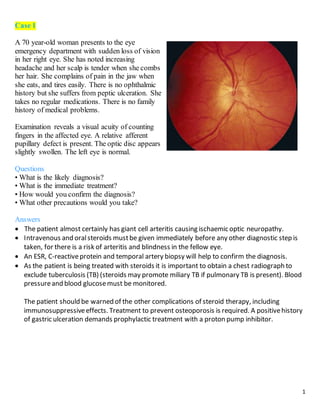
- 1. 1 Case 1 A 70 year-old woman presents to the eye emergency department with sudden loss of vision in her right eye. She has noted increasing headache and her scalp is tender when she combs her hair. She complains of pain in the jaw when she eats, and tires easily. There is no ophthalmic history but she suffers from peptic ulceration. She takes no regular medications. There is no family history of medical problems. Examination reveals a visual acuity of counting fingers in the affected eye. A relative afferent pupillary defect is present. The optic disc appears slightly swollen. The left eye is normal. Questions • What is the likely diagnosis? • What is the immediate treatment? • How would you confirm the diagnosis? • What other precautions would you take? Answers The patient almost certainly has giant cell arteritis causing ischaemic optic neuropathy. Intravenous and oralsteroids mustbe given immediately before any other diagnostic step is taken, for there is a risk of arteritis and blindness in the fellow eye. An ESR, C-reactiveprotein and temporal artery biopsy will help to confirm the diagnosis. As the patient is being treated with steroids it is important to obtain a chest radiograph to exclude tuberculosis (TB) (steroids may promote miliary TB if pulmonary TB is present). Blood pressureand blood glucosemust be monitored. The patient should be warned of the other complications of steroid therapy, including immunosuppressiveeffects. Treatment to prevent osteoporosis is required. A positivehistory of gastric ulceration demands prophylactic treatment with a proton pump inhibitor.
- 2. 2 Case 2 A 40 year-old man presents with sudden onset of a drooping left eyelid. When he lifts the lid with his finger, he notices that he has double vision. He has a severe headache. He is otherwise fit and well with no past ophthalmic history. He is on no regular medication. There is no family history of medical problems. Examination reveals normal visual acuity in both eyes. A left ptosis is present. The left pupil is dilated. The left eye is abducted in the primary position of gaze. Testing eye movements reveals reduced adduction, elevation, and depression of the left eye. The remainder of the eye examination is normal. Questions • What nerve palsy is present? • What is the most likely cause? • What is the management? Answers The man has a third nervepalsy. An aneurysmfromthe posterior communicating artery pressing on the third nerve mustbe the initial diagnosis in a painful third nervepalsy. The patient requires urgent neurosurgical investigation with a magnetic resonanceangiogram (MRA) and possibly angiography. Urgent treatment may be required. Itis also important to check blood pressureand blood glucose. Diabetics may develop a painful third nerve palsy, but the pupil is not always affected (a “pupil-sparing third nerve palsy “).
- 3. 3 Case 3 A 55 - year - old man presents to his family doctor with a 5- day history of the suddenonset of floaters in the left eye. These were accompanied by small flashes of light. He has treated hypertension but no other medical problems. The family doctor examines the eye and finds a normal visual acuity. Dilated fundoscopy reveals no abnormality. Questions • What should the family doctor advise? • What is the diagnosis? • What are the associated risks? Answers As the symptoms areacute, the family doctor should arrangefor an urgent ophthalmic assessment. The man has a posterior vitreous detachment. With careful ophthalmoscopy it will be possible to identify vitreous opacities in keeping with this diagnosis. Theflashing lights are caused by traction of the detached vitreous gel on the retina. A specialized examination of the peripheral retina is needed. A tear may occur in the retina, which in turn may lead to a retinal detachment. Laser applied around the tear while it is flat can prevent retinal detachment.
- 4. 4 Case 4 A 75 year-old woman attends the emergency department with nausea and vomiting. She says that her right eye is painful and red. Vision is reduced. She is longsighted and wears glasses for near and distance vision. She is generally fit. There is no family history of medical problems. On examination the emergency doctor finds the vision to be reduced to counting fingers in the right eye. The eye is red, the cornea appears cloudy and edematous, and the pupil is oval and dilated on the affected side. No view of the fundus is obtained. Questions • What is the diagnosis? • How might it be confirmed? • What is the treatment? Answers The lady is longsighted and has acute angle closureglaucoma. Tonometry would reveal an intraocular pressureof about 65 mm Hg. If possible, through the cloudy cornea, gonioscopy would confirm the presenceof a closed angle and a narrow angle in the fellow eye. This can also be seen using high resolution ultrasound or confocal microscopy. The pressuremustbe lowered with intravenous acetazolamide and topical hypotensivedrops, including pilocarpine, to producemiosis. A peripheral iridotomy is then performed, usually with a YAG laser, in both eyes, to prevent further attacks.
- 5. 5 Case 5 A 28 year-old man presents to his ophthalmologist with a painful, red right eye. The vision has becomeincreasingly blurred over the last 2 days. He wears softcontact lenses, and contact lens tolerance has decreased in the preceding days. The ophthalmologist notes that the vision is reduced to 6/60 in the right eye, the conjunctiva is inflamed, and there is a central opacity on the cornea. A small hypopyon is present. Questions • What is the likely diagnosis? • What should the optician do? Answers The man has a bacterial corneal ulcer secondary to contact lens wear. The ulcer will be scraped for culture and the contact lens and any lens containers cultured. Intensive, topical, broad-spectrumantibiotics are administered as an inpatient pending the result of the microbiological investigation. Case 6 A mother takes her 8 month-old baby to her ophthalmologist. He has had a persistently watery eye since birth. Intermittently there is a yellow discharge surrounding the eye. The white of the eye has never been red. The baby is otherwise healthy. Examination reveals a white, quiet, normal eye. Slight pressure over the lacrimal sac produces a yellowish discharge from the normal puncta. Questions • What is the diagnosis? • What advice would you give the mother? Answers Itis likely that the child has nasolacrimalobstruction due to an imperforate nasolacrimalduct. The mother should be reassured thatthis often resolves spontaneously. Thelids should be kept clean and the region overlying the lacrimal sac massaged gently daily. Antibiotics are generally not effective. If the symptoms persistafter the child’s firstbirthday the child can be referred for syringing and probing of the nasolacrimal duct.
- 6. 6 Case 7 A 14 year-old complains of intermittent redness and soreness of the right eye. He has noticed a small lump on the upper lid. The vision is unaffected. Examination reveals a small, raised, umbilicated lesion on the skin of the upper lid, associated with a follicular conjunctivitis below. Questions • What is the likely diagnosis? • What is the treatment? Answers Itis likely that the lid lesion is a molluscumcontagiosum. Itis treated by excision. Case 8 A 35 year-old man presents to his family doctorwith erythematous, swollen right upper and lower eyelids, worsening over the previous 2 days. He is unable to open them. He feels unwell and has a temperature. Examination reveals marked lid swelling, a tender globe and, on manual opening of the lids, a proptosis with chemotic injected conjunctiva. Eye movements are limited in all directions. Visual acuity and color vision are normal, and there is no relative afferent pupillary defect. The optic disc and retina also appear normal. Questions • What is the diagnosis? • What is the management? Answers The man has orbital cellulitis. Blood cultures and a high nasal swab should be performed, together with an orbital CT scan, to confirm the diagnosis and delineate any abscess.
- 7. 7 He requires admission to hospital for intravenous antibiotics and close monitoring of his vision, color vision and pupillary reflexes, as he is at risk of severeoptic nervedamage. The ENT surgeons should beinformed, as they may be required to drain an abscess. The normal acuity and color vision suggestthat the optic nerveis not compromised at present, but should these signs worsen, urgentsurgicaldrainagewill be required. Case 9 While working in the laboratory a colleague inadvertently sprays his eyes with an alkali solution. Questions • What is the immediate treatment? • What should you do next? Answers Anesthesia with Benoxinate then irrigate for at least 30 minutes (Normal saline, Lactated ringer, BSS) until pH returns normal(7). Evert/double evert lids and removeparticulate matter. If unable to remove particulate matter, consider general anesthesia. Debridement of necrotic corneal epithelium, necrotic conjunctival& sub-conjunctivaltissue. Do not patch the eye. Take visualacuity for both eyes (with correction, unaided and pinhole) Start treatment according to grading. Case 10 A 27 year-old man presents with a 2-day history of a painful red right eye; the vision is slightly blurred and he dislikes bright lights. He is otherwise fit and well, but complains of some backache. He wears no glasses. Questions • What is the likely diagnosis? • What would you expect to find on examination of the eye? • What treatment would you give? • What is the eye condition likely to be associated with? Answers The patient has iritis. Examination would reveal a reduction in visualacuity, redness of the eye that is worseat the limbus, cells in the anterior chamber and possibly on the cornea (keratic
- 8. 8 precipitate) or a collection at the bottom of the anterior chamber (hypopyon). Theiris may be stuck to the lens (posterior synechiae). Theremay be inflammation of the vitreous and retina. The patient is treated with steroid eye drops to reduce the inflammation and dilating drops to prevent the formation of posterior synechiae. The history of backachesuggests that the patient may haveankylosing spondylitis. Case 11 A 68 year-old lady presents with a mildly painful red eye and some blurring of vision. One year previously she had a corneal graft. She is on no medications and is otherwise well. Questions • What is the possible diagnosis? • What treatment should the patient be given? Answers A diagnosis of graft rejection mustbe considered first. Intensivetreatment with topical steroids to savethe graft. Case 12 A 68 year-old hypertensive man noted a fleeting loss of vision in one eye lasting for about a minute. He described it as a curtain coming down over the vision. Recovery was complete. There was no pain. Examination reveals no abnormality. Questions • What is the diagnosis? • What treatment would you advise? Answers The patient has had an episode of amaurosis fugax, mostlikely caused by the passageof a fi brin-platelet embolus through the retinal circulation. The patient requires treatment with antiplatelet drugs and a cardiovascular work - up. The most likely abnormality is a plaque on the carotid artery, which may require surgery.
- 9. 9 Case 13 A 60 year-old lady presented to her family doctor with gradual loss of vision over some months. She noticed that the problem was particularly bad in bright sunshine. The eye was not painful or red. She was otherwise well. Questions • What is the probable diagnosis? • How can the diagnosis be confirmed? • What treatment may be advised? Answers Itis likely that the lady has a cataract. These can be readily seen with a slit lamp, but are also well visualized with the direct ophthalmoscopein the red reflex. The advantages and possiblecomplications of cataractsurgery should be discussed with her once the diagnosis has been confirmed. Case 14 An 80 year-old lady who has already lost the vision in one eye develops distortion and reduction of vision over a few days in her good eye. Examination reveals an acuity of 6/12, an early cataract, and an abnormality at the macula. Questions • What is the likely diagnosis? • What treatment may be helpful? Answers The rapid onset suggests thatthe cataract has little to do with the new visual disturbance. Itis most likely due to age-related macular degeneration (AMD). In somepatients, with wet AMD, where a fibrovascular membrane grows beneath the macula that can be shown on a fluorescein angiogram, anti-VEGF injections into the vitreous may be helpful in preventing further progression and in somecases improving vision.
- 10. 10 Case 15 A 30 year-old builder was using a hammer to hit a steel chisel. He felt something hit his eye and the vision became blurred. He is fit and well and there is no history of medical problems. On examination, vision was reduced to 6/12. A fluorescein staining lesion was seen on the cornea but this appeared Seidel-negative. A small hyphema was seen in the anterior chamber, and in the red reflex observed with a direct ophthalmoscopea well delineated lens opacity was seen. The retina appeared normal. Questions • What is the cause of the reduced acuity? • What is the likely origin of the lens opacity? • What is the possible management of the patient? Answers The relatively good acuity suggests that there has been no damage to the macula. Itis likely that a piece of steel travelling at high velocity has penetrated the cornea, traversed the iris (resulting in the hyphema) and passed into or through the lens (causing the opacity). The corneal wound, if self-sealing, will probably not require suturing. The exact location of the foreign body must be determined. Although it is unlikely to causean infection (heat generated by the impact of the hammer on the metal may effectively sterilize the fragment) it may causeretinal toxicity if it has entered the vitreous cavity or retina. If it is enclosed in the lens there is less chance of retinal toxicity developing but the patient is at high risk of developing a subsequentcataractthat may require an operation. A foreign body that impacts on the retina or the vitreous body requires a vitrectomy to remove it, with careful examination of the retina for tears. Case 16 A 2 year-old child was thought to have a squint by her parents. The finding was confirmed by her family doctor and she was referred to hospital. Question • What examination must be conducted in hospital? Answer Having taken a full history, an orthoptistwill measurethe visualacuity of the child, examine the range of eye movements, determine the presence type of squintwith a cover test, trying to assess thedegree of binocular vision present.
- 11. 11 The child will have a refraction performed and glasses prescribed if there is a significant refractiveerror or a difference in the strength of the lens needed between the two eyes (anisometropia). An ophthalmologistwill examine the eye to check that there is no ocular or neurological condition that may account for the squintand can discuss any futureneed for surgery. Case 17 A 26 year-old lady presents with a 3-day history of blurring of vision in the right eye. This has become progressively worse. She also has pain caused by moving the eye. She has previously had an episode of weakness in the right arm 2 years ago, but this settled without treatment. She is otherwise fit and well. On examination the vision was 6/60, with no improvement on looking through a pinhole. A central scotomawas present on confrontation. The eye was white and quiet with no abnormality noted except for a right relative afferent pupillary defect. Questions • What is the diagnosis? • How could this be confirmed? • What are the management options? • What is the prognosis? Answers The patient has the typical symptoms and signs of optic neuritis. The diagnosis can be supported by an MRI scan to look for additional plaques of demyelination and a visual evoked potential to examine the functioning of the optic nerve. A neurologistmay also suggestperforming a lumbar puncture, particularly if there is any doubt about the diagnosis. With the possibility of a previous neurological episode, it is likely that the patient has multiple sclerosis. Itis of great importance that appropriatecounselling is given. Steroid treatment may speed up the recovery of vision. The prognosis for recovery of vision over a few months is good.
- 12. 12 Case 18 A 79 year-old man presents with a lesion on his right lower lid. It has been there for some months and has gradually grown bigger. It is ulcerated and the ulcer shows a pearly margin. Questions • What is the lesion? • How should it be treated? Answers This is a basalcell carcinoma. Itrequires local excision. There is no problem with metastatic spread but local extension could causesevere problems as the tumor grows and infiltrates surrounding structures. Case 19 A 60 year-old man presents with tired sore eyes. He has noted that the eyelids may crust in the morning. Sometimes the white of the eye is red. The vision is unaffected. He is otherwise fit and well. Questions • What is the probable diagnosis? • What signs would you look for? • How can this condition be treated? Answers The patient has blepharitis. Scaling of the lid margins and at the base of the lashes, together with inflammation of the lid margins and plugging of the meibomian glands, may be present. Lid cleaning, along with the useof local antibiotic ointment and possibly topical steroids (supervised by an ophthalmologist), will improve, if not alleviate, the symptoms. Heat and lid massagecan restore oil flow. Courses of systemic tetracycline may be beneficial in more advanced cases and are in any case used in the treatment of acne rosacea which may be an associated condition.
- 13. 13 Case 20 A 30 year-old man developed an acute red eye first on the right and then in the left eye, associated with a watery discharge. Vision is unaffected but the eye irritates. He is otherwise fit and well. Questions • What is the diagnosis? • What confirmatory signs would you look for on examination? • What precautions would you take following your examination? Answers The patient has viral conjunctivitis – probably adenovirus. Examination for a preauricular lymph node and conjunctivalfollicles on the lower tarsus would confirm the diagnosis. This formof conjunctivitis is highly contagious; it is important to ensurethat hands and equipment arethoroughly cleaned following the examination, and that the importance of good hygieneis emphasized to the patient.