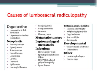
Low back pain is one of the most common musculoskeletal complaints encountered in clinical practice. It is the leading cause of disability in the developed world and accounts for billions of dollars in healthcare costs annually. Although epidemiological studies vary, the incidence of low back pain is estimated to be anywhere between 5% to more than 30% with a lifetime prevalence of 60% to 90%. Most occurrences of low back pain are self-limited and resolve without intervention. Approximately 50% of cases will resolve within one to two weeks. 90% of cases will resolve in six to 12 weeks. The differential for low back pain is broad, and amongst other diagnoses, should include lumbosacral radiculopathy. Lumbosacral radiculopathy is a term used to describe a pain syndrome caused by compression or irritation of nerve roots in the lower back. It can be caused by lumbar disc herniation, degeneration of the spinal vertebra, and narrowing of the foramen from which the nerves exit the spinal canal. Symptoms include low back pain that radiates into the lower extremities in a dermatomal pattern. Other accompanying symptoms can include numbness, weakness, and loss of reflexes, although the absence of these symptoms does not exclude a diagnosis of lumbosacral radiculopathy.
2. • Lumbosacral radiculopathy is one of the most common disorders
evaluated by neurologists and is a leading referral diagnosis for the
performance of electromyography.
• Degenerative spondyloarthropathies are the principal underlying
cause of these syndromes and are increasingly common with age.
3. Epidemiology
• Although precise epidemiologic data are difficult to establish, the
prevalence of lumbosacral radiculopathy is approximately 3% to 5%,
distributed equally in men and women.
• Men are most likely to develop symptoms in their 40s, whereas
women are affected most commonly between ages 50 and 60.
4. History and physical examination
• Performance of a careful history and physical examination is the
initial and integral step in the diagnosis and management of
lumbosacral radiculopathy.
• Lesion localization depends on demonstration of a segmental
myotomal or dermatomal distribution of abnormalities.
• Sciatica, the classic presenting symptom of lumbosacral
radiculopathy, is characterized by pain in the back radiating into the
leg. Patients variably describe this pain as sharp, dull, aching,
burning, or throbbing. Pain related to disk herniation is exacerbated
by bending forward, sitting, coughing, or straining and relieved by
lying down or sometimes walking.
5. • Conversely, pain related to lumbar spinal stenosis characteristically
is worsened by walking and improved by forward bending.
• Pain that is exacerbated by or fails to respond to the recumbent
position is a distinctive feature of radiculopathy produced by
inflammatory or neoplastic lesions and other nonmechanical causes
of back pain.
• The distribution of pain radiation along a dermatome may be
helpful in localizing the level of involvement; when present, the
dermatomal distribution of paresthesias is more specific.
6. • In the majority of cases, lumbosacral radiculopathy is caused by
compression of nerve roots from pathology in the intervertebral disk
or associated structures.
• The differential diagnosis of lesions producing lumbosacral
radiculopathy, however, is broad and includes neoplastic, infectious,
and inflammatory disorders.
• Important risk factors for serious underlying disease that should be
sought in the history include age greater than 50, previous history of
cancer, unexplained weight loss, and failure to improve after 1
month of conservative therapy.
7. • Aside from assessment of potential serious disease, the history is
geared toward establishing the involvement of nerve roots and their
anatomic level.
• Similarly, the aim of the physical examination is to elucidate motor,
sensory, or reflex abnormalities in a radicular distribution relevant
to the suspected clinical level.
• Sciatic nerve tension signs may provide supporting evidence of L5-
S1 radiculopathy; however, they may be present with lesions of the
lumbosacral plexus, sciatic nerve, or hip joint and in mechanical
lower back pain .
8. • With the patient supine and one hand on the iliac crest of the
affected side, an examiner passively elevates the heel slowly while
keeping the knee straight; the angle at which pain or paresthesias
are produced and the distribution are noted. Dorsiflexion of the foot
may increase symptoms. The straight leg raise test is positive if
symptoms are produced between 30 and 70.
• In a similar fashion, the femoral nerve stretch test produces tension
on the L3, L4 nerve roots. With a patient lying on the asymptomatic
side and the lower limb flexed at the hip and knee, the symptomatic
knee is extended passively at the hip .
• Pain radiating into the anterior thigh with this maneuver suggests
L3 or L4 radiculopathy.
10. L1 radiculopathy
• Disk herniation at this level is rare; consequently, L1 radiculopathy
is extremely uncommon.
• The typical presentation is one of pain, paresthesias, and sensory
loss in the inguinal region, without significant weakness.
Infrequently, subtle involvement of hip flexion is noted.
11. L2 radiculopathy
• L2 radiculopathy produces pain, paresthesias, and sensory loss in
the anterolateral thigh. Weakness of hip flexion may occur.
• Lateral femoral cutaneous neuropathy (meralgia paresthetica) may
mimic L2 radiculopathy; the presence of hip flexor weakness
suggests radiculopathy rather than meralgia. Femoral neuropathy
and upper lumbar plexopathy may present similarly.
12. L3 radiculopathy
• Pain and paresthesias involve the medial thigh and knee, with
weakness of hip flexors, hip adductors, and knee extensors; the knee
jerk may be depressed or absent.
• L3 radiculopathy may be confused with femoral neuropathy,
obturator neuropathy, diabetic amyotrophy, or upper lumbar
plexopathy.
• Combined weakness of hip adduction and hip flexion differentiates
L3 radiculopathy from femoral and obturator mononeuropathies.
13. L4 radiculopathy
• Unlike the higher lumbar levels,L4 radiculopathy is produced
most commonly by disk herniation.
• Spinal stenosis frequently involves this nerve root in
conjunction with roots at adjacent spinal levels.
• Sensory symptoms involve the medial lower leg in the
distribution of the saphenous nerve.
• As with L3 radiculopathy, knee extension and hip adduction
may be weak; additionally, foot dorsiflexion weakness
uncommonly may be observed.
• When present, ankle dorsiflexion weakness generally is less
prominent than in L5 radiculopathy. The knee jerk may be
depressed or absent.
14. L5 radiculopathy
• The most common cause of L5 radiculopathy is intervertebral disk
herniation. Foot drop is the salient clinical feature, with associated
sensory symptoms involving the anterolateral leg and dorsum of the
foot.
• In addition to weakness of ankle dorsiflexion, L5 radiculopathy
commonly produces weakness of toe extension and flexion, foot
inversion and eversion, and hip abduction.
• Common peroneal neuropathy closely mimics and must be
distinguished from L5 radiculopathy.
15. • Physical examination is helpful in localization as weakness of foot
eversion (mediated by the L5/peroneal innervated peroneus
muscles) in conjunction with inversion (mediated by the L5/tibial-
innervated tibialis posterior) places the lesion proximal to the
peroneal nerve.
• Lumbosacral plexopathy and sciatic neuropathy are important
differential diagnostic considerations.
• The involvement of hip abductors (gluteus medius and minimus)
indicates a lesion proximal to the sciatic nerve
but does not differentiate L5 radiculopathy from lumbosacral
plexopathy.
16. S1 radiculopathy
• S1 radiculopathy also is caused commonly by intervertebral disk
herniation, with associated weakness of foot plantar flexion, knee
flexion, and hip extension.
• Sensory symptoms typically involve the lateral foot and sole. The
ankle jerk is depressed or absent.
• Sciatic neuropathy and lower lumbosacral plexopathy may mimic S1
radiculopathy. Both of these conditions, however, also are expected
to affect L5- innervated muscles.
18. • Multiple, contiguous nerve roots may be involved by compressive
lesions affecting several individual nerve roots, either in the
vertebral canal or the neural foramina; less frequently, infiltrating
or inflammatory processes spreading along the meninges produce
similar clinical syndromes.
• Lesions involving the cauda equina should be considered when
nerve roots at more than two neighboring levels are involved,
developing acutely or gradually.
• Acute cauda equina syndrome most often is the result of
compression of the lower lumbar and sacral nerve roots by a large,
central disk herniation, usually at L4-5.
19. • Sacral nerve roots, which lie medially in the cauda equina, often are
affected disproportionately, leading to sacral polyradiculopathy with
prominent bowel and bladder dysfunction and characteristic saddle
anesthesia.
• Lumbar nerve roots also may be involved, resulting in leg weakness
that can progress to paraplegia, depending on the extent of nerve
root compromise.
• A true neurologic emergency, prompt recognition of cauda equina
syndrome is necessary to preserve sphincter function and
ambulation.
20. • A more insidious manifestation of cauda equina dysfunction results
from central spinal stenosis, creating a clinical syndrome of
intermittent neurogenic claudication.
21. Differential diagnosis
• The majority of lesions causing lumbosacral radiculopathy are
compressive in nature and result from disk herniation or
spondylosis with entrapment of nerve roots. It is important,
however, to recognize a variety of other lesions that may produce
lumbosacral radiculopathy, including several neoplastic, infectious,
and inflammatory disorders.
23. Degenerative spine disorders
Acute disk herniation
• Intervertebral disk herniation is the most common cause of
lumbosacral radiculopathy in patients under age 50.
• Acute disk herniation produces symptoms by direct compression of
the nerve roots and by inflammatory and ischemic mechanisms
involving the roots and dorsal root ganglia.
• The intervertebral disks affected most frequently are L4-5 and L5-
S1, leading to L5 or S1 radiculopathies. Pain characteristically is of
abrupt onset and intense, often precipitated by bending over or
lifting.
24. • Pain characteristically is of abrupt onset and intense, often
precipitated by bending over or lifting.
• Patients may report of sciatica without back pain.
• Aggravation of pain with movement, particularly forward or lateral
bending, or with Valsalva’s maneuver is typical; usually, pain is
relieved with recumbency. In addition to pain, patients frequently
report paresthesias in the involved dermatome.
• Cauda equina syndrome with prominent bowel and bladder
involvement may be the presenting syndrome with large, central
disk herniations.
25. Degenerative spondylosis
• After age 50, acute disk herniation is a less common cause of
lumbosacral radiculopathy, and chronic lesions related to degenerative
spinal arthropathy predominate.
• With advancing age, intervertebral disks desiccate and flatten,
transferring increasing axial load to the facet joints, with resultant facet
joint hypertrophy, osteophyte formation, and thickening of the
ligamentum flavum. These changes contribute to narrowing of the
central spinal canal, lateral recesses, and neural foramina. L4-5 and L5-
S1 levels in particular are affected.
• Chronic radiculopathy may result from entrapment of nerve roots in the
lateral recess, intervertebral foramen, or central canal, involving single
or multiple nerve roots.
26. • Clinical syndromes of radicular pain involving buttock, hip, or posterior
thigh and intermittent neurogenic claudication are more common than
back pain.
• MRI frequently is performed in the evaluation of these lesions, although
bony pathology is demonstrated better by CT. Because degenerative
changes are commonplace in older patients, electrodiagnostic studies
frequently are necessary to establish the relevance of neuroimaging
abnormalities.
• Initial management involves pain control with analgesic medications
and physical therapy to strengthen supporting musculature and
improve postural mechanics. Surgical decompression is considered for
progressive or recalcitrant symptoms or worsening neurologic deficits.
27. Neoplasms
• Radiculopathy may result from tumor in various locations within
the spinal canal;usually, these lesions are extramedullary.
• Primary tumors tend to be intradural, whereas metastatic lesions
are extradural.
Furthermore, primary lesions tend to be solitary ,
whereas metastatic lesions frequently are multiple.
28. Primary tumors
• Primary nerve root tumors are a rare cause of lumbosacral
radiculopathy.
• Most primary spinal tumors are benign and slow growing, and their
clinical manifestations may be difficult to distinguish from more
common causes of radiculopathy, such as disk herniation. Both are
characterized by back pain; however, the nature of pain related to
tumor is distinctive, as it becomes increasingly severe over time and
is worse when lying down, often interfering with sleep.
29. • Primary tumors producing lumbosacral radiculopathy most
frequently are neurofibromas and ependymomas; less common are
schwannomas, meningiomas, lipomas and dermoids.
• Ependymomas and neurofibromas typically affect the filum
terminale, producing a cauda equina syndrome. Diagnosis of
primary tumors is established by MRI, and their definitive
treatment is surgical.
30. Epidural and vertebral metastases
• Metastatic tumor is the most common type of neoplasm
involving the spinal canal, it is rare in the general
population.
• These lesions chiefly are seen in patients who have a known
Malignancy and, in A small percentage, are the presenting
feature.
• Approximately 30% of epidural metastases occur in the
lumbar spine, and radicular pain is an initial symptom in
approximately half.
• Metastases typically invade the spinal column and extend
from there into the epidural space .Metastases seed the
vertebrae by way of batson’s venous plexus (which drains
the vertebrae and anastomoses with veins draining the
viscera
31. • Less commonly, paravertebral lesions spread directly to nerve roots
through the Intervertebral neural foramina.
• The three most common cancers involving the lumbosacral spine
are breast, lung, and prostate cancer, each accounting for
approximately 10% to 20% of cases.
• Virtually all cancers, however, may produce metastatic spinal cord
compression, and in 20%, spinal cord compression is the initial
feature.
• Tumors of the pelvic region, including colon and prostate,
preferably metastasize to the lumbosacral region.
• Back pain is the most common initial complaint and, as with
primary tumors, is unremitting and characteristically worse with
recumbency; radicular pain is more variable.
32. • Percussion tenderness at the site of the lesion is noteworthy.
• Bowel and bladder disturbances occur in a minority of patients at
onset but tend to be more common as disease progresses.
• The neurologic prognosis of patients who have radiculopathy as the
sole symptom of metastatic disease is good; most patients likely
maintain ambulation after treatment with radiotherapy.
• Prognosis is correlated closely with the degree of neurologic
dysfunction at diagnosis, so early recognition is crucial.
33. Leptomeningeal metastases/meningeal
carcinomatosis
• Cancer cells may infiltrate the leptomeninges and subarachnoid space
diffusely, leading to a syndrome reflecting involvement of cranial
nerves, spinal nerve roots, and brain.
• Manifestations include radiculopathy, cranial polyneuropathy,
headache, memory loss, seizures, and gait disturbances .
• Radicular discomfort is the most common presenting symptom, usually
involving lumbosacral levels resulting from involvement of the cauda
equina.
• Although all cancers have the potential to produce this condition, the
most likely primary tumors to do so are leukemia, lymphoma, and
breast carcinoma . Other tumors that may produce leptomeningeal
metastasis include melanoma, lung cancer, gastrointestinal cancers,
and sarcoma.
34. Spinal epidural abscess
• Spinal epidural abscess (SEA) most commonly involves the thoracic and
lumbar spine. Risk factors for development of SEA include diabetes
mellitus, history of intravenous drug abuse, spinal surgery, spinal or
paraspinal injection, epidural catheter placement, an immunocompromised
status.
• Severe back pain, often with a radicular component, is the presenting
complaint . Fever is a common, but not universal, sign.
• Leukocytosis and elevation of the erythrocyte sedimentation rate are typical
and in the presence of fever and back pain, the diagnosis should be
straightforward.
• Only 20% of patients, however, have the classical clinical triad of fever,back
pain, and neurologic deficits, so a high index of suspicion should be
maintained .
• the diagnostic test of choice is contrast-enhanced MRI.
35. • Treatment of SEA must be initiated urgently with surgical
debridement generally the treatment of first choice.
• Antibiotic treatment should be directed to treat the most common
infecting organisms, which include Staphylococcus aureus, other
gram-positive cocci, gram-negative rods, and anaerobes.
• There is increasing evidence, however, that management with 6 to 8
weeks of intravenous antibiotics with or without oral antibiotics
may result in similar outcomes.
• Close monitoring is necessary, and urgent surgical decompression
must be considered strongly if neurologic compromise develops.
36. Polyradiculopathy in HIV and AIDS
• Polyradiculopathy secondary to HIV infection is uncommon,
accounting for only 2% of HIV-related neurologic consultations .
• The majority of patients have an AIDS-defining illness before the
development of radiculopathy, and the CD4 count is less than 100
cells per mL in almost all patients.
• Polyradiculopathy in AIDS tends to involve the lumbo-sacral nerve
roots, producing a rapidly progressive cauda equina syndrome with
severe low back pain.
• Cytomegalovirus accounts for most HIV-related radiculopathy.
Other causes of HIV-radiculopathy include herpes simplex virus,
lymphomatous meningitis, mycobacteria, Cryptococci, and
treponemal infection .
37. • Examination of CSF demonstrates pleocytosis, with
polymorphonuclear predominance and, in some patients, decreased
glucose.A positive CSF polymerase chain reaction for cytomegalovirus
also is supportive of the diagnosis.
• Development of polyradiculopathy in AIDS generally prtends a poor
prognosis, with minimal functional recovery after treatment and a
median survival time of 2.7 months.
38. Diabetes (diabetic amyotrophy)
• Diabetes may cause a syndrome of severe lower extremity pain and
weakness, commonly referred to as diabetic amyotrophy. This
syndrome usually involves multiple lumbosacral nerve roots but
rarely presents as a monoradiculopathy.
• Patients typically have well controlled type 2 diabetes and are
middle aged or older. In some patients, the neurologic impairment
heralds the onset of diabetes .
• Sudden onset, unilateral lower extremity pain variably involves the
groin, anterior thigh, and lower leg; weakness follows shortly.
• Proximal muscles, in particular quadriceps, tend to be affected
first and most conspicuously, but the majority of patients also
develop distal and bilateral symptoms .
39. • Weight loss is a frequent accompanying symptom . The precise
pathophysiology of diabetic radiculopathy is controversial, with
nerve ischemia, inflammation, and metabolic causes implicated.
• EMG is helpful in diagnosing diabetic amyotrophy.
• There is evidence of subacute polyradiculopathy with prominent
denervation changes involving limbs and multiple, bilateral
paraspinal regions. Underlying axonal polyneuropathy may also be
present. CSF protein is elevated without pleocytosis.
40. • Diabetic radiculopathy is a monophasic illness that improves with
time. Improvement, however, often is incomplete and prolonged,
with motor symptoms slower to resolve than sensory symptoms .
• Pain control in the early stages can be challenging. Standard agents
for neuropathic pain, such as anticonvulsants or tricyclic
antidepressants, are beneficial, but narcotic analgesics also may be
needed temporarily .
• Physical therapy and orthoses should be provided as indicated . Up
to 20% of patients may suffer a recurrence on the same side.
41. Spinal cysts
• Cystic lesions in the sacral spine are common, with an incidence ranging
from 4.6% to 17% on imaging studies .
• Most sacral meningeal cysts are dural diverticula (Tarlov cysts) produced by
fluctuations in CSF pressure .
• There is little to differentiate the presentation of meningeal sacral cysts
from other causes of lumbosacral radiculopathy .
• Radicular pain often is relieved or disappears when patients are recumbent
and is aggravated by Valsalva’s maneuver . Because they are common and
not necessarily the cause of symptoms, establishing a cyst as the cause of
lumbosacral radiculopathy involves eliminating other causes first.
• MRI is the diagnostic procedure of choice for demonstrating lumbosacral
cysts; however, clinical relevance of the imaging findings must still be
established.
• Although analgesic medications may reduce pain, relief of symptoms with
fluoroscopic-guided aspiration and surgical treatment is definitive .
42. Spinal hematomas
• Hematomas are uncommon causes of lumbosacral radiculopathy.
• Epidural and subdural spinal hematomas occur most frequently in
patients who have coagulopathies, who are taking anticoagulants, or
who recently have undergone epidural injections or instrumentation
of the lumbosacral spine.
• Spinal subarachnoid hemorrhage is uncommon. Unlike its
intracranial counterpart, spinal subarachnoid hemorrhage is caused
most commonly by arteriovenous malformation rupture rather than
aneurysmal rupture.
• Hemorrhage into synovial cysts or the ligamentum flavum also may
produce hematomas and lumbosacral radiculopathy .