More Related Content
Similar to Definitions of acute coronary syndromes.pdf (20)
Definitions of acute coronary syndromes.pdf
- 1. Definitions of acute
coronary syndromes
Retesh Bajaj
Ajay K Jain
Charles Knight
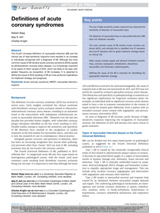
Abstract
The Fourth Universal Definition of myocardial infarction (MI) and the
clinical use of high-sensitivity troponins have resulted in an increase
in individuals recognized with a diagnosis of MI. Although the most
common cause of MI remains acute coronary syndrome (ACS) caused
by atherosclerotic coronary artery disease, it is increasingly important
to be aware of other causes of ACS, which are likely to be seen with
greater frequency as recognition of MI increases. It is essential to
define the cause of ACS resulting in MI as it has profound implications
for treatment strategy and prognosis.
Keywords Acute coronary syndrome; MRCP; myocardial infarction;
troponin
Background
The definition of acute coronary syndrome (ACS) has evolved in
recent years. Early insights correlated the clinical syndrome
with thrombotic coronary artery occlusion related to obstructive,
atherosclerotic coronary artery disease, resulting in an ischaemic
insult to the myocardium with subsequent cardiomyocyte ne-
crosis or myocardial infarction (MI).1
Research over the last two
decades has provided further insights, with vulnerable coronary
plaque disruption identified as the key event resulting in ACS.
Parallel studies aiming to improve the sensitivity and specificity
of MI detection have resulted in the recognition of cardiac
troponins as the best marker for myocardial injury, and their use
is now the standard of care in contemporary clinical practice.
Alongside greater accuracy and precision of detection, there
has been an increasing recognition that myriad other patholog-
ical processes other than ‘classic’ ACS can lead to MI, including
processes that do not involve the coronary arteries.
The Fourth Universal Definition of MI, published in 2019,
reflected this.2
It categorized acute MI into five types based on
heterogenous pathological causes, with the classic (and most
common) cause resulting from thrombotic coronary occlusion
being just one type. It is important to note that although the terms
ACS and MI have evolved to be used interchangeably, the cause of
troponin leak or MI may not necessarily be ACS, and ACS may not
strictly be caused by occlusive epicardial coronary artery disease.
This distinction and specificity in pathophysiology is essential as it
has significant implications for long-term treatment plans: for
example, an individual with no significant coronary artery disease
noted to have a rise in troponin concentration in the context of
sepsis would be treated quite differently from a patient with an
occluded coronary artery treated with angioplasty and percuta-
neous coronary intervention (PCI).
As rates of diagnosis of MI increase, partly because of high-
sensitivity troponins improving the recognition of myocardial
necrosis, the definition of ACS will become ever more critical to
guide treatment.
Types of myocardial infarction (based on the Fourth
Universal Definition)
MIs can be divided into five types based purely on pathological
causes, as suggested by the Fourth Universal Definition
published in 2019 (Table 1).2
Type 1 MI is caused by the classically recognizable clinical
entity of ACS related to atherosclerotic plaque rupture or erosion.
A decrease in epicardial blood supply to a region of myocardium
results in hypoxic damage and, ultimately, tissue necrosis and
infarction. Type 1 MI is clinically subdivided based on symp-
toms, electrocardiograph (ECG) changes and biomarker evalua-
tion to expedite appropriate triage and management. This can
include early invasive coronary angiography and intervention
with angioplasty and coronary stent insertion.
Type 2 MI results in myocardial necrosis and troponin leak
caused by an imbalance between myocardial oxygen demand
and supply. The underlying pathological mechanisms are heter-
ogenous and include coronary dissection or spasm, emboliza-
tion, anaemia, tachy- or brady-arrhythmia, hypertension or
hypotension, coronary endothelial dysfunction and respiratory
failure.
Key points
C The use of high-sensitivity cardiac troponins has improved the
sensitivity of detection of myocardial injury
C The detection of myocardial injury or myocardial infarction (MI)
does not implicate causality
C The most common cause of MI remains acute coronary syn-
drome (ACS), and clinically this is classified into ST elevation
and non-ST elevation ACS to guide treatment strategy based
on clinical risk
C Other causes include supply and demand mismatch (arrhyth-
mias, coronary vasospasm, embolization, dissection),
myocarditis and Takotsubo cardiomyopathy
C Defining the cause of the MI is essential for identifying the
appropriate treatment strategy
Retesh Bajaj MB BS BSc MRCP is a Cardiology Specialist Registrar at
Barts Health, London, UK. Competing interests: none declared.
Ajay K Jain BSc MRCP MD is a Consultant Cardiologist at Barts Health
and Clinical Lead of the Barts Heart Attack Centre, London, UK.
Competing interests: none declared.
Charles Knight OBE MD FRCP FESC is a Consultant Cardiologist and
Chief Executive of St Bartholomew’s Hospital, London, UK.
Competing interests: none declared.
ISCHAEMIC HEART DISEASE
MEDICINE xxx:xxx 1 Ó 2022 Published by Elsevier Ltd.
Please cite this article as: Bajaj R et al., Definitions of acute coronary syndromes, Medicine, https://doi.org/10.1016/j.mpmed.2022.04.005
Descargado para Ronald Eduardo Lozano Acosta (loacro@yahoo.com) en Cayetano Heredia Pervuvian University de ClinicalKey.es por Elsevier en mayo 30,
2022. Para uso personal exclusivamente. No se permiten otros usos sin autorización. Copyright ©2022. Elsevier Inc. Todos los derechos reservados.
- 2. A rise in troponin concentration caused by myocardial
necrosis is also recognized in critically ill individuals and patients
undergoing major non-cardiac surgery; here it can be related to
cell dysfunction and damage caused by toxins, sepsis syndrome
or pharmacological agents. Notably, in these patients, the
epicardial vessels may be patent, but there is a ‘supply and
demand’ mismatch leading to myocardial necrosis. Coronary
artery disease may be worsening this mismatch, so a careful
assessment of risk factors, cardiac symptom history and current
clinical context is essential in guiding management.
Type 3 MI is defined as a typical presentation of myocardial
ischaemia/infarction in which the person dies before it is
possible to detect biomarker elevation.
Two types of type 4 MI have been described: 4a, in which
serum troponin rises after PCI, and type 4b, which relates to
acute stent thrombosis.
Finally, critical elevation of troponin in association with
coronary artery bypass surgery is known as type 5 MI.
The impact of ACS
Coronary artery disease causing ACS and MI remains one of the
leading causes of death worldwide, irrespective of socioeconomic
status. World Health Organization statistics estimate that 13% of
the 57.1 million global deaths recorded in 2015 resulted from
coronary artery disease, with over three-quarters of these taking
place in low- and middle-income countries.3
This agrees with
British Heart Foundation statistics from 2015 suggesting that
14% of all deaths in men and 9% in women in the UK resulted
from coronary artery disease.4
Troponins in recognizing myocardial infarction versus
diagnosing ACS
It is difficult to overstate the key role that troponins, especially
high-sensitivity troponins, have played as sensitive and specific
biomarkers of myocardial necrosis in recognizing MI; this has led
to their incorporation as key to its definition (Table 2).5
Apart
from being needed for governance and epidemiological accuracy,
the correct diagnosis of MI is essential to guide appropriate pa-
tient management and indicate risk, as myocardial necrosis por-
tends greater morbidity and mortality across all patient groups.
However, even though positive troponin results help in
identifying MI, correlation with ACS or an alternative cause can
be clinically challenging. MI can be the first presentation of
significant coronary artery disease and the result of an ACS;
alternatively, it can be a coincidental finding in an alternative
pathology and unrelated to ACS.
The clinical presentation of ACS is known to be variable and
can include so-called ‘silent’ events (without overt chest pain)
and classic anginal chest pains at rest, through to sudden death
from catastrophic cardiac ischaemia causing heart failure or
cardiac arrest.
Clinical diagnosis of ACS: ST and non-ST elevation
As occlusive coronary artery disease is the most common cause
of ACS, clinical classification aims at rapid identification and
treatment, informed by established data and guidelines to
minimize morbidity and mortality.
ACS can usually be identified clinically by a patient’s history,
ECG findings and highly sensitive cardiac biomarkers. Rapid
diagnosis allows for earlier identification of high-risk patients
and revascularization to minimize MI. Classically, individuals
presenting with persistent (>20 minutes) cardiac-sounding chest
pain at rest who have persistent ST segment elevation in two or
more contiguous leads are designated as having ‘ST elevation
ACS’ or ‘ST elevation MI’ (STEMI). The degree of ST elevation
depends on age and sex. Acute or evolving changes in STeT
waveforms can be highly valuable in locating the responsible
artery and the timing of the event, and in estimating the pro-
portion of the myocardium at risk.
ACS can be the result of plaque rupture and thrombosis
(most commonly) impairing coronary blood supply to the
myocardium, with myriad causes that can include coronary
vasospasm caused by cocaine or coronary dissection, which can
effectively do the same. Rarer causes include embolization of
clot or septic embolization down a coronary artery related to
infective endocarditis.
Patients diagnosed with STEMI are treated as a medical
emergency, ideally with primary PCI; rapid intervention aiming
to restore blood flow is the gold standard of treatment, unless
contraindicated.5
Patients presenting with continuing cardiac chest pains
without persistent ST elevation are designated as having non-ST
elevation ACS. Where biomarker values indicating car-
diomyocyte necrosis are raised, patients are said to have had a
non-STEMI (NSTEMI). In these patients, new ST depression or T
wave changes can occur, or alternatively the ECG can be normal.
Patients with high-risk features such as significant increases in
troponins and dynamic ECG changes are generally treated with
an early interventional approach. Continuing chest pain re-
fractory to medical therapy, for example intravenous nitrates, is
treated with emergency angiography and PCI, if appropriate (see
Further reading).
Types of MI
C Type 1 e spontaneous MI related to ischaemia caused by a primary coronary event, such as plaque fissuring or rupture
C Type 2 e MI secondary to ischaemia resulting from an imbalance between oxygen supply and demand
C Type 3 e sudden death from cardiac disease with symptoms of myocardial ischaemia, accompanied by new ST elevation or left bundle branch
block, or verified coronary thrombus at angiography and/or autopsy
C Type 4 e MI associated with PCI
C Type 5 e MI associated with coronary artery bypass grafting
Table 1
ISCHAEMIC HEART DISEASE
MEDICINE xxx:xxx 2 Ó 2022 Published by Elsevier Ltd.
Please cite this article as: Bajaj R et al., Definitions of acute coronary syndromes, Medicine, https://doi.org/10.1016/j.mpmed.2022.04.005
Descargado para Ronald Eduardo Lozano Acosta (loacro@yahoo.com) en Cayetano Heredia Pervuvian University de ClinicalKey.es por Elsevier en mayo 30,
2022. Para uso personal exclusivamente. No se permiten otros usos sin autorización. Copyright ©2022. Elsevier Inc. Todos los derechos reservados.
- 3. Patients without elevated biomarkers and symptoms at rest
are labelled as having unstable angina. With the advent of
high-sensitivity troponins, this group has become smaller, but
current research indicates that, compared with NSTEMI pa-
tients, they have a substantially lower risk of death (see Further
reading).
It is important to interpret ECGs within the clinical context
and, where possible, compare them with previous ECGs as ST
deviation can be observed in other conditions, including left
ventricular hypertrophy, Brugada syndrome, acute pericarditis,
myocarditis, Takotsubo cardiomyopathy and early repolarization
e some of these mimics are discussed below.
Clinical classification of ACS allows triaging and selection of
the first investigative step, namely coronary angiography, to
potentially define the ACS and, with atherosclerotic disease,
allow potential treatment.
ACS defined by pathophysiology
Although it is valuable to recognize MI and clinically diagnose
ACS, it is vital to be aware of the various causes of ACS, some of
which are described below.
Plaque disease and thrombosis: coronary atherosclerotic plaque
narrows the coronary artery lumen. Plaque rupture can become a
nidus for thrombosis and vessel occlusion, resulting in myocar-
dial ischaemia and infarction. Acute stent thrombosis is a well-
recognized phenomenon of clot formation within a previous
coronary artery stent; its incidence has markedly reduced with
the advent of newer drug-eluting stents and more advanced
antiplatelet therapy regimens.
Coronary artery dissection: this can result from a type A aortic
dissection involving a coronary artery and is a recognized mimic
of ACS. Clinical suspicion can be raised by differential pulses
and blood pressures in the limbs, haemodynamic instability and
occasionally bedside echocardiography; however, the definitive
investigation is contrast-enhanced computed tomography (CT)
aortography.
Spontaneous coronary artery dissection is a recognized entity
classically affecting women who may be young with no coronary
atherosclerosis. This diagnosis is generally made on angiog-
raphy, and clinically these patients can present as having classic
ACS.
Coronary vasospasm: this can be associated with cocaine abuse
and must be considered, particularly, although not exclusively,
in younger age groups. A thorough history is essential. b-adre-
noceptor blockers must be avoided in this group, and treatment
with nitrates is indicated.
Prinzmetal or variant angina: this results in coronary vaso-
spasm and is diagnosed angiographically with ST elevation
revealed using intracoronary acetylcholine. It is treated with
calcium channel blockers and nitrates.
Coronary embolization: this very rare condition can be either
septic embolization from infective endocarditis, resulting in
vessel occlusion and STEMI, or clot embolization related to a
patent foramen ovale or atrial septal defect. The result can be
coronary occlusion, and clinically it can be indistinguishable
from STEMI because of this e the clinical context (e.g. sepsis in
infective endocarditis) and single-vessel isolated disease/occlu-
sion should raise suspicion.
Takotsubo cardiomyopathy: this can present as STEMI and is
recognized in postmenopausal women predominantly after
Criteria for acute MI
C Detection of a rise and/or fall of cardiac biomarkers (preferably troponin (cTn)) with at least one value above the 99th percentile of the upper
reference limit (URL) together with evidence of myocardial ischaemia (at least one of the following):
e symptoms of ischaemia
e new ischaemic ECG changes
e development of pathological Q waves in the ECG
e imaging evidence of new loss of viable myocardium or new regional wall motion abnormality in a pattern consistent with an ischaemic
aetiology
e identification of an intracoronary thrombus by angiography or autopsy
C Cardiac death with symptoms suggestive of myocardial ischaemia and presumed new ischaemic ECG changes, but death occurred before blood
samples could be obtained or before cardiac biomarker values would be increased
C PCI-related MI is arbitrarily defined by an elevation of cTn values (>5 99th percentile URL) in patients with normal baseline values (99th
percentile URL) or a rise of cTn values of 20% if the baseline values are elevated and are stable or falling. In addition, either (i) symptoms
suggestive of myocardial ischaemia or (ii) new ischaemic ECG changes or (iii) angiographic changes consistent with a procedural complication or
(iv) imaging demonstrating new loss of viable myocardium or new regional wall motion abnormality are required
C Stent thrombosis associated with MI detected by coronary angiography or autopsy in the setting of myocardial ischaemia and a rise and/or fall of
cardiac biomarker values with at least one value above the 99th percentile URL
C Coronary artery bypass grafting-related MI is arbitrarily defined by an elevation of cardiac biomarkers (10 99th percentile URL) in patients
with normal baseline cTn values (99th percentile URL). In addition, either (i) new pathological Q waves, or (ii) angiographically documented
new graft or native coronary artery occlusion, or (iii) imaging evidence of new loss of viable myocardium or new regional wall motion abnormality
Adapted from Thygesen et al. (2019).2
Table 2
ISCHAEMIC HEART DISEASE
MEDICINE xxx:xxx 3 Ó 2022 Published by Elsevier Ltd.
Please cite this article as: Bajaj R et al., Definitions of acute coronary syndromes, Medicine, https://doi.org/10.1016/j.mpmed.2022.04.005
Descargado para Ronald Eduardo Lozano Acosta (loacro@yahoo.com) en Cayetano Heredia Pervuvian University de ClinicalKey.es por Elsevier en mayo 30,
2022. Para uso personal exclusivamente. No se permiten otros usos sin autorización. Copyright ©2022. Elsevier Inc. Todos los derechos reservados.
- 4. physical or emotional stress. The exact cause is unclear and has
been speculated to be multivessel epicardial spasm,
catecholamine-induced myocardial stunning or microvascular
spasm. A classic picture of left ventricular contraction seen on
invasive angiography is said to resemble an ‘octopus pot’ (its name
being a direct translation from the Japanese for this), and is
pathognomonic. Troponin concentrations are likely to be elevated.
The acute phase can involve dramatic and life-threatening heart
failure that requires supportive management and sometimes crit-
ical care. However, myocardial recovery and prognosis are
excellent.
Mimics of ACS
The following conditions can present to hospital acutely and be
mistaken clinically for ACS. However, they are strictly speaking
not coronary syndromes even though they can result in ECG
changes and myocardial necrosis. Some also result in troponin
leak and therefore show evidence of myocardial necrosis or MI.
The following classic findings are noted.
Pericarditis: associated with widespread saddle-shaped ST
elevation. PR depression, particularly in leads II and V1 (and
reciprocal PR elevation in aVR), is relatively specific for this.
Troponin concentrations can increase if there is coexisting
myocarditis.
Myocarditis: the umbrella term for heterogenous conditions that
can be mild and self-limiting or catastrophic and life-threatening,
resulting in severe and rapid heart failure with a large increase in
troponins. Specialist input to guide the management of these
often complex and unwell patients should be sought early.
Normal variant/benign early repolarization: sometimes
referred to as a ‘high take-off’ pattern. This is associated with a
lack of cardiac symptoms and normal troponin concentrations.
Brugada syndrome: ST elevation in a pathognomonic shape
only in leads V1eV3, related to a channelopathy. The ST eleva-
tion may only be precipitated during a fever, and a lack of chest
pain should arouse suspicion in these patients. Troponin con-
centrations should not be elevated.
Diagnosis e defining ACS
The advent of high-sensitivity troponin use and the Fourth Universal
Definition of MI have increased the number of patients recognized
as having had an MI e most are either type 1 or type 2. Most cases
are diagnosed as ACS and, given the multiple causes of ACS, accu-
rate diagnosis is key to the appropriate management of this heter-
ogenous group. Generally, most patients with clinical ACS undergo
coronary angiography invasively as a diagnostic test to assess
epicardial coronary blood flow; this allows for simultaneous ther-
apeutic treatment if there is vessel occlusion. An alternative mo-
dality is CT coronary angiography; this may be preferable in a subset
of patients to exclude significant epicardial coronary artery disease.
Contrast-enhanced cardiac magnetic resonance imaging is
extremely valuable if there is diagnostic uncertainty, and pro-
vides insights into myocardial damage and aetiology. It can help
to identify myocarditis and MI, and characterize cardiomyopa-
thy, with great specificity.
As MI and ACS become more widely recognized, the multiple
potential causes of ACS must be appreciated, as must the mimics
of ACS, to guide appropriate management. A
KEY REFERENCES
1 Libby P. Current concepts of the pathogenesis of the acute coro-
nary syndromes. Circulation 2001; 104: 365e72.
2 Thygesen K, Alpert JS, Jaffe AS, et al. Fourth universal definition of
myocardial infarction (2018). Eur Heart J 2019; 40: 237e69.
3 World Health Organization. Cardiovascular diseases (CVDs). http://
www.who.int/mediacentre/factsheets/fs317/en/ (accessed 4 Jul
2018).
4 British Heart Foundation. Cardiovascular disease statistics. 2017.
https://www.bhf.org.uk/what-we-do/our-research/heart-statistics/
heart-statistics-publications/cardiovascular-disease-statistics-
2017 (accessed 13 March 2022).
5 Ibanez S, James S, Agewall S, et al. 2017 ESC Guidelines for the
management of acute myocardial infarction in patients presenting
with ST-segment elevation. Eur Heart J 2018; 39: 119e77.
FURTHER READING
Roffi M, Patrono C, Collet JP, et al. 2015 ESC Guidelines for the man-
agement of acute coronary syndromes in patients presenting with-
out persistent ST-segment elevation. Eur Heart J 2016; 37: 267e315.
ISCHAEMIC HEART DISEASE
MEDICINE xxx:xxx 4 Ó 2022 Published by Elsevier Ltd.
Please cite this article as: Bajaj R et al., Definitions of acute coronary syndromes, Medicine, https://doi.org/10.1016/j.mpmed.2022.04.005
Descargado para Ronald Eduardo Lozano Acosta (loacro@yahoo.com) en Cayetano Heredia Pervuvian University de ClinicalKey.es por Elsevier en mayo 30,
2022. Para uso personal exclusivamente. No se permiten otros usos sin autorización. Copyright ©2022. Elsevier Inc. Todos los derechos reservados.