
Recommended
More Related Content
What's hot
What's hot (20)
Similar to Pressure syringe
Similar to Pressure syringe (20)
More from Pooja Jayan
Recently uploaded
Recently uploaded (20)
Pressure syringe
- 2. CONTENTS • INTRODUCTION • ENDODONTIC PRESSURE SYRINGE • INDICATIONS • TECHNIQUE • LITERATURE REVIEW • DRAWBACKS • CONCLUSION • REFERENCES
- 3. INTRODUCTION • The ultimate goal of endodontic obturation is to create a fluid-tight seal along the length of the root canal system from the coronal opening to the apical termination. • Several techniques have been used to fill the material into the root canals. • An ideal filling technique should assure complete filling of the canal without overfill and with minimal or no voids. • Endodontic pressure syringe is one of the techniques. • Greenberg and Katz designed the Endodontic Pressure Syringe in 1963 which was manufactured by Pulpdent Corporation.
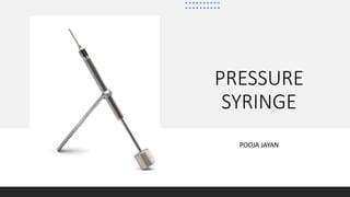
- 4. ENDODONTIC PRESSURE SYRINGE • Using the Pressure Syringe, the practitioner fills the apex first and then back fills the remaining root canal space. • The Pressure Syringe slowly extrudes the filling material, and the flow of material can be stopped instantly. • The needle was inserted into the simulated canal until wall resistance was encountered. • Using a slow, withdrawing-type motion the needle was withdrawn in 3-mm intervals with each quarter turn of the screw until the canal was visibly filled at the orifice.1 1.Hiremath MC, Srivastava P. Comparative evaluation of endodontic pressure syringe, insulin syringe, jiffy tube, and local anesthetic syringe in obturation of primary teeth: An in vitro study. J Nat Sc Biol Med 2016;7:130-5.
- 5. ENDODONTIC PRESSURE SYRINGE • It also eliminates the problems associated with the inaccessibility of posterior teeth and the difficulties encountered when filling narrow and tortuous canals.2 • A 30- gauge needle was selected for filling primary teeth if the canal was able to accommodate a standard size 15-30 endodontic file. 2.Jha M, Patil SD, Sevekar S, Jogani V, Shingare P. Pediatric Obturating Materials And Techniques. Journal of Contemporary Dentistry 2011;1(2):2732.
- 6. INDICATIONS • For the obturation of areas of internal resorption in permanent and primary teeth. • For the obturation of large canals in traumatized or incompletely formed teeth with large apices. • For complete obturation of the root canal space from the retrograde approach.
- 7. ADVANTAGES • The screw type syringe provides complete control and accuracy, assuring precise placement at the apex and total obturation of the root canal.
- 8. TECHNIQUE FILE SIZE IN THE APICAL 1-2 mm PRESSURE SYRINGE NEEDLE GAUGE 15-30 30 ( DARK BLUE) 40 27 (YELLOW) 50 25 ( RED) 60-80 22 (LIGHT BLUE) 90-110 18 (GREEN)
- 15. LITERATURE REVIEW • Dandashi et al. reported fewer voids with pressure syringe technique when compared to the lentulospiral, tuberculin syringe, and packing method.3 • The discrepancies in the results among the studies probably reflects differences in the type of teeth, sample size, needle thickness, and technique used to quantify voids and operator experience. • The endodontic pressure syringe also has been found to be effective in placing ZOE into root canals or primary teeth.4 • Pressure syringes can also be used to deliver intraligamentary local anaesthesia. 3.Dandashi MB, Nazif MM, Zullo T, Elliott MA, Schneider LG, Czonstkowsky M. An in vitro comparison of three endodontic techniques for primary incisors. Pediatr Dent 1993;15:254-6. 4.Berk H, Krakow AA. The endodontic pressure syringe. CDS Rev. 1975;68(9):21‐24.
- 17. LITERATURE REVIEW • Endodontic pressure syringe produced best results in terms of length of root canal obturation when compared with jiffy tube, insulin syringe and local anesthetic syringe (Vashista et al, 2015). • There were no under filled canals by using endodontic pressure syringe. The possible reason could be the design of the pressure syringe in which hub of the syringe being small and thin needle provided a better reach in the middle one third of the canals.5 • More number of overfilled canals were observed with pressure syringe than under filled canals. 5. Dr. Nagarathna, C., Dr. Veena, R. L. Dr. Jaya Agali Ramachandra and Dr. Umapathy Thimmegowda syringe obturation technique – pulpectomy procedures in necrotic primary molars” International Journal of Current Research Available 9, (11), 61473-61477
- 19. LITERATURE REVIEW • According to Grover et al the highest number of overfilled canals was observed with pressure syringe when comparing with lentulospiral, and bi-directional spiral (Subba et al., 1997). This might be due to excessive pressure placed while placing the material into the canal, when the quarter turn of the screw was made. • The consistency of the materials and displacement of rubber stopper, unprecise working length measurement, over instrumentation, resistance disappearance due to inadequate root canal preparation also could be factors for overfilled canals in the study.6 6.Greenberg M: Filling root canals by an injection technique. Dent Dig 67:61-63, 1963
- 20. DRAWBACKS • Overfill is a common clinical finding when using pressure syringe especially in cases of apical resorption. • Difficulties in placing the rubber stop correctly and removing the needle because of the need to refill the hub of the syringe several times during the procedure may lead the clinician to remove and reinsert the syringe repeatedly, which in turn may displace the paste, create voids and thus decrease filling quality. • The need to clean the syringe immediately after use makes this method complex and time consuming. • Needles are flexible and can break when used in tortuous canals.7 • The application of endodontic pressure syringe system in routine dental practice is hampered due to the initial practical difficulty of handling this system by an inexperienced operator.5 7.Memarpour M, Shahidi S, Meshki R. Comparison of different obturation techniques for primary molars by digital radiography. Pediatr Dent. 2013;35(3):236‐240 5. Dr. Nagarathna, C., Dr. Veena, R. L. Dr. Jaya Agali Ramachandra and Dr. Umapathy Thimmegowda syringe obturation technique – pulpectomy procedures in necrotic primary molars” International Journal of Current Research Available 9, (11), 61473-61477
- 21. CONCLUSION Canal filling techniques utilizing the endodontic pressure syringe and the lentulospiral were found to be superior when filling straight canals. The lentulospiral filling method was superior for filling curved canals.Therefore, the canal shape governed the selection of the filling technique.
- 22. REFERENCES 1. Hiremath MC, Srivastava P. Comparative evaluation of endodontic pressure syringe, insulin syringe, jiffy tube, and local anesthetic syringe in obturation of primary teeth: An in vitro study. J Nat Sc Biol Med 2016;7:130-5. 2. Jha M, Patil SD, Sevekar S, Jogani V, Shingare P. Pediatric Obturating Materials And Techniques. Journal of Contemporary Dentistry 2011;1(2):2732. 3. Dandashi MB, Nazif MM, Zullo T, Elliott MA, Schneider LG, Czonstkowsky M. An in vitro comparison of three endodontic techniques for primary incisors. Pediatr Dent 1993;15:254-6. 4. Berk H, Krakow AA. The endodontic pressure syringe. CDS Rev. 1975;68(9):21‐24. 5. Dr. Nagarathna, C., Dr. Veena, R. L. Dr. Jaya Agali Ramachandra and Dr. Umapathy Thimmegowda syringe obturation technique – pulpectomy procedures in necrotic primary molars” International Journal of Current Research Available 9, (11), 61473-61477 6. Greenberg M: Filling root canals by an injection technique. Dent Dig 67:61-63, 1963. 7. Memarpour M, Shahidi S, Meshki R. Comparison of different obturation techniques for primary molars by digital radiography. Pediatr Dent. 2013;35(3):236‐240. 8. Aylard SR, Johnson R. Assessment of filling techniques for primary teeth. Pediatr Dent 1987;9:195-8.