

Recommended
Recommended
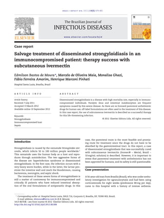
More Related Content
What's hot
What's hot (20)
Viewers also liked
Similar to Strongyloidiasis
Similar to Strongyloidiasis (20)
Recently uploaded
Recently uploaded (20)
Strongyloidiasis
- 1. braz j infect dis. 2012;16(5):479–481 The Brazilian Journal of INFECTIOUS DISEASES www.elsevier.com/locate/bjid Case report Salvage treatment of disseminated strongyloidiasis in an immunocompromised patient: therapy success with subcutaneous ivermectin Edmilson Bastos de Moura∗, Marcelo de Oliveira Maia, Monalisa Ghazi, Fábio Ferreira Amorim, Henrique Marconi Pinhati Hospital Santa Luzia, Brasília, Brazil a r t i c l e i n f o Article history: Received 7 July 2011 Accepted 13 March 2012 Available online 10 September 2012 Keywords: Strongyloidiasis Immunocompromised host Sepsis a b s t r a c t Disseminated strongyloidiasis is a disease with high mortality rate, especially in immuno-compromised individuals. Paralytic ileus and intestinal malabsorption are frequent symptoms caused by this severe disease. As there are no licensed parenteral anthelmintic drugs for human use, off-label formulations are often used in the treatment of this disease. In this case report, the use of subcutaneous ivermectin is described as a successful therapy for this life-threatening infection. © 2012 Elsevier Editora Ltda. All rights reserved. Introduction Strongyloidiasis is caused by the nematode Strongyloides ster-coralis, which infects 50 to 100 million people worldwide.1 This nematode uses the human body as a host and repro-duces through autoinfection. The two aggressive forms of the disease are: hyperinfection syndrome or disseminated strongyloidiasis. In the first case, the infection occurs with a very heavy worm burden, while in the latter, the larvae pen-etrate the intestine wall and reach the bloodstream, causing bacteremia, meningitis, and septic shock. The treatment of these severe forms of strongyloidiasis is still a matter of controversy for immunocompromised and critically ill patients who have reduced intestinal absorp-tion of the oral formulations of antiparasitic drugs. In this case, the parenteral route is the most feasible and promis-ing route for treatment since the drugs do not have to be absorbed by the gastrointestinal tract. In this report, a case of disseminated strongyloidiasis that was successfully cured with subcutaneous ivermectin (Ivomec® – Merial, Brazil – 10mg/mL solution) is described. However, it is important to stress that parenteral treatment with anthelmintics has not been approved for humans, and its safety is still questionable. Case presentation A56-year-oldmanfromBrasília (Brazil),whowas under ambu-latory investigation for agranulocytosis and had been using corticosteroids for eight weeks (prednisone 80mg per day), came to this hospital with a history of intense asthenia. ∗ Corresponding author at: Hospital Santa Luzia, SHLS 716, Conjunto E, Brasília, DF, 70390-903, Brazil. E-mail address: ebmoura@terra.com.br (E.B. Moura). 1413-8670/$ – see front matter © 2012 Elsevier Editora Ltda. All rights reserved. http://dx.doi.org/10.1016/j.bjid.2012.08.008
- 2. 480 braz j infect dis. 2012;16(5):479–481 Fig. 1 – Bronchoalveolar lavage sample demonstrating S. stercoralis larvae. Ziehl-Neelsen stain, x 400. At admission, he was afebrile, hypotensive and, accord-ing to primary examination, presented with leukopenia and eosinophilia, which were treated with 2 g IV cefepime q8h after collecting blood for cultures. On the third day after admission, the patient’s respiratory status deteriorated, and he was transferred to the intensive care unit. Despite non-invasive ventilation, he required intu-bation, mechanical ventilation, and vasopressor support by his fifth day in the hospital. Therefore, antibiotic escalation was prescribed to broaden the drug spectrum. Additionally, computed tomography scans of the thorax and abdomenwere performed. The results of the former exam indicated bilateral areas of pulmonary condensation, and the latter was normal. Lastly, a pulmonary catheterwas placed, which showed hemo-dynamic parameters indicating septic shock. The patient’s blood tests demonstrated a decreased hematocrit value, and therefore he underwent an esopha-gogastroduodenoscopy to rule out bleeding. In this exam, multiple vegetative lesions were observed, which were associ-ated with erosions and diffuse enanthema in the duodenum. On the tenth day of hospitalization, infection with S. stercoralis was diagnosed based on the duodenal biopsy and the bron-choalveolar lavage fluid (Fig. 1), which were both performed simultaneously. As soon as the nematode was detected, treatment was immediately initiated with ivermectin via the nasogastric tube, using 18mg daily, for two days, along with albenda-zol 400mg q12 h. However, on the second day of treatment it became clear that this treatment was not as effective as expected and, with the consent of the patient’s family, the treatment was continued with a veterinary formulation of subcutaneous ivermectin (Ivomec®; Merial, Brazil – 10mg/mL solution), and oral use of the drug was discontinued. This therapy was very successful. On the third day of anthelmintic treatment, the patient was completely removed from vasopressor support. On the tenth day, mechanical ven-tilation was discontinued, and ten days thereafter, the patient was discharged from the ICU. Discussion The patient’s final diagnosis was disseminated strongyloidi-asis with associated septic shock and acute respiratory distress syndrome. The key indicators leading to this diagno-sis were the results from the bronchoalveolar lavage and the esophagogastroduodenoscopy. There have been similar symp-toms related to this disease based on upper gastrointestinal endoscopy.2,3 However, these results are not specific to dis-seminated strongyloidiasis and are also associated with other pathological diseases. Recently, reports4 have indicated that treatment of dis-seminated strongyloidiasis in immunocompromised patients with ivermectin is effective, although it is still a matter of controversy. It has been shownthat enteral use of this medica-tion could cause pharmacokinetic changes in case of paralytic ileus,5 jeopardizing its bioavailability and leading to lower con-centrations of the drug than in its subcutaneous formulation (0.8 vs. 11.4-17.2 ng/mL).6 Conversely, it has also been argued that the oral route provides the appropriate plasma and cere-brospinal fluid concentration of the drug,7 contradicting the previously mentioned findings. Furthermore, the pharmacokinetic properties of iver-mectin may be modified in critically ill patients, because the drug is highly bound to human serum albumin.8 Systemic inflammation causes hypoalbuminemia, and therefore, both free drug concentration and therapeutic action are elevated.6 Nevertheless, a recent study, which was the first to document total and free levels of subcutaneous ivermectin,3 surprisingly found less than 1% of free ivermectin located in the plasma.3 A possible explanation for this finding is the strong binding between the drug and high alpha-1 acid glycoprotein concen-tration, which reduces the drug’s distribution to tissues and contributes to poor therapeutic outcomes.3 Successful7 and unsuccessful3 treatments of disseminated strongyloidiasis were found. In a case report described by Rose et al.,8 the patient was treated exclusively with oral iver-mectin, and this therapy was ineffective; the nematode was not eliminated, and the patient died. In other reports, however, enteral use of this drug successfully eradicated S. stercoralis, but the patient still died due to toxicity complications6,9 or due to the severity of the underlying disease.10,11 In the case described in the present article, favorable outcomes were doc-umented, as no indication of larva were found in the stool or bronchoalveolar lavage fluid, and moreover, the patient sur-vived and was discharged from the intensive care unit and the hospital. The dosage of subcutaneous ivermectin usedwas 15mg per day for the first four days (214g/kg) and then, five days after discontinuation of the parenteral ivermectin, an additional seven-day therapy was initiated (20mg per day; 285g/kg) because the patient presented with worsening neurological status and fever. Additionally, there was evidence indicating that the central nervous systemwas compromised. A fewdays later, the patient’s symptomatologywas diagnosed as herpetic encephalitis. The following table shows both successful and unsuccessful therapeutic procedures. However, because they were performed in different situations, they cannot be directly compared (Fig. 2).
- 3. braz j infect dis. 2012;16(5):479–481 481 Moura et al.∗ Leung et al.3 Salluh et al.9 Hauber et al.10 Turner et al.6 Pacanowski et al.12 Marty et al.2 Ivermectin SC Ivermectin (enteral) Albendazol 2 4 6 8 10 12 14 16 18 Days of treatment Fig. 2 – Therapeutic regimens for disseminated strongyloidiasis.*present study. Daily monitoring of the ivermectin concentration in the serum was essential, as the drug can be toxic to the cen-tral nervous system. The patient’s persistent coma could have been confused with symptoms of toxicity even though the dose (285g/Kg) was eight times lower than what can be toler-ated by humans.12 It could also be due to herpetic encephalitis, but because the concentration of drug in the plasma could not be assessed, this hypothesis could not be confirmed. With this study, the best treatment for disseminated strongyloidiasis cannot be definitely determined. Enteral use of ivermectin can result in therapeutic failure; in addition, in some situations, it does achieve adequate plasma levels. In this patient, parenteral use of this drug was essential for therapeutic success. Considering that data extrapolated from animal experiments are insufficient, it is extremely impor-tant that reports about the usage of parenteral ivermectin in humans are discussed, along with its viability and its toxicity, in order to continue to improve treatment for this devastating form of strongyloidiasis. Conflict of interest All authors declare to have no conflict of interest. Acknowledgements The authors would like to thank Paulo de Oliveira Martins Jr. from the Parasitology Laboratory of the Santa Luzia Hospital for his valuable cooperation. references 1. Genta RM. Global prevalence of strongyloidiasis: critical review with epidemiologic insights into the prevention of disseminated disease. Rev Infect Dis. 1989;11:755–67. 2. Marty FM, Lowry CM, Rodriguez M, et al. Treatment of human disseminated strongyloidiasis with a parenteral veterinary formulation of ivermectin. Clin Infect Dis. 2005;41:e5–8. 3. Leung V, Al-Rawahi GN, Grant J, Fleckenstein L, Bowie W. Failure of subcutaneous ivermectin in treating Strongyloides hyperinfection. Am J Trop Med Hyg. 2008;79:853–5. 4. Luna OB, Grasselli R, Ananias M, et al. Estrongiloidíase disseminada: diagnóstico e tratamento. Rev Bras Ter Int. 2007;19:463–8. 5. Nonaka D, Takaki K, Tanaka M, et al. Paralytic ileus due to strongyloidiasis: case report and review of the literature. Am J Trop Med Hyg. 1998;59:535–8. 6. Turner SA, Maclean JD, Fleckenstein L, Greenaway C. Parenteral administration of ivermectin in a patient with disseminated strongyloidiasis. Am J Trop Med Hyg. 2005;73:911–4. 7. Salluh JIF, Feres GA, Velasco E, Holanda GS, Toscano L, Soares M. Successful use of parenteral ivermectin in an immunosuppressed patient with disseminated strongyloidiasis and septic shock. Intensive Care Med. 2005;31:1292. 8. Rose CE, Paciullo CA, Kelly DR, Dougherty MJ, Fleckenstein LL. Fatal outcome of disseminated strongyloidiasis despite detectable plasma and cerebrospinal levels of orally administered ivermectin. J Parasitol Res. 2009:818296. 9. Hauber HP, Galle J, Chiodini PL, et al. Fatal outcome of a hyperinfection syndrome despite successful eradication of Strongyloides with subcutaneous ivermectin. Infection. 2005;33:383–6. 10. Chiodini PL, Reid AJ, Wiselka MJ, Firmin R, Foweraker J. Parenteral ivermectin in Strongyloides hyperinfection. Lancet. 2000;355:43–4. 11. Pacanowski J, Santos MD, Roux A, et al. Subcutaneous ivermectin as a safe salvage therapy in Strongyloides stercoralis hyperinfection syndrome: a case report. Am J Trop Med Hyg. 2005;73:122–4. 12. Guzzo CA, Furtek CI, Porras AG, et al. Safety, tolerability, and pharmacokinetics of escalating high doses of ivermectin in healthy adult subjects. J Clin Pharmacol. 2002;42:1122–33.