College Call Girls Vyasarpadi Whatsapp 7001305949 Independent Escort Service
Csf ada hiv tbm
1. The validity of cerebrospinal fluid parameters for the diagnosis of
tuberculous meningitis§
Lely Solari a,b,
*, Alonso Soto a,c
, Juan Carlos Agapito f
, Vilma Acurio c
, Dante Vargas c
,
Tulia Battaglioli a
, Roberto Alfonso Accinelli d,e
, Eduardo Gotuzzo e,f
, Patrick van der Stuyft a,g
a
Unit of General Epidemiology and Disease Control, Institute of Tropical Medicine of Antwerp, Nationalestraat 155, B-2000 Antwerp, Belgium
b
Unidad de Ana´lisis y Generacio´n de Evidencias en Salud Publica (UNAGESP), Instituto Nacional de Salud del Peru, Lima, Peru
c
Department of Medicine, Hospital Nacional Hipolito Unanue, Lima, Peru
d
Instituto de Investigaciones de la Altura, Universidad Peruana Cayetano Heredia, Lima, Peru
e
Hospital Nacional Cayetano Heredia, Lima, Peru
f
Universidad Peruana Cayetano Heredia, Lima, Peru
g
Department of Public Health, Ghent University, Ghent, Belgium
1. Introduction
The diagnosis of tuberculous meningitis (TBM) continues to be a
clinical challenge, even after the introduction of molecular tests.1
The physiopathology of this condition, in which disproportionate
inflammatory phenomena rather than numbers of circulating
bacteria play a role, hinders bacteriological diagnosis, and the
available microbiological tests fail to attain the accuracy standards
required.2
As a result, most guidelines for the diagnosis and management
of TBM agree on the use of simple cerebrospinal fluid (CSF)
analyses, such as determining glucose and protein levels and the
number and formula of leukocytes, to guide decision-making.3,4
Computed tomography (CT) scans and magnetic resonance
imaging (MRI)5
and other biochemical analyses of CSF, in particular
adenosine deaminase activity (ADA),6,7
have also been advocated.
Peruvian guidelines recommend the use of changes in protein,
glucose, chloride, and ADA levels and the presence of lymphocytic
pleocytosis in CSF as key elements for guiding the diagnosis of
TBM.8
However, evidence on the utility of these tests for decision-
making in the first hours after admission, when appropriate
initiation of anti-tuberculous treatment can prevent disability and
mortality, is quite limited.9
The few studies that have addressed
the predictive value of these tests or their combinations have
primarily focused on differentiating between TBM and acute
bacterial meningitis.10–12
The objective of this study was to evaluate the validity of these
laboratory tests in CSF, in isolation or in combination, for the
diagnosis of TBM in patients with a clinical suspicion of meningitis.
International Journal of Infectious Diseases 17 (2013) e1111–e1115
A R T I C L E I N F O
Article history:
Received 18 February 2013
Received in revised form 3 June 2013
Accepted 4 June 2013
Corresponding Editor: Eskild Petersen,
Aarhus, Denmark
Keywords:
Tuberculous meningitis
Adenosine deaminase
Sensitivity and specificity
Diagnosis
Neurological
S U M M A R Y
Objectives: To assess the diagnostic validity of laboratory cerebrospinal fluid (CSF) parameters for
discriminating between tuberculous meningitis (TBM) and other causes of meningeal syndrome in high
tuberculosis incidence settings.
Methods: From November 2009 to November 2011, we included patients with a clinical suspicion of
meningitis attending two hospitals in Lima, Peru. Using a composite reference standard, we classified
them as definite TBM, probable TBM, and non-TBM cases. We assessed the validity of four CSF
parameters, in isolation and in different combinations, for diagnosing TBM: adenosine deaminase
activity (ADA), protein level, glucose level, and lymphocytic pleocytosis.
Results: One hundred and fifty-seven patients were included; 59 had a final diagnosis of TBM (18
confirmed and 41 probable). ADA was the best performing parameter. It attained a specificity of 95%, a
positive likelihood ratio of 10.7, and an area under the receiver operating characteristics curve of 82.1%,
but had a low sensitivity (55%). None of the combinations of CSF parameters achieved a fair performance
for ‘ruling out’ TBM.
Conclusions: Finding CSF ADA greater than 6 U/l in patients with a meningeal syndrome strongly
supports a diagnosis of TBM and permits the commencement of anti-tuberculous treatment.
ß 2013 International Society for Infectious Diseases. Published by Elsevier Ltd. All rights reserved.
§
This study was presented in part as an abstract at the 43rd
Union World
Conference on Lung Health, Kuala Lumpur, Malaysia, November 13–17, 2012.
* Corresponding author. Tel.: +32 3 247 62 55; fax: +32 3 247 66 58.
E-mail address: lelysol@hotmail.com (L. Solari).
Contents lists available at ScienceDirect
International Journal of Infectious Diseases
journal homepage: www.elsevier.com/locate/ijid
1201-9712/$36.00 – see front matter ß 2013 International Society for Infectious Diseases. Published by Elsevier Ltd. All rights reserved.
http://dx.doi.org/10.1016/j.ijid.2013.06.003
2. 2. Materials and methods
2.1. Setting
The study was performed in Lima, Peru. Peru is a country with a
high incidence of tuberculosis (101/105
) and a concentrated HIV
epidemic.13
Adult cases with a clinical suspicion of meningitis are
routinely referred to third-level hospitals where they undergo a
lumbar puncture. The most frequent causative agents in this
context are considered to be Mycobacterium tuberculosis, Crypto-
coccus neoformans, common bacteria, and enteroviruses.14
2.2. Diagnostic parameters evaluated
The diagnostic parameters in CSF considered in the Peruvian
national guidelines were evaluated at their respective cut-off
points: elevated proteins (>50 mg/dl), decreased glucose
(<50 mg/dl), decreased chloride (<100 mg/dl), lymphocytic pleo-
cytosis (CSF white cell count of >10 cells/mm3
, with lymphocyte
predominance >50%), and elevated ADA level (>6 U/l). The
chloride level was not included in this study as it is neither
routinely performed nor readily available at referral hospitals.
2.3. Patient recruitment and procedures
The sample size needed was 139, considering an overall
accuracy of the best combination of predictors of 90% and a
precision of 5%. All patients older than 18 years with a clinical
suspicion of meningitis, hospitalized in one of two third-level
hospitals (Hipolito Unanue and Cayetano Heredia) from November
2009 to November 2011 were invited to participate in the study. A
clinical suspicion of meningitis was defined as having any
combination of the following symptoms: headache, irritability,
vomiting, fever, neck stiffness, convulsions, focal neurologic deficit,
and altered consciousness or lethargy, with no other general
medical condition explaining them. Patients already receiving
specific treatment were excluded (for instance patients with
cryptococcal meningitis attending with recurrence of their
symptoms, patients already being treated for pulmonary tubercu-
losis who had developed neurologic symptoms, etc.).
All included patients underwent a lumbar puncture using
standard procedures. CSF samples were sent within 1 h to the
laboratory to perform microbiological (acid-fast bacillus stain
(AFB), culture for mycobacteria in Ogawa medium, Gram stain,
culture for common bacteria, cryptococcal antigen agglutination
test), molecular (PCR for Mycobacterium tuberculosis; IS6110 PCR,
Qiagen Multiplex PCR),15
cytological (total white cell count and
determination of the percentage of lymphocytes), and biochemical
(glucose, protein, ADA) analyses. According to the clinical findings,
the attending physicians requested further tests/procedures
(biopsy or culture of other body fluids, lymph node aspiration, etc.).
2.4. Definition of TBM
Our reference standard for the diagnosis of TBM contemplated
two categories: ‘definite’ TBM and ‘probable’ TBM. All cases were
assigned to one of these categories by a data analyst who was
blinded to the results of the evaluated CSF parameters. Definite
TBM was defined as the presence of AFB in CSF smears, or positive
CSF culture for M. tuberculosis, or positive CSF PCR test.7
Probable
TBM was defined as a clinical suspicion of meningitis (as described
above), with negative Gram stain and cultures for bacteria,
negative cryptococcal latex agglutination test and cultures for
fungi, and at least one of the following: (1) bacteriological evidence
of tuberculosis in other organs (positive culture for M. tuberculosis
in other body fluids or tissues or biopsies, with histopathological
findings of caseous necrosis or granulomas); (2) good response to
anti-tuberculous therapy, defined as complete resolution of the
constitutional signs at 1 month after treatment initiation. For
patients not completing 1 month of follow-up due to death, an
expert panel defined whether the case was probable TBM or not.
TBM was defined as definite or probable TBM. All other patients
were classified as non-TB, and a diagnosis was reached according
to each etiology, for instance: bacterial and fungal meningitis were
microbiologically confirmed by cultures or presence of antigen;
viral meningitis was defined as a compatible clinical presentation,
an abnormal CSF, and complete resolution of symptoms without
antibiotic, antifungal, or anti-tuberculous treatment, or a positive
PCR for viruses in CSF; metabolic conditions were diagnosed on the
basis of laboratory blood tests, etc.
2.5. Analysis
Patients found to have more than one diagnosis (for example
tuberculous and bacterial meningitis, or tuberculous and fungal
meningitis) were excluded from the analysis. Differences between
TBM patients and non-TBM patients with regard to the CSF
parameters were compared using their actual values and
dichotomized according to the cut-off points suggested in the
Peruvian guidelines. To test for significance, we used the Mann–
Whitney test and Chi-square test for numerical and categorical
variables, respectively. We calculated areas under the receiver
operating characteristic (ROC) curve for each parameter, and
sensitivity, specificity, and positive and negative likelihood ratios
at the suggested cut-off levels. Ninety-five percent confidence
intervals (95% CI) were constructed for all estimates. A positive
likelihood ratio of 10 and a negative likelihood ratio of 0.10
were considered to provide convincing evidence in favor or against
the diagnosis of TBM, respectively.16
As a second step, the
diagnostic accuracy of all possible combinations of two, three,
or four parameters were evaluated, as well as having one, two,
three, or four positive parameters present. All statistical analyses
were performed with STATA version 11.0 (Stata Corp., College
Station, TX, USA).
2.6. Ethical aspects
All included patients, or a direct relative for those with altered
consciousness, gave informed consent to participate in the study.
The ethics committees of the Universidad Peruana Cayetano
Heredia, both participating hospitals, and the Institute of Tropical
Medicine, Antwerp, approved the study.
3. Results
One hundred and fifty-seven patients fulfilled the inclusion
criteria and agreed to participate in the study. Two patients were
excluded from the analysis given co-infection with two pathogens
(they had HIV/AIDS, TBM confirmed by a positive PCR in CSF, and
meningeal cryptococcosis confirmed by a positive culture). The
median age of the 155 patients constituting the study group was 35
years (interquartile range 26–54 years) and 109 (70.3%) were male.
Fifty-nine (38.1%) had a diagnosis of TBM. Eighteen (30.5%) were
definite TBM and 41 (69.5%) were probable TBM. Of the latter, nine
had M. tuberculosis isolated in specimens from another body site,
28 had a good response to tuberculosis treatment, and four died
before the 1 month of treatment follow-up but were identified as
TBM by the expert panel. The most frequent diagnoses in non-TBM
cases (n = 96) were viral meningitis in 19 (19.7%), cryptococcal
meningitis in 12 (12.5%), liver and other metabolic encephalopa-
thies in eight (8.3%), bacterial meningitis in six (6.3%), and other
causes of meningeal syndrome (meningeal carcinomatosis, sepsis
L. Solari et al. / International Journal of Infectious Diseases 17 (2013) e1111–e1115e1112
3. of other origin, toxoplasmic encephalitis, subarachnoid hemor-
rhage, epilepsy) in 51 (53.1%) patients. HIV infection was present in
22 (38%) of the patients in the TBM group and 33 (34%) of the
patients in the non-TBM group.
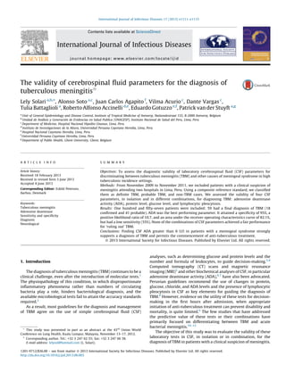
Figure 1 shows the distribution of the three investigated
diagnostic parameters, which were continuous variables: ADA,
protein level, and glucose level in TBM (probable and definite) and
non-TBM patients. All of them differed significantly between the
TBM and non-TBM groups at a level of p 0.001. Although a
comparison between definite and probable TBM was not an
objective of the study (and power was limited for a comparison
between the subgroups), there were no significant differences
between the two groups except in the glucose level (median of
32.5 mg/dl in the definite TBM group vs. 44 mg/dl in the probable
TBM group).
The diagnostic performance of the evaluated parameters for the
diagnosis of TBM is shown in Table 1. ADA was the parameter that
in isolation performed the best, followed by protein level (area
under the ROC curve 82.1% and 75.5%, respectively). Protein level
was the most sensitive test, while ADA was the most specific one.
The latter attained a positive likelihood ratio of 10.7. Noteworthy,
four out of the 34 (12%) patients who did not fulfill any of the four
criteria had a final diagnosis of TBM.
When the 41 probable TBM cases were excluded from analysis,
this did not affect the diagnostic accuracy estimates for ADA: it had
a sensitivity of 55.6%, a specificity of 94.9%, a positive likelihood
ratio of 10.9, and a negative likelihood ratio of 0.5.
Although the study was underpowered for comparisons
between subgroups, we analyzed the performance of the four
parameters according to the HIV status of the patients. Glucose
levels and lymphocytic pleocytosis performed less well in HIV
patients, with a significantly decreased area under the ROC curve.
However, ADA and protein levels did not have a significantly
decreased area under the ROC curve (85% vs. 76% for ADA in HIV-
negative and HIV-positive patients, respectively).
We evaluated the performance of all possible combinations of
the CSF parameters. We considered as patients fulfilling a
combination, those who tested positive for at least one of the
parameters included in it, and as patients not fulfilling it, those
testing negative for all the parameters included in the combina-
tion. Table 2 shows the results of the best performing combinations
in terms of positive and negative likelihood ratios. None of the
combinations attained our definition of fair evidence of the
presence or absence of TBM. We also assessed the performance of
combinations defined as having at least three positive parameters
(any of them) and of having all four parameters positive. The
sensitivity, specificity, positive likelihood ratio, and negative
likelihood ratio were 64%, 82%, 7.66, and 0.4, respectively, for
having at least three positive parameters, and 32%, 100%, 61, and
0.7, respectively, for having all four parameters positive. Nineteen
patients (32.2% of TBM patients) belonged to this last category.
4. Discussion
Our most relevant finding was that out of the four evaluated CSF
parameters, the best performing one for ‘ruling in’ TBM was ADA,
with a specificity of 95% at a cut-off point of 6 U/l. However, its
sensitivity was low (55%). No combinations of the parameters
reached the accepted standard for ‘ruling out’ TBM (a negative
likelihood ratio of 0.10).
A limitation of our study is that only 30% of our TBM cases were
bacteriologically confirmed. However, this proportion lies in the
expected range according to the literature.17
Our definition of
‘probable’ TBM discarded patients with other likely diagnoses, but
the response to anti-tuberculous therapy (and expert consensus
for patients under therapy dying before 1 month of follow-up),
could be judged as suboptimal evidence for TBM diagnosis. This is
mainly because conditions such as viral meningitis could mimic a
good response to TB therapy. However, this would not change our
main conclusion concerning the high specificity of ADA: viral
meningitis does not usually elevate ADA levels,18,19
and if we had
classified patients with viral meningitis as TBM, we would have
underestimated its specificity for TBM. Furthermore, it is worth
highlighting that, despite our study being underpowered for a
subgroup analysis, we found no differences between the ‘con-
firmed’ and the ‘probable’ TBM subgroups except in the glucose
level. Finally, the study was performed in Peru, a country with a
low HIV prevalence, a high tuberculosis incidence, and decreasing
0
20
40
60
80
No TBM Probable TBM Definite TBM
ADA levels in CSF according to TBM category
0
100
200
300
400
No TBM Probable TBM Definite TBM
Protein levels in CSF according to TBM category
0
50
100
150
No TBM Probable TBM Definite TBM
Glucose levels in CSF according to TBM category
ADAlevelinCSF(U/L)ProteinlevelinCSF(mg/dl)GlucoselevelinCSF(mg/dl)
Figure 1. Distribution of diagnostic parameters among TBM (confirmed and
probable) and non-TBM patients.
L. Solari et al. / International Journal of Infectious Diseases 17 (2013) e1111–e1115 e1113
4. morbidity due to bacterial and fungal meningitides. Our results
may not be generalizable beyond settings with comparable
characteristics.
The study has an important strength: the wider clinical spectrum
of patients included in comparison to previously published works.
Other studies have focused on distinguishing TBM from specific
meningitides, generally acute bacterial meningitis,11,12
excluding
patients with uncertain diagnoses. The latter are precisely the ones
that could benefitfromthe use ofclinicaltools.20
Our study makesan
attempt to capture the real diagnostic challenge in clinical practice,
where TBM is suspected in a wide array of clinical presentations and
clinicians have to make quick decisions concerning treatment.
Additionally, although TBM is a condition with decreasing preva-
lence, we managed to include a fair number of patients, which gave
us power for statistical comparisons.
Our main finding, the utility of ADA for the diagnosis of TBM,
has been the subject of debate for many years, and there are two
meta-analyses published on the subject. Tuon et al.21
found results
similar to ours: a specificity of 96% and a sensitivity of 59% for ADA
values higher than 8U/l. Xu et al.22
reported a sensitivity of 79% and
a specificity of 91% by pooling studies with various ADA cut-off
points. Studies published after these meta-analyses have also
found that the use of ADA for the diagnosis of TBM has significant
clinical utility.23,24
However, accuracy estimates vary according to
the setting, the patient mix, cut-off point, and laboratory
specifications.6
In settings with a high prevalence of acute bacterial
meningitis, ADA can give false-positive results, and most studies
assessing clinical predictors for TBM have been conducted in such
populations.10,11
This is possibly the reason why a group of experts
who have developed a definition for TBM have refrained from the
use of this test.7
Actually, Peruvian guidelines are amongst the few
existing ones that explicitly advocate the determination of ADA in
CSF to diagnose TBM.8
We feel, in the light of new evidence18
and
our present findings, that the use of this test should be further
evaluated. Compared to other tools such as CT scans25
and MRI,
which have a specificity up to 90%,26
and appraisal of macroscopic
CSF appearance, which as part of a score achieves a specificity of
77%,12
ADA can make a more important contribution to the
diagnosis of TBM. With a positive likelihood ratio of 10.7 and a
pretest probability of 38% as in our setting, the post-test
probability of having TBM becomes 87%, and thus treatment
initiation should be offered. In our series, 56% of the TBM cases
would have benefited from a decision made on this basis. However,
the low sensitivity (55%) precludes decision-making in patients
with a negative ADA test, and the addition of the other parameters
evaluated only marginally increased the number of TBM patients
correctly diagnosed.
In recent years, new diagnostic assays, in particular molecular
techniques like GeneXpert MTB/RIF, have been developed, and
these could contribute to the diagnosis of extrapulmonary forms of
tuberculosis;27–30
however their clinical utility needs further
assessment. Furthermore, rolling out these tests will take time and
consume considerable resources. Therefore, the use of diagnostic
tools that are already implemented, cheap, and easy to perform
should be optimized. Our study shows that ADA in CSF above 6 U/l
in patients with a meningeal syndrome has a high positive
likelihood ratio for TBM. Taking into account the specific clinical
context, this should permit the decision to initiate treatment,
particularly in settings like ours, with a high prevalence of TBM and
low prevalence of acute bacterial meningitis. On the other hand,
none of the evaluated combinations of CSF parameters allows the
condition to be ‘ruled out’. More research should be done on the
clinical utility of including ADA alongside other simple diagnostic
tests in diagnostic algorithms for TBM.
Acknowledgements
To Francine Matthysy
for her contribution to the development of
the study protocol.
Table 1
Diagnostic accuracy of the cerebrospinal fluid parameters for the diagnosis of tuberculous meningitis
CSF parameter Sensitivity
(95% CI)
Specificity
(95% CI)
Positive likelihood
ratio (95% CI)
Negative likelihood
ratio (95% CI)
Area under the ROC
curve (95% CI)
ADA 6 U/l 55.9%
(43.3–67.9%)
94.8%
(88.4–97.8%)
10.7
(4.4–26.0)
0.5
(0.4–0.6)
82.1%
(75.0–87.6%)
Lymphocytic pleocytosisa
62.7%
(50.0–73.9%)
77.1%
(67.7–84.4%)
2.74
(1.8–4.2)
0.5
(0.3–0.7)
54.6%
(42.8–65.7%)
Protein level 45 mg/dl 81.4%
(70.0–89.3%)
53.1%
(43.2–62.8%)
1.74
(1.4–2.2)
0.4
(0.2–0.6)
75.5%
(67.9–82.0%)
Glucose level 50 mg/dl 69.5%
(56.9–79.8%)
63.5%
(53.6–72.5%)
1.91
(1.4–2.6)
0.5
(0.3–0.7)
70.5%
(62.5–77.4%)
CSF, cerebrospinal fluid; CI, confidence interval; ROC, receiver operating characteristics; ADA, adenosine deaminase activity.
a
Defined as 10 cells/mm3
and 50% lymphocytes.
Table 2
Diagnostic accuracy of the combinations of cerebrospinal fluid parameters for the diagnosis of tuberculous meningitis
Combination of CSF laboratory parameters (tests) Sensitivity
(95% CI)
Specificity
(95% CI)
Positive likelihood
ratio (95% CI)
Negative likelihood
ratio (95% CI)
Area under the ROC
curve (95% CI)
Combination of two parameters
ADA 6 U/l or lymphocytic pleocytosis 81.4%
(69.6–89.3%)
71.9%
(62.2–79.9%)
2.9
(2.1–4.1)
0.3
(0.2–0.5)
75.1%
(67.2–81.5%)
Lymphocytic pleocytosis or protein 45 mg/dl 89.8%
(80.0–95.3%)
45.8%
36.2–55.8%)
1.7
(1.4–2.0)
0.2
(0.1–0.5)
69.2%
(61.1–76.2%)
Combinations of three parameters
ADA 6 U/l or lymphocytic pleocytosis or
glucose 50 mg/dl
89.8%
(79.5–95.3%)
50.0%
40.2–59.8%)
1.8
(1.4–2.2)
0.2
(0.1–0.5)
70.1%
(63.1–78.0%)
Combination of four parameters
ADA 6 U/l or lymphocytic pleocytosis or protein
45 mg/dl or glucose 50 mg/dl
93.2%
(83.8–97.3%)
31.3%
22.9–41.1%)
1.4
(1.2–1.6)
0.2
(0.1–0.6)
66.8%
(59.1–74.4%)
CSF, cerebrospinal fluid; CI, confidence interval; ROC, receiver operating characteristics; ADA, adenosine deaminase activity.
L. Solari et al. / International Journal of Infectious Diseases 17 (2013) e1111–e1115e1114
5. To Daniela Salazar and Yenny Bravo for their active participa-
tion in the patient recruitment. This study was funded by the
Damien Foundation. LS holds a PhD scholarship from the Belgian
Development Cooperation.
Conflict of interest: No conflict of interest to declare.
References
1. Takahashi T, Tamura M, Takasu T. The PCR-based diagnosis of central nervous
system tuberculosis: up to date. Tuberc Res Treat 2012;2012:831292.
2. Thwaites GE, Simmons CP, Than Ha QN, Thi Hong CT, Phuong MP, Thi DN, et al.
Pathophysiology and prognosis in Vietnamese adults with tuberculous menin-
gitis. J Infect Dis 2003;188:1105–15.
3. NICE. Tuberculosis. Clinical diagnosis and management of tuberculosis, and
measures for its prevention and control. UK: National Institute for Health and
Care Excellence; 2011. Available at: http://www.nice.org.uk/cg033 (accessed
September 15, 2012).
4. Improving the diagnosis and treatment of smear-negative pulmonary
and extrapulmonary tuberculosis among adults and adolescents. Recommen-
dations for HIV-prevalent and resource-constrained settings. WHO/HTM/TB/
2007.379 WHO/HIV/2007.1. Geneva: World Health Organization;
2007. Available at: http://www.who.int/tb/publications/2007/en/index.html
(accessed July 8, 2012).
5. Semlali S, El Kharras A, Mahi M, Hsaini Y, Benameur M, Aziz N, et al. [Imaging
features of CNS tuberculosis]. J Radiol 2008;89:209–20.
6. Belagavi AC, Shalini M. Cerebrospinal fluid C reactive protein and adenosine
deaminase in meningitis in adults. J Assoc Physicians India 2011;59:557–60.
7. Marais S, Thwaites G, Schoeman JF, Torok ME, Misra UK, Prasad K, et al.
Tuberculous meningitis: a uniform case definition for use in clinical research.
Lancet Infect Dis 2010;10:803–12.
8. Norma tecnica de salud para el control de la tuberculosis, 2006. Direccion
General de Salud de las Personas y Estrategia Sanitaria Nacional de Prevencion y
Control de la Tuberculosis MdSdP. Peru: Ministerio de Salud; 2006. Available at:
http://www.minsa.gob.pe/portada/esntbc_tbnormas.asp (accessed September
15, 2012).
9. George EL, Iype T, Cherian A, Chandy S, Kumar A, Balakrishnan A, et al. Predictors
of mortality in patients with meningeal tuberculosis. Neurol India 2012;60:
18–22.
10. Moghtaderi A, Alavi-Naini R, Izadi S, Cuevas LE. Diagnostic risk factors to
differentiate tuberculous and acute bacterial meningitis. Scand J Infect Dis
2009;41:188–94.
11. Thwaites GE, Chau TT, Stepniewska K, Phu NH, Chuong LV, Sinh DX, et al.
Diagnosis of adult tuberculous meningitis by use of clinical and laboratory
features. Lancet 2002;360:1287–92.
12. Youssef FG, Afifi SA, Azab AM, Wasfy MM, Abdel-Aziz KM, Parker TM, et al.
Differentiation of tuberculous meningitis from acute bacterial meningitis using
simple clinical and laboratory parameters. Diagn Microbiol Infect Dis
2006;55:275–8.
13. Global tuberculosis report 2012. Geneva: World Health Organization; 2012.
Available at: http://www.who.int/tb/publications/global_report/en/ (accessed
November 17, 2012).
14. Concepcio´n Urteaga LA, Alquı´zar Horna O, Correa Aldave J, Zavaleta Gutie´rrez F,
Zavaleta Gutie´rrez J, Concepcio´n Urteaga R. Caracterı´sticas clı´nicas de la
meningoencefalitis tuberculosa/Clinic characteristics of the tubercular menin-
goencephalitis. Bol Soc Peru Med Interna 1996;9:140–7.
15. Deshpande PS, Kashyap RS, Ramteke SS, Nagdev KJ, Purohit HJ, Taori GM, et al.
Evaluation of the IS6110 PCR assay for the rapid diagnosis of tuberculous
meningitis. Cerebrospinal Fluid Res 2007;4:10.
16. Deeks JJ, Altman DG. Diagnostic tests 4: likelihood ratios. BMJ 2004;329:168–9.
17. Kumar R, Singh SN, Kohli N. A diagnostic rule for tuberculous meningitis. Arch
Dis Child 1999;81:221–4.
18. Karsen H, Koruk ST, Karahocagil MK, Calisir C, Baran FC. Comparative analysis of
cerebrospinal fluid adenosine deaminase activity in meningitis. Swiss Med Wkly
2011;141:w13214.
19. Choi SH, Kim YS, Bae IG, Chung JW, Lee MS, Kang JM, et al. The possible role of
cerebrospinal fluid adenosine deaminase activity in the diagnosis of tubercu-
lous meningitis in adults. Clin Neurol Neurosurg 2002;104:10–5.
20. Knottnerus JA, van Weel C, Muris JW. Evaluation of diagnostic procedures. BMJ
2002;324:477–80.
21. Tuon FF, Higashino HR, Lopes MI, Litvoc MN, Atomiya AN, Antonangelo L, et al.
Adenosine deaminase and tuberculous meningitis—a systematic review with
meta-analysis. Scand J Infect Dis 2010;42:198–207.
22. Xu HB, Jiang RH, Li L, Sha W, Xiao HP. Diagnostic value of adenosine deaminase
in cerebrospinal fluid for tuberculous meningitis: a meta-analysis. Int J Tuberc
Lung Dis 2010;14:1382–7.
23. Gupta BK, Bharat A, Debapriya B, Baruah H. Adenosine deaminase levels in CSF
of tuberculous meningitis patients. J Clin Med Res 2010;2:220–4.
24. Sun Q, Sha W, Xiao HP, Tian Q, Zhu H. Evaluation of cerebrospinal fluid
adenosine deaminase activity for the differential diagnosis of tuberculous
and nontuberculous meningitis. Am J Med Sci 2012;344:116–21.
25. Pienaar M, Andronikou S, van Toorn R. MRI to demonstrate diagnostic features
and complications of TBM not seen with CT. Childs Nerv Syst 2009;25:941–7.
26. Pui MH, Memon WA. Magnetic resonance imaging findings in tuberculous
meningoencephalitis. Can Assoc Radiol J 2001;52:43–9.
27. Hillemann D, Rusch-Gerdes S, Boehme C, Richter E. Rapid molecular detection
of extrapulmonary tuberculosis by the automated GeneXpert MTB/RIF system.
J Clin Microbiol 2011;49:1202–5.
28. Ioannidis P, Papaventsis D, Karabela S, Nikolaou S, Panagi M, Raftopoulou E,
et al. Cepheid GeneXpert MTB/RIF assay for Mycobacterium tuberculosis detec-
tion and rifampin resistance identification in patients with substantial clinical
indications of tuberculosis and smear-negative microscopy results. J Clin Micro-
biol 2011;49:3068–70.
29. Kosters K, Nau R, Bossink A, Greiffendorf I, Jentsch M, Ernst M, et al. Rapid
diagnosis of CNS tuberculosis by a T-cell interferon-gamma release assay on
cerebrospinal fluid mononuclear cells. Infection 2008;36:597–600.
30. Shao Y, Xia P, Zhu T, Zhou J, Yuan Y, Zhang H, et al. Sensitivity and specificity of
immunocytochemical staining of mycobacterial antigens in the cytoplasm of
cerebrospinal fluid macrophages for diagnosing tuberculous meningitis. J Clin
Microbiol 2011;49:3388–91.
L. Solari et al. / International Journal of Infectious Diseases 17 (2013) e1111–e1115 e1115