2. General Principles
84
Deep transverse friction
Deep transverse friction (although the word friction is techni-
cally incorrect and would be better replaced by ‘massage’) is a
specific type of connective tissue massage2
developed in an
empirical way by Cyriax.3
Transverse massage is applied by the finger(s) directly to the
lesion and transverse to the direction of the fibres. It can be
used after an injury and for mechanical overuse in muscular,
tendinous and ligamentous structures.4–6
In many instances the
friction massage is an alternative to infiltrations with steroids.
Friction is usually slower in effect than injections but leads to
a physically more fundamental resolution, resulting in more
permanent cure and less recurrence. Whereas steroid injection
is usually successful in 1–2 weeks, deep friction may require
up to 6 weeks to have its full effect.
The technique is often used before and in conjunction with
mobilization techniques. In minor muscular tears, friction is
usually followed by active movement, in ligamentous tears by
passive movement and in tendinous lesions by active unloaded
movements until full resolution has been achieved.
It is vital that transverse massage is performed only at the
site of the lesion. The effect is so local that, unless the finger
is applied to the exact site and friction given in the right direc-
tion, relief cannot be expected.
Over the years, and unfortunately enough, the technique
has developed a reputation for being very painful for the
patient. However, pain during friction massage is usually the
result of a wrong indication, a wrong technique or an unac-
customed amount of pressure. Friction massage applied cor-
rectly will quickly result in an analgesic effect over the treated
area and is seldom a painful experience for the patient.
Mode of action
Transverse massage should be used empirically for what it is
and what it achieves; there is no scientific proof for any pos-
tulates about the underlying mechanism of action.
Only a few studies exist,7,8,9
and more research is urgently
needed. However, experienced therapists know in what
kind of soft tissues they can expect good results with trans-
verse massage and where the technique does not work. Trans-
verse massage either is effective quickly (after 6–10 sessions)
or not at all. Advice on indications, contraindications and
modalities of the technique that are given in this book rely
solely on the experiences of the authors and not on scientific
research.
However, although the exact mode of action is not known,
some theoretical explanations have been put forward. It has
been hypothesized that friction has a local pain-diminishing
effect and results in better alignment of connective tissue
fibrils.
Relief of pain
It is a common clinical observation that application of local
transverse friction leads to immediate pain relief – the patient
cure? And what is the patient’s attitude towards certain thera-
peutic methods such as corticosteroids and manipulation?
Orthopaedic medicine based on a detailed functional exami-
nation requires more knowledge, skill, time and effort from
the physician than just to order technical investigations, but
leads to greater professional interest, more appropriate diagno-
sis and a higher degree of patient satisfaction. Clear diagnosis
and consequent selection of treatment on logical grounds also
leads to better understanding between doctors and therapists.
Because the two groups work with the same types of patient,
they must share a common approach. Therapists should no
longer be regarded purely as technicians who listen to the
physician and carry out orders. On the contrary, they should
be aware that they have diagnostic and therapeutic responsi-
bilities. Their opinion must be taken seriously and is important
to avoid unnecessary delay in achieving a satisfactory outcome.
Techniques
The treatment techniques used in orthopaedic medicine thus
depend entirely on the type of disorder. The different types
of treatment we describe are:
• Manipulation techniques (rapid, small-amplitude, thrusting
passive movement – also called ‘grade C mobilization’) are
used to reduce small cartilaginous displaced fragments
both in the spine and in peripheral joints (loose bodies).
Manipulation is also called for to restore normal mobility
in a joint restricted by ligamentous adhesion and in
subluxation of bones.
• Gentle passive mobilizations (grade A and B
mobilizations) are used to stretch capsular adhesions and
to improve the function of ligaments and tendons. In the
treatment of traumatic injuries they are often used in
combination with deep transverse massage.
• Active movements and proprioceptive training are needed
in the treatment of functional disorders and instability.
In the treatment of minor muscular tears they are very
useful in avoiding the formation of abnormal intralesional
adhesion formation.
• Injection and infiltration techniques are used to reduce
traumatic or rheumatoid inflammation. They are most
valuable in arthritis, bursitis, ligamentous and tendinous
lesions and in neurocompression syndromes.
• Deep friction is a very useful technique in treating
traumatic and overuse soft tissue lesions. The rationale for
using deep friction (which is in fact a form of soft tissue
mobilization) is supported by experimental studies of
the past several decades that confirm and explain the
beneficial effects of activity on the healing musculoskeletal
tissues (see Connective tissue).
Repair and remodelling of healing tissues respond to cyclic
loading and motion.1
Early motion and loading of injured
tissues is not without risk, however, and excessive loading can
inhibit or stop healing. Deep transverse friction imposes cyclic
loading without bringing too much tension on the healing lon-
gitudinal structures of tendon or ligament and can therefore
be considered as beneficial.
3. C H A P T E R 5Principles of treatment
85
It is now generally recognized that internal and external
mechanical stress applied to the repair tissue is the main stimu-
lus for remodelling immature and weak scar tissue – with fibres
that are oriented in all directions and through several planes
– into linearly rearranged bundles of connective tissues.16
Therefore, during the healing period, the affected structures
should be kept mobile by normal use. However, because of
pain, the tissues cannot be moved to their full extent. This
problem can be solved by friction. Transverse friction massage
imposes rhythmical stress transversely to the remodelling col-
lagenous structures of the connective tissue and thus reorients
the collagen in a longitudinal fashion. Friction is thus a useful
treatment to apply early in the repair cycle (granulation and
beginning of remodelling stage): the cyclic loading on and
motion of the healing connective tissues stimulates formation
and remodelling of the collagen.17
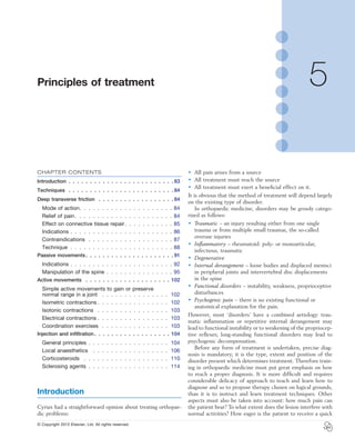
Friction prevents adhesion formation and
ruptures unwanted adhesions (Fig. 5.1)
In that transverse friction aims to achieve transverse movement
of the collagen structure of the connective tissue, crosslinks
and adhesion formation are prevented. In the early stages of
proliferation when crosslinks are absent or still weak, friction
must be very light so as to cause only minimal discomfort.
Therefore, in the first day or two following an injury, friction
is given with slight pressure only and over a short duration, e.g.
1 minute.
At a later stage when strong crosslinks or adhesions have
formed, more intense friction is needed to break these
experiences a numbing effect during the friction and reassess-
ment immediately after the session shows reduction in pain
and increase in strength and mobility. The time to produce
analgesia during the application of transverse friction is a few
minutes and the post-massage analgesic effect may last more
than 24 hours.10
The temporary relief at the end of a session
may prepare the patient for treatment with mobilization not
otherwise possible, such as selective rupture of unwanted
adhesions.
A number of hypotheses to explain the pain-relieving effect
of transverse massage have been put forward:
• Pain relief during and after friction massage may be the
result of modulation of the nociceptive impulses at spinal
cord level: the gate control theory (see Ch. 1). The
centripetal projection into the dorsal horn of the spinal
cord from the nociceptive receptor system is inhibited by
the concurrent activity of the mechanoreceptors located
in the same tissues. Selective stimulation of the
mechanoreceptors by rhythmical movements over the
affected area thus ‘closes the gate for pain afference’.
• According to Cyriax, friction also leads to increased
destruction of pain-provoking metabolites, such as Lewis’s
substances. This metabolite, if present in too high a
concentration, provokes ischaemia and pain.3
• It has also been suggested that prolonged deep friction of
a localized area may give rise to a lasting peripheral
disturbance of nerve tissue, with local anaesthetic effect.
• Another mechanism through which reduction in pain may
be achieved is through diffuse noxious inhibitory controls,
a pain-suppression mechanism that releases endogenous
opiates. The latter are inhibitory neurotransmitters that
diminish the intensity of the pain transmitted to higher
centres.11–13
Effect on connective tissue repair
Connective tissue regenerates largely as a consequence of the
action of inflammatory cells, vascular and lymphatic endothe-
lial cells and fibroblasts. Regeneration comprises three main
phases: inflammation, proliferation (granulation) and remodel-
ling. These events do not occur separately but form a continu-
ous sequence of changes (cell, matrix and vascular changes)
that begins with the release of inflammatory mediators and
ends with the remodelling of the repaired tissue (see Ch. 3).
Friction massage may have a beneficial effect on all three
phases of repair.
Friction stimulates phagocytosis
It has been suggested that gentle transverse friction, applied in
the early inflammatory phase enhances the mobilization of
tissue fluid and therefore increases the rate of phagocytosis.14
Friction stimulates fibre orientation .
in regenerating connective tissue
During maturation, the scar tissue is reshaped and strength-
ened by removing, reorganizing and replacing cells and matrix.15
Fig 5.1 • Friction achieves a transverse movement between
longitudinally arranged collagenous fibres.
4. General Principles
86
prevent (in the early stage) or to break down (in the chronic
stage) adhesion formation between the individual fibres and
between individual fibres and the surrounding connective
tissue. It is obvious that to break down crosslinks in a chronic
stage, the friction can be given forcefully and for a duration of
15–20 minutes, whereas in more recent lesions the technique
must be applied more gently and for a shorter duration. Fric-
tion to a muscle belly is always given with the muscle well
relaxed.
In recent tears, especially in the large muscles of the lower
limb, friction is followed by active or electrical contractions
with the muscle in a position of maximal relaxation and without
weight bearing, so that tension does not fall on the healing
breach.
To avoid early recurrence, friction is given for 1 week after
all clinical tests have become negative. During the period of
treatment, all movements or activities that bring on pain should
be avoided by the patient.
Theoretically, friction can be used for all muscle belly
lesions. However, some lesions respond so well to local anaes-
thetic infiltration that friction is not used. This is the case in
type IV tennis elbow (lesion at the muscle belly of the extensor
carpi radialis). On the other hand, sometimes no alternatives
exist to treatment with deep transverse friction (Box 5.1). A
lesion of the subclavius or intercostal muscles for instance can
be treated only by deep transverse friction.
Musculotendinous junctions
It is a common clinical experience that all musculotendinous
junctions (containing both muscular and tendinous fibres)
throughout the whole body can be treated only by deep trans-
verse friction. It would seem that no alternatives exist: local
anaesthetics, so curative for some lesions of muscle bellies, and
steroids, so effective for tenoperiosteal lesions, have not the
slightest effect on musculotendinous lesions, whereas deep
transverse friction usually has.
Tendons
All overuse tendinitis can be treated by deep massage except
for the tenoperiosteal origin of the extensor carpi radialis brevis
(type II tennis elbow), which is best treated by an infiltration
with corticosteroid or, in refractory circumstances, sometimes
by manipulations.
down.18–21
The technique is then used to soften the scar tissue
and to mobilize the crosslinks between the collagen fibres and
the adhesions between healing connective tissue and surround-
ing tissues. This, together with the local anaesthesia produced,
prepares the structures for mobilizations that apply longitudi-
nal stress to the structures and rupture the larger adhesions.
Friction induces traumatic hyperaemia
Forceful deep friction produces vasodilatation and increased
blood flow to the area. It may be hypothesized that this facili-
tates the removal of chemical irritants and increases the trans-
portation of endogenous opiates, so causing a decrease in pain.
Such a forceful friction, resulting in hyperaemia is only desir-
able in chronic, self-perpetuating lesions.
Indications
Diagnosis
The reduction in pain achieved after a few minutes of localized
transverse friction may be very helpful to define the exact
location of the lesion. In muscular, tendinous or ligamentous
lesions, a few minutes of massage on the suspected area results
in diminished pain on testing immediately thereafter, so con-
firming the diagnosis as accurately as an infiltration with local
anaesthesia.
Preparation for mobilizations and manipulation
Transverse massage is often applied before and in conjunction
with other mobilizing techniques. In muscular lesions, friction
is given before active or electrical contractions on an unloaded
muscle. The purpose is to allow broadening of the muscle and
so the prevention of adhesion formation between adjacent
muscle fibres and/or bundles.
For reasons of pain relief, transverse massage is usually
required before manipulative breakage of ligamentous adhe-
sions is performed. This may be indicated in chronic ligamen-
tous lesions at knee and ankle.
Deep and thorough friction also precedes manipulation of
the elbow in type II tennis elbow. The technique is used for
its desensitizing and softening effect which makes the manipu-
lation more tolerable.
Therapy
Muscle bellies
Friction is given to a healing muscle belly after contusion, in
minor muscular tears and in so-called ‘myosynovitis’. In minor
muscular tears the friction is often part of combined treatment
because it is usually applied after an infiltration with local
anaesthesia and is followed by active contractions.
The aim of treatment in muscular tears is to allow the torn
fibres to heal in such a way that normal increase in breadth
on contraction remains possible, a characteristic that can be
disturbed by abnormal adhesion formation. Transverse friction
aims to achieve a transverse sweeping movement over the
longitudinal muscular fibres without pulling on the tear, so to
Box 5.1
Muscle belly disorders that can be treated only
by deep transverse friction
Subclavius
Brachialis
Supinator
Adductors of the thumb
Interosseus muscles of the hand
Intercostal muscles
Oblique muscles of the abdomen
Interosseus muscles of the foot
5. C H A P T E R 5Principles of treatment
87
eases the pain and the tissue can be moved to and fro in an
imitation of its normal behaviour.
In recent cases the friction need not last long nor be very
vigorous – 1 or 2 minutes of daily gentle transverse sweeping
movement over the regenerating fibrils is enough. As pain
diminishes over subsequent days friction is progressively
increased to about 4–5 minutes for 2 or 3 days and finally to
a full duration of 15–20 minutes. From the third day, friction
is followed by passive and active movements within the limits
of pain to maintain normal gliding of the ligament over adjacent
bones. When the lower limb is involved, the patient should be
instructed to walk as normally as possible but without provok-
ing too much pain.
In chronic ligamentous lesions, frictions are also used but in
a totally different way. Here adherent scar tissue has formed
abnormal attachments as the result of healing during a period
of insufficient movement. As a result of the reduced mobility
of the ligament, vigorous use of the joint re-sprains the liga-
ment and in due course leads to recurrent sprains.
Treatment will consist of rupturing the adhesions by manip-
ulation, for which vigorous deep friction to the site of the
adhesions prepares the ligaments. The massage weakens and
desensitizes the structure, making the forced movement prac-
ticable and painless.
Experience has shown that particular ligamentous lesions
can be treated only by friction. This is the case for the posterior
carpal ligaments at the wrist and the tibiotalar ligaments.
Joint capsules
Deep transverse friction can be applied to the capsules of the
trapezium–first metacarpal joint, the temporomandibular joint
and the cervical facet joints. The indication is traumatic arthri-
tis or osteoarthrosis. Results are fair, provided the arthrosis is
not too advanced. Indications and contraindications to friction
are outlined in Table 5.1.
Contraindications
Ossification and calcification of soft tissues
Extensive ossification in muscles, tendons, ligaments or cap-
sules is a bar to all active treatment. However the minor cal-
cifications that may occur after a sprain can be managed by
Tenosynovitis also usually responds well to deep transverse
massage. In this condition, occurring in long tendons with a
sheath, inflammation and roughening of the gliding surfaces of
both tendon and sheath give rise to pain and sometimes to
crepitus. Friction rolls the sheath around the stretched tendon,
so facilitating functional movement between the tendon and
its sheath. The technique is useful in both acute and chronic
lesions.
Lesions at the tenoperiosteal insertion can be treated either
with corticosteroid infiltrations or with deep transverse
massage. Corticosteroid suspension quickly converts an
inflamed and painful scar into one free of inflammation.
However, the recurrence rate is rather high, between 20%
and 25%.3
The aim of the massage is to get rid of the self-
perpetuating inflammation by breaking up the disorderly scar
tissue and adhesion formation by converting it into properly
arranged longitudinal connective fibres. This takes longer but
once cure is achieved there will be less of a tendency towards
recurrence. It may therefore be a policy to start treatment with
infiltrations but if the trouble recurs after a few months to
substitute with massage.
As a rule, however, friction is always selected as the
treatment of choice in athletes or when the tendon is weak-
ened (partial rupture). It cannot be denied that repeated use
of corticosteroids, even in small doses and correctly applied,
temporarily weakens a tendinous structure. Steroids also take
away inflammation and pain, so giving the patient the false
impression of being cured. The combination of a weakened
tendon and abolition of pain can be disastrous – rupture
may ensue.
There exist also a few conditions that seem to respond only
to deep transverse friction. Steroid infiltrations are useless
here. This is so in tendinous lesions of the interosseus in the
hand and at the quadriceps expansion at the patella.
Lesions in the tendinous body, either traumatic or resulting
from overuse, are contraindications for infiltration with corti-
costeroids. Ruptures have been reported after intralesional
steroid infiltrations of long tendons and therefore deep fric-
tions are the treatment of choice here.22,23
It is obvious that during the whole period of treatment of
tendinitis, tenosynovitis or tenovaginitis, the patient must
avoid all activities that provoke the pain, especially the loading
of the affected contractile tissue.
Ligaments
Transverse massage is an excellent treatment in acutely sprained
ligaments, especially in ligaments of the knee and ankle. The
background, mode of action and technique differ considerably
and depend on the stage of the lesion.
It has been explained (see Ch. 3) that early mobilization is
extremely important for swift and full recovery of ligamentous
sprains. However, in advocating this, one main difficulty is
encountered: the intensity of the initial inflammatory reaction.
The slightest movement causes pain which forces the patient
to immobilize the joint and the ligaments. However, during
immobilization, regenerating fibrils quickly start to form ran-
domly organized scar tissue, leading to crosslinks and adhesion
formation. This problem can be solved by gentle transverse
frictions. Rhythmic movement across the inflamed ligament
Table 5.1 Indications and contraindications to friction
Indications Contraindications
Diagnostic difficulties
Preparative massage
Therapeutic massage:
• To muscle bellies
• To musculotendinous
junctions
• To tendons
• To ligaments
• To joint capsules
Ossification and calcification of soft tissues
Bacterial and rheumatoid-type tendinitis,
tenosynovitis and tenovaginitis
Skin problems such as ulcers, psoriasis or
blisters
Neighbouring bacterial infection
Bursitis and disorders of nerve structures
Haematoma, if large
6. General Principles
88
fingers are used, the amount of pressure, the duration and
frequency of the sessions. The patient’s skin and the therapist’s
finger must move as one, so that the deep layers of the skin
move over the affected fibres. Therefore all cream, ointments,
powder or any other procedure, such as previous heat, that
makes the skin sweat, must be avoided. Six to 12 treatments
are normally necessary. Except in acute ligamentous disorders
they are not given more often than every other day because
otherwise the site of the lesion may still be too tender from
the previous treatment to permit adequate massage.
Position of the patient
The patient’s position must be comfortable because it must
be maintained for up to 15–20 minutes. Sitting or lying is
preferable.
The lesion must be brought within finger’s reach. In some
structures this can be easily attained but others such as the
supraspinatus insertion and the anterior aspect of Achilles
tendon, require more specific positioning of the patient.
In addition, positioning must place the affected structure
under the required amount of tension. Full relaxation is neces-
sary for a muscle belly in order not only to treat its surface but
also to access a deeply seated lesion. Tendons with a sheath
must be kept taut otherwise friction will be ineffective between
tendon and sheath. The same applies in ligamentous lesions,
which are also placed in tension but within the limits of pain.
Position of the therapist and the hands
The bodily position of the patient should be the most comfort-
able and least tiring for the therapist. Working height is of chief
importance, so an adjustable high–low couch is ideal. To have
some economy of effort the therapist should adopt a position
that utilizes body weight to a maximum. Usually this is stand-
ing and with the patient on a slightly lower plane. The therapist
should avoid flexed positions. The shoulder should also not be
in abduction because this quickly leads to pain and cramp in
the neck and shoulder girdle.
Massage is performed by the whole arm and is not just an
activity of hand and digits. Movement is generated in the
shoulder and conducted via elbow and forearm to the digits.
One set of muscles is used to apply force and another to
provide movement, for example pressure with the fingers,
movement with the arms. Digits, hand and forearm should
generally form a straight line and are kept parallel to the direc-
tion of movement.
The majority of friction techniques are performed in two
phases: an active movement, usually as a result of flexor mus-
cular activity and a passive movement, when the arm and hand
are returned to the starting position. At the end of the passive
phase there should also be a moment of rest during which the
therapist fully relaxes the muscles.
The hands can be used in a variety of ways depending on
the tissues to be treated and the surface worked on. The wrist
and metacarpophalangeal joints should be kept in an almost
neutral position. The interphalangeal joints are slightly flexed
to avoid traumatic arthritis.
Three main techniques can be distinguished.
friction. In supraspinatus tendinitis, calcification is regarded as
responsible for complaints when the insertion is very tender to
touch and a radiograph shows calcification. These findings are
a contraindication to friction. In contrast, when calcification is
present in the absence of severe tenderness, transverse massage
can be given.
Bacterial and rheumatoid-type tendinitis,
tenosynovitis, tenovaginitis
All types of bacterial and rheumatoid disorders, no matter at
what stage of inflammation, are absolute contraindications to
friction.
Skin problems such as ulcers, psoriasis and blisters
When normal skin has been abraided – sometimes by friction
– massage should not be given. In skin disorders, it must be
abandoned when stable skin–finger contact becomes impossi-
ble and friction aggravates the skin problem.
Neighbouring bacterial infection
Because these may be reactivated by or may extend if
friction is used, it must be postponed until the infection
has resolved.
Bursitis and disorders of nerve structures
When bursitis is mistaken for a tendinous or ligamentous dis-
order and friction is given, the problem will either increase or,
at best, the pain will remain unchanged – it certainly will not
improve. Friction to a nerve is also harmful.
Haematoma
A haematoma in a muscle belly or after an ankle sprain is not
a contraindication to friction. Even if the haematoma is the
result of deep friction, treatment may be continued unless the
effusion is large.
Technique
Introduction
Transverse massage is not an easy technique. In order to
produce results, three conditions must be satisfied.
First, the therapeutic movement should be applied to the
exact site of the lesion which may occupy only a very small
volume of tissue. In other words, an identification of the site
to within 1 cm must be achieved which relies entirely on clini-
cal diagnosis and palpation of the lesion, based in turn on
anatomical knowledge. In some instances it will be necessary
to palpate carefully the entire structure at fault so as to find
the point that reproduces the patient’s pain.
Secondly, friction should be applied transversely across the
longitudinally orientated fibres, with sufficient sweep to reach
all the affected tissue and firmly enough to produce movement
between the individual connective tissue fibres of the affected
structure.
Third, the movement can only reach deeply seated struc-
tures if the deep friction technique of Cyriax is used; that
implies attention must be paid to different elements such as
the position of the patient and of the therapist’s hand, which
7. C H A P T E R 5Principles of treatment
89
middle finger or the middle finger aided by the index finger.
When the thumb does the massage, counterpressure is from
the fingers (Fig. 5.3). The most common way of applying fric-
tion around a round edge on a flat surface is to use the index
reinforced by the middle finger. Sometimes the opposite is
done: the middle finger is reinforced by the index. Sometimes
counterpressure is not given, for example in friction to the
quadriceps expansion or intercostal muscles.
Pronation–supination
This technique is often used where the lesion is difficult to
reach: the anterior aspect of the Achilles tendon, popliteus
tendon and the dorsal interossei of the metacarpals. Massage
To-and-fro movements
These are used in the treatment of dense, round or flat colla-
genous bundles (tendons or ligaments) and in the treatment of
tenosynovitis. The active phase is a sweep with the tip(s) of
one or two digits across the tendinous structure. During the
passive relaxation phase the finger is returned to the starting
position, without losing contact between finger and skin.
Movement is with the arm; friction is given by use of the pulpy
part of the finger (Fig. 5.2). In large lesions, as in peroneal
tendinitis, two or three adjacent fingers are used together.
In deep-seated lesions as in tendinitis of the long head of
biceps in the bicipital groove or at its insertion on the
radius or in infraspinatus tendinitis, the thumb performs
friction.
Counterpressure is usually provided to enable a good sweep.
The finger(s) applying counterpressure and stabilization are
most important in bringing those applying friction into the
right position and also determining the direction of the friction.
The thumb is used (to give counterpressure) when the sweep
is performed by a movement of the index reinforced by the
Fig 5.2 • Friction to the supraspinatus tendon: counterpressure is
by the thumb.
Fig 5.3 • Friction to the infraspinatus tendon: counterpressure is by
the fingers.
8. General Principles
90
Sometimes the same technique is used in tendinous lesions,
for example, at the sides of the Achilles tendon.
No movement between finger and skin .
is allowed
Deep friction can only be effective when skin and subcutane-
ous fascia are moved over tendon ligaments or muscles. No
movement is allowed between the therapist’s finger and the
is performed with the pulpy part of the third finger (long
finger), reinforced by the index finger. The long finger is used
because its long axis is the prolongation of the axis of pronation–
supination rotation of the forearm (Fig. 5.4).
The active phase is usually on supination. No counterpres-
sure is given. Caution is taken not to move the finger on the
skin but rather to move the skin and the fingertip as a whole.
The passive phase is the pronation movement that brings the
frictioning finger back to the starting position without losing
contact with the skin.
Pinch grip
This is the normal technique for a muscle belly. The pinch is
between the thumb and the other fingers. The muscle is fully
relaxed. The fingers are placed at one side of the affected area
and the thumb at the opposite side (Fig. 5.5). By drawing the
fingers upwards over the affected area, the therapist feels the
muscle fibres escape from the grip until only skin and subcu-
taneous tissue remain.
During the passive phase the fingers are slightly relaxed and
moved downwards into the previous deep position where the
same movement starts again.
Fig 5.4 • Active phase of pronation–supination friction technique
to the anterior aspect of the Achilles tendon. a, starting position;
b, end of supination (active) movement.
(a) (b)
Fig 5.5 • Pinch grip friction to the Achilles tendon. a, starting
position; b, end of active phase.
(a) (b)
9. C H A P T E R 5Principles of treatment
91
have not had time to form. In long-standing cases more
pressure is needed to get rid of these. However, pressure
should always be associated with movement and should
not replace it because pressure alone is both painful and
ineffective.
• The tenderness of the lesion: in severely inflamed lesions
that are very tender to touch, friction with the usual
amount of force may be very painful. Pain can be avoided
by starting with a minimal amount of pressure – just
enough to reach the lesion – and progressively increasing
the force as treatment proceeds.
In order to avoid painful sessions of deep transverse friction it
is good practice to grade its application. Begin with a sweep
that is gentle and continue this for a few minutes; some numb-
ness of the treated area follows which allows slight intensifica-
tion of the amount of pressure, which in turn leads to more
numbness. Finally, it will be possible to give effective massage
that is practically painless to the patient.
Duration and frequency
Friction is usually given for about 10–20 minutes and, because
of tenderness, on every second day. The ideal timing of the
next treatment is when local tenderness caused by the previous
session has resolved. If tenderness persists after 2 days, the
pressure used during friction should not be diminished but the
interval between sessions must be increased.
Massage immediately after a ligamentous sprain or a minor
muscular rupture may be applied daily for the first week but
should be of very low intensity and short duration.
Treatment is stopped once the patient is pain-free during
daily activities and functional tests are totally negative. Local
tenderness may persist longer but disappears spontaneously
because it is the outcome of repetitive hard pressure. However,
in a minor lesion of a muscle belly, massage is continued for
1 week after full clinical recovery to prevent recurrence (see
Table 5.2; see also Box 5.2).
Passive movements
Treatment by passive movement is otherwise known as mobi-
lization. It cannot be performed by the patient and requires
the intervention of a therapist. Depending on its velocity and
the range of movement that is aimed for, it can be graded as
A, B and C mobilization:
• Grade A mobilization is a passive movement performed
within the pain-free range.
• Grade B mobilizations are passive movements performed
to the end of the possible range. The latter is indicated by
an end-feel. All stretching and traction techniques are
grade B mobilizations.
• Grade C mobilization is a minimal thrust with a high
velocity and over a small amplitude. It is performed at the
end of the possible range, i.e. the moment the therapist
has reached the end-feel. Another word for grade C
mobilization is manipulation.
patient’s skin. If movement occurs between finger and skin,
blistering soon takes place and usually indicates faulty tech-
nique. Sometimes it can be avoided by keeping the skin dry by
the use of 95% alcohol in water and/or by placing a piece of
cotton in between the finger and the skin.
In the obese, subcutaneous soreness and/or ecchymosis may
occasionally occur and sometimes a nodule may form. For this
reason the finger should not be in continuous contact with the
same area but should displace the skin slightly to one or other
side, before pressure is applied.
Direction of friction must be transverse .
to the tissue fibres
Longitudinal massage improves the circulation of blood and
lymph but has no effect on musculoskeletal lesions. On the
contrary, because lesions of tendons, muscles and ligaments are
normally caused by a longitudinal force, longitudinal massage
can possibly be harmful in that it may separate the ruptured
ends further. To restore and/or maintain full mobility of a
lesion, massage must be given across the fibres, so moving all
fibres in relation to each other. To achieve this, the therapist
must have a good anatomical knowledge of the direction of the
fibres.
Sweep
The main goal of friction is to move fibres in relation to
each other and adjacent structures. Enough sweep must
be given to the friction for this purpose, so the frictioning
finger starts at the far side of the lesion, glides over it and ends
at the near edge. Pressure alone, however hard and painful it
may be, is totally ineffective. Adequate sweep is sometimes
limited by the amount and elasticity of the overlying skin.
Initial displacement of skin over the lesion from the near to
the far side may help increase sweep and reduce the risk
of blistering.
Amount of pressure
Over recent decades, friction has been held in some disrepute
in that it was perceived by some as synonymous with very
painful treatment. Though it cannot be claimed as wholly pain-
free, the pain should not be unbearable. When excessive pain
is provoked, this is usually the result of a failure to understand
the meaning of the term ‘deep’, which means ‘as deep as
needed to reach the lesion’. Many therapists misinterpret this
in such a way that they feel that they always have to work hard
physically, which obviously leads to pain and may do more
harm than good.
The amount of pressure applied depends on three
elements:
• The depth of the lesion: that friction must always reach
sufficient depth to move the affected fibres in relation to
their neighbours and sometimes the underlying bone or
capsule, increased pressure must be applied to deeper
structures.
• The ‘age’ of the lesion: recent sprains and injuries require
only preventive friction because crosslinks or adhesions
10. General Principles
92
Indications
Grade A mobilizations
To promote healing of injured connective tissue
Passive movements within the pain-free range are usually
called for in the treatment of injured connective tissue. A
comprehensive literature evaluation and meta-analysis of
experimental studies of the past several decades have demon-
strated that regeneration of injured connective tissue is signifi-
cantly better with the application of continuous passive motion.
If the healing tissues are not loaded, regeneration results in
unstructured scar tissue. Under functional load, the collagen
fibres are oriented in a longitudinal direction and the mechani-
cal properties are optimized.24
Grade A mobilizations are therefore applied early in the
treatment of sprained ligaments to promote orientation of the
regenerating fibres. They are given in conjunction with gentle
transverse massage and within the pain-free range. Care should
be taken not to bring the fibres under longitudinal stress in
order not to disrupt the healing breach. The movements are of
short duration but repeated often.
Distractions at the shoulder
Grade A mobilizations are also used on the capsule of the
shoulder in stage III arthritis when stretching and intra-articular
steroids are contraindicated (see Ch. 14). In this condition,
long-standing stimulation of the nociceptors has increased
neuro-sympathetic activity, giving rise to vasoconstriction,
muscle spasm and pain.
Gentle and rhythmical grade A movements are performed
in such a way that the fibres are stretched longitudinally, stimu-
lating the mechanosensor mechanisms in the joint and so
Table 5.2 Transverse massages/modalities
Indication Duration (min) Pressure Frequency Combined treatment
Diagnostic 15–20 High Once
Acute ligamentous 30 sec Very low Daily Effleurage before and active movement after the treatment
Subacute ligamentous 3–10 Low Daily–3 times/week Passive grade B movements after
Chronic ligamentous 15–20 High 3 times/week Passive grade B movements after
Ligamentous adhesions 15–20 High Once Before manipulation (grade C mobilization)
Tendinitis – tenoperiosteal 15–20 Grading 3 times/week Relative rest – unloaded active movement
Tenosynovitis 15–20 Grading 3 times/week Relative rest – unloaded active movement
Musculotendinous 15–20 Grading 3 times/week Relative rest – unloaded active movement
Myosynovitis 15–20 Grading 3 times/week Relative rest – unloaded active movement
Muscular tear – acute 5–10 Low Daily Procaine infiltration before and active unloaded
contractions after treatment
Muscular tear – chronic 10–15 High 3 times/week Active unloaded contractions after treatment
Capsular lesions 15–20 Grading 3 times/week
Box 5.2
Summary of deep friction technique
1 Position of the patient must:
be comfortable
bring lesion within finger’s reach
be appropriate for the type of structure at fault:
• tendon/ligament under tension
• muscle belly: relaxed
2 Position of the therapist must:
be comfortable
facilitate economy of effort:
• alternating active and passive phases
• using large muscles
3 Use of the hand:
To-and-fro movement
Pronation/supination
Pinch grip
4 Use of the fingers:
Counterpressure
Friction using the fingers
5 Other points:
Fingers and skin move as one unit
Direction of friction must be transverse
Sufficient sweep must be used
The pressure must be appropriate
Duration and frequency must be appropriate
11. C H A P T E R 5Principles of treatment
93
In the very beginning of arthritis, muscle spasm forces the joint
to be held in a position of ease, so restricting movement in
some directions more than in others (see Capsular pattern, Ch.
4). Immobilization and inflammation cause disordered deposi-
tion of collagen fibres in the joint capsule and lead to the
formation of capsular adhesions, which in turn are responsible
for more restriction of movement and pain. Stretching aims at
restoring mobility and function by breaking micro-adhesions
and producing elongation of the shortened capsule. To be appli-
cable, however, the ligamentous end-feel must be reached
before the protective muscle spasm begins. To be successful,
the therapist should therefore be able to differentiate between
an elastic and a spastic end-feel.
The technique is a slow and steady pressure, performed at
the end of range over about 30 seconds to 1 minute with as
much force as is reasonable for the patient to bear. Tension is
slightly diminished for a few seconds, so affording the patient
some respite, and then again increased. From time to time
the procedure is completely interrupted. If tension is released
too quickly, some pain may be felt and it is therefore wise
to bring the limb back into neutral position under traction.
The technique is not painless. The stretching causes some
micro-ruptures, which result in an inflammatory response and
after-pain that lasts for a few hours.
Normally, capsular stretching is given for 15–20 minutes,
three times a week. The therapeutic effect is slow.
Capsular stretching can be preceded by application of
heat, either through short-wave diathermy or ultrasound.
This can relieve some pain and seems to lower the viscosity
of the collagenous tissue, allowing more movement for less
force. In vivo studies on the effects of heat on ligament
extensibility have shown that sustained force applied after
elevating tissue temperature produced significantly greater
residual elongation.25,26
Manipulation of a joint capsule under anaesthesia is a grade
C mobilization and is only considered for postoperative intra-
articular adhesions. A joint that has been manipulated under
anaesthesia requires daily intensive mobilization immediately
afterwards in order to prevent the formation of new intra-
articular adhesions.
To stretch a muscle
Children with short calf muscles can be helped by sustained
stretching. The procedure consists of a series of alternating
passive stretchings and active contractions. Stretching is main-
tained for about 8–10 seconds and is followed by full relaxation
and active contraction of the muscle. These alternating move-
ments are performed six to eight times per session, preferably
daily but at a minimum of three times a week. The earlier the
stretching is started, the better the result. Above the age of 15
not much improvement can be expected.
Traction
Traction is used to separate articular surfaces from each other
and can be employed in two ways: as an accessory to manipula-
tion or as the sole treatment. Reducing a displaced fragment is
obviously easier when the bone ends between which it lies are
pulled apart. If the fragment projects beyond the articular
edge, tautening of the ligaments and capsule also provides a
inhibiting somatosympathetic reflexes that are co-responsible
for the increased inflammation of the joint.
Deformity correction
Some cases of lumbago show persistent spinal deviation even
after the pain has ceased. A quick thrust manipulation so effec-
tive in relief of pain is not effective in correcting the remaining
deformity, but sustained translatory gliding in the opposite
direction is most helpful. The movement is performed slowly
and care is taken to keep the gliding within the pain-free range
(see Ch. 40).
Reduction of an intra-articular displacement
in a peripheral joint
When a meniscus or some other piece of intra-articular carti-
lage (with or without an osseous nucleus) becomes displaced
and locks a joint, the logical treatment is either to remove it
or manœuvre it into such a position that the joint can again
move freely over a normal range. The technique needed for
the latter is usually a series of manipulative movements which
normally contain elements of traction combined with move-
ments of rotation and flexion or extension. In general, these
are first performed in the less painful direction of movement
and repeated several times with progressively increasing force.
Unlike manipulations in the spine, the manœuvre to reduce
an intra-articular loose body is not a grade C mobilization
because the movement is not performed at the end of range
nor does it contain a ‘thrust’ element. The flexion–extension
movement is over a wide range and stops before the end-feel
is reached. The rotation movements are performed to the
end of range where end-feel is sensed by the therapist. The
‘manipulation of an intra-articular displacement in a peripheral
joint’ is therefore a combination of grade A and grade B
mobilizations.
Grade B mobilizations
To maintain a normal range at the joint
Paralysed muscles may lead to a loss of normal range of motion
of the corresponding joint. This can be avoided by gently
stretching the capsule, starting as soon as possible after the
onset of paralysis. The approach should also be considered for
joints that have been injured or subjected to surgery. In such
circumstances, there may be the paradox that immobilization
is needed for a fracture to heal but that movement is required
to prevent loss of capsular elasticity. Often the problem can be
solved by adapting the technique of capsular stretching so that
it does not influence the site of the fracture.
To stretch the capsule of a joint
Grade B mobilizations may be required to stretch the joint
capsule in non-acute arthritis and in early osteoarthrosis. The
technique will be further referred to as capsular stretching.
Capsular stretching is particularly useful in shoulder and hip
joints but is applicable in all ‘non-irritable’ capsulitis. The con-
dition is characterized by:
• a limitation in the capsular pattern (see Ch. 4)
• demonstration of a hard-elastic end-feel to restricted
movements (see Ch. 4).
12. General Principles
94
Rupture of tenoperiosteal adhesions
In type II tennis elbow (tendinitis of the attachment of the
extensor carpi radialis brevis), adherent and disorganized scar
tissue causes a self-perpetuating inflammation. The manipula-
tion aims to rupture the adhesions and produce a permanent
elongation of the tendon. The high-velocity manœuvre is pre-
ceded by thorough deep transverse friction in order to numb
and to weaken the spot. The manipulation is performed only
once per session; 10–15 sessions may be required to achieve a
result.
To reduce a bony subluxation
A subluxation of one of the carpal bones or of the cuboid bone
can easily be reduced by digital pressure combined with trans-
latory movement during traction.
Manipulation of the spine
Spinal manipulative therapy is a major part of treatment tech-
niques in orthopaedic medicine and is discussed thoroughly
below.
Contraindications to forced movements
Contraindications to spinal manipulations are discussed later
in this chapter.
Capsular inflammation
Forced movements should not be performed when signs and
symptoms of capsular inflammatory activity are present. These
are spontaneous pain, pain especially at night, wide reference
of pain, inability to lie on the affected side at night or to bear
weight on the affected side. Local warmth and effusion are
other pointers of a highly inflamed joint. However, if these
symptoms and signs are present but the rest of the clinical
examination demonstrates internal derangement (e.g. knee,
hip, ankle), manipulation is indicated and can safely be
performed.
Muscle spasm
Grade C mobilizations should never be applied to a joint that
is protected by a muscle spasm. Grade B mobilizations may be
used unless the end-feel of the movement that is intended to
be forced through is also spastic.
Severe osteoporosis
Grade B mobilizations, for instance stretching of the shoulder
or hip joint in elderly people, should always be carried out with
caution for fear of fracturing the humerus or the neck of the
femur.
Joints and ligaments not under voluntary
tension control
Mobilization is also contraindicated for those joints and liga-
ments on which the tension is not under voluntary control.
This is the case for the acromioclavicular, the sternoclavicular
and the sacroiliac joints and the sacrococcygeal ligament.
centripetal force. In that traction diminishes the pressure on
the fragment, pain decreases, which allows the patient to relax
the muscles more.18
In the cervical and thoracic spines, traction
is a built-in safety measure for protecting the spinal cord during
manipulation (see below) although the use of traction for this
purpose and at these sites does not imply that manipulation
can be performed on a basis of ‘try and see what happens’
without a proper diagnosis.27
In the spine, traction is used as the sole treatment only in
nuclear disc protrusions, which are rare at the cervical and
thoracic levels but are more common in the lumbar area. Spinal
traction is always mechanical and is performed with the help
of a harness (lumbar or low thoracic) or a sling (cervical or
upper thoracic). Spinal traction distracts the intervertebral disc
spaces. It also pulls the apophysial joints apart and slightly
widens the intervertebral foramina.27–31
At the same time,
negative intradiscal pressure is produced with centripetal
‘suction’ on any protrusion. The posterior longitudinal ligament
is tightened, which may help reduce a displaced fragment. All
these elements are helpful in the progressive reduction of a
nuclear disc protrusion. Reduction of herniated bulges has been
demonstrated on epidurography31–33
and on CT scan34
during
and after traction. The effect of traction depends on the
amount of force applied, the length of time per session, the
interval between each session and the total number of
sessions.35
Grade C mobilizations
Grade C mobilizations or manipulations are forceful passive
movements, performed at the end of range. Spinal manipula-
tions are mainly to interrupt discodural or discoradicular
contact. At the peripheral joints the purpose of a manipulation
is to rupture unwanted adhesions between bone and ligament
or bone and tendon or to reduce small bony subluxations in
the wrist or foot.
Rupture of ligamentous adhesions
Small ligamentous adhesions sometimes develop between a
healing ligament and bone. They usually result from a sprained
ligament that has been immobilized during the healing process.
The usual presentation is at the lateral ligaments of the ankle
and at the medial collateral ligament of the knee. The clinical
features are local pain during exertion and a small limitation
of movement in one direction only. The adhesions can be rup-
tured by a high-velocity, small-amplitude thrust manipulation,
after preparation of the affected ligament with intensive deep
transverse friction.
The joint is stretched as far as possible in the limited direc-
tion and manipulated with a single firm thrust, during which a
typical ‘snap’ is often heard. Harm is not caused to the liga-
ment nor to the other parts of the joint because the adhesions
bear the brunt of the force. The manipulation is almost painless
and after-pain is not to be expected. A successful manipulation
should achieve an immediate result. Active movements during
the following days to maintain function should be highly
encouraged.
13. C H A P T E R 5Principles of treatment
95
Diagnosis is mainly based on palpation for restricted spinal
mobility and treatment consists of a manipulative system in
which joints are forced by a distant leverage. Cure is sought
for all kinds of visceral and musculoskeletal disorders.27
Chiropraxy
This method was started in 1885 by D. Palmer. It is based on
a revision of techniques that originated with Hippocrates and
is also influenced by osteopathy. Chiropraxy was long regarded
as maintaining osteopathic dogma in its most primitive form
and having a strong commercial character.
Chiropractors also claim to cure visceral diseases via the
musculoskeletal system. Diagnosis is made on palpation for
vertebral displacement and manipulative pressure is applied
directly to the bone.
Orthopaedic medicine
This term describes the system of diagnosis and treatment of
musculoskeletal lesions introduced by J.H. Cyriax. It is the
system on which this book is based. Diagnosis rests on careful
history and functional examination. Treatment depends mainly
on the type of lesion, and manipulation is applied only when
indicated. In spinal manipulation, Cyriax proposed a fixed set
of high-velocity, small-amplitude thrusts performed at a certain
distance from the lesion and, characteristically for this method,
usually under strong traction. The objective of Cyriax’s spinal
manipulative techniques is to alter the discodural or disco-
radicular interaction by moving a displaced cartilaginous frag-
ment away from the sensitive dura mater and dural nerve
sleeve. Spinal rotation manipulations apply a torsion stress
throughout a whole part of the spine, not at just one level.
With an intact posterior longitudinal ligament and annulus
fibrosus, some of this torsion force exerts a centripetal force
by suction on the protruding disc material.42
This effect is not
confined to one level and full reduction is not absolutely neces-
sary for pain relief, in that when contact between dura and disc
has ceased the problem is frequently solved.
Manual therapy
Treatment is characterized by rhythmic repeated movements
within the physiological range. Oscillatory techniques had
already been used by E. Cyriax (father of J.H. Cyriax) but
were more widely employed by Maitland and later slightly
changed by the different schools of manual therapy (Cyriax:3
p. 40). Pressure is applied to what is believed to be the appro-
priate level.
Orthopaedic medicine technique
Before any manipulation is done an exact diagnosis must be
made. The decision to manipulate is followed by choice of the
correct manœuvre. The patient is put in a comfortable position
and the manipulator adopts a stable stance. The floor and shoes
should not be slippery, so that there is no risk of inappropriate
movement.
Attention must be given to the following general matters,
which are important for all manipulations.
Manipulation of the spine
Introduction
Spinal manipulative therapy includes all procedures of mobiliz-
ing or adjusting the spine by means of the hands. As in the
peripheral joints, grade A and B mobilizations are movements
of low velocity with varying amplitude but remaining within
physiological limits and within the patient’s tolerance and
control.
A manipulation or grade C mobilization usually implies a
single thrust of high velocity performed at the end of a passive
movement after the ‘slack’ has been taken up, and over a small
amplitude. It goes beyond the physiological limit but remains
within the anatomical range. Precision of the movement and
control of the applied force are required.36
Spinal manipulative
therapy is a valuable method in the treatment of mechanical
spinal disorders. Although it has not been scientifically vali-
dated, some studies have shown beneficial effect.37–40
However,
its potential benefit should not be overestimated and the indi-
cations must be well defined and based on a sound clinical
diagnosis. It must never be done as a test to see if it is effec-
tive. Therefore it should not be used on all those with back
and neck pain although it may well cure a proportion who
actually require it. To use McKenzie’s words:
Even if you have a hammer in your hand not everything you see
is a nail. Therefore indiscriminate use of spinal manipulative
therapy must not be made if the criticisms that have been
justifiably levelled at chiropractice and osteopathy are to be
avoided. The development of postgraduate courses in
manipulation is welcome, although some have overvalued the
benefits of manipulative therapy. All who undertake
manipulation have experienced the feeling of pride and joy in
producing cure. It is the duty of those who have more experience
of the benefits and limitations of manipulative therapy to
moderate the understandable enthusiasm of those entering the
field – a few successes may quickly lead to the temptation to
manipulate every patient for any disorder.41
Manipulation either helps quickly or not at all. Therefore if
improvement does not occur after one or two sessions, manip-
ulation is not likely to be successful and it is pointless to
continue with it.
Historical note
Manipulation is as old as medicine and embraces both
medicine and mankind in general. In recent times, the
medical aspect has become structured and different methods
have been developed which are subject to controversy and
competition.
Osteopathy
The concept of osteopathy was introduced by A. T. Still
(1828–1917) and developed out of frustration with traditional
medicine. His ideas were based on two principles: (1) the body
has within itself the processes to combat all disease, and (2)
the cause of all disease is dislocated bones, abnormal ligaments
or contracted muscles with consequent mechanical pressure on
blood vessels and nerves.
14. General Principles
96
pain-free movement. The patient and not the manipulator is
the arbiter.49
Depending on the immediate outcome, the thera-
pist decides whether to repeat the same manipulation, prob-
ably with increased strength, to try another manœuvre or to
refrain from further manipulation.
It should be appreciated that after successful manipulation
the anatomical lesion is still present: a piece of cartilage,
although put back in place or into a neutral position, persists
and may redisplace. For this reason, those who undertake
manipulation should note the results obtained and what
manipulations were used, in case of recurrence.
Figure 5.6 outlines the assessment of spinal lesions and their
manipulation.
Selectivity of a manipulative treatment
Selectivity must be considered both in diagnosis and therapy.
Selectivity of diagnosis
Osteopaths and manual therapists claim to have developed the
clinical skills to localize by palpation the exact site of fixation,
and are therefore able to perform the manipulation at the
required level. Diagnosis is mainly based on segmental mobility
tests: joint play, springing test or tests of passive physiological
movements. Movement can be tested by exerting local pres-
sure at one side of a vertebra while counterpressure is applied
to the contralateral side of the vertebra above or below. For
the lumbar spine, it can be done with the patient on the side
with both hips flexed to 90°. Small movements of the thighs
cause the lumbar spine to flex or extend which can be detected
by palpation of the spinous processes.
Other practitioners look mainly for palpable soft tissue
changes, such as local subcutaneous thickening or exquisite
tender spots (trigger points) in muscles, ligaments (iliolumbar,
Traction during manipulation
Most types of spinal manipulation in orthopaedic medicine are
performed under traction. For the cervical and thoracic spine,
traction is applied by the manipulator with the help of a fixing
belt or by one or two assistants. At the lumbar level, traction
is usually already built into the manœuvre. Traction facilitates
the reduction of a displaced fragment and provides an impor-
tant safety element against the possibility of a protrusion con-
tacting the spinal cord during manipulation.
End-feel on taking up the ‘slack’
All spinal manipulations are performed over only a small ampli-
tude. Therefore all ‘slack’ must be taken up by moving the
vertebral joints passively to the end of the normal passive range
of movement. At this stage it is absolutely necessary to have a
clear idea of the end-feel, which is nominally elastic for the
entire spine. An end-feel that does not correspond with this
– muscle spasm, or hard or empty end-feel – is an absolute
contraindication to any manipulation and the manœuvre is not
continued.
Final thrust
Immediately after the slack has been taken up in the surround-
ing tissues, a minimal amplitude, high-velocity thrust is given
to affect the target tissue. The velocity is of great importance
because tissues loaded quickly are stiffer so that the manœuvre
will affect only the displaced fragment of disc and will not
damage the surrounding structures.43
The amount of force used for the final thrust depends
mainly on the patient and manipulator in that a tall manipula-
tor will have to use less force in a small patient and vice versa.44
The length of the lever (see later) is also important. The force
should always be kept reasonable and may be progressively
increased, according to the immediate result.
The manipulation thrust is often accompanied by an audible
‘pop’.29,45
Although it is a common belief that pops or clicks
are provoked by the formation of a temporary vacuum, as
occurs in small peripheral joints put under traction,46–48
this is
not definitely established for the spine. An alternative and more
likely explanation is movement of cartilaginous fragments, as
may be heard during manipulation for a loose body in the knee
or hip. If the clicks were simply the result of the collapse of a
vacuum they should also be – but are not – heard during
mechanical traction, in which the traction force is much higher.
Leverage
The amount of force used depends on the length of the lever.
If for example a rotation of the lumbar spine is forced via the
shoulder and pelvis, the lever offered by the shoulder is the
same length as that offered by the pelvis, so an equal amount
of force must be used by both hands. But if the femur is used
instead of the pelvis, the length of the pelvic lever doubles.
The hand on the shoulder must apply double the amount of
force that is used on the knee. The longer the lever, the less
force is needed.
Reassessment
After each manœuvre the patient is assessed, the criteria of
success being the absence of symptoms and the restoration of
Fig 5.6 • Spinal manipulation.
Is the lesion discodural or discoradicular?
Is the lesion an indication for manipulation?
Are there contraindications?
Does the patient have a positive attitude?
Decision to manipulate
Choice of manœuvre
Positions of patient and
manipulator determined
Technique
Take up the slack
Check end-feel
Thrust
Reassess
15. C H A P T E R 5Principles of treatment
97
In orthopaedic medicine most manœuvres used are non-
specific long-lever manipulations. These include all procedures
in which a force is exerted on a part of the body some distance
away from the area where it is expected to have its beneficial
effect. Levers may include the shoulder, transverse processes
and parts of the skull, pelvis or thigh (Frymoyer et al:45
p. 1594). Although some criticize the crudity of long-lever
high-velocity manipulation it should be realized that it is not
elegance, impressiveness, specificity or technical difficulty
which count but effectiveness and safety. Furthermore, the use
of a lever enables the manipulator to reach the lesion more
effectively. During the preparative phase – on taking up the
slack – all the normal joints are brought to their anatomical
end position except for the joint that is blocked. When the
additional thrust is given, the final extra pressure falls inevita-
bly first and to the greatest degree on the deranged joint. The
manœuvre thus becomes specific even though in general the
techniques are regarded as non-specific.
Long-lever manipulations are in full contrast to what are
called ‘specific’ short-lever high-velocity manipulations. Here
the goal is to act specifically at what is believed to be the level
of the lesion. The spinal segment and the facet joints adjacent
to the lesion are locked by moving the spine to the physiologi-
cal limit of passive movement and a high-velocity small-
amplitude thrust is given to the short vertebral lever (transverse
process or spinous process) in the specific direction that will
liberate the restricted movement. However, it is technically
not possible to lock all other joints and then to manipulate at
just one level (Cyriax:64
p. 108). It was even demonstrated that
by mobilizing the sacroiliac joints after locking of the lumbar
spine, the largest movement took place between L4 and L5.66
Furthermore, if diagnosis fails to be absolutely right, how can
there be certainty as to the exact level of the lesion to work
on? Fortunately for those who employ ‘specific’ short-lever
manipulations, these are much less specific than they think,
because the manipulations actually cover a much larger part of
the spine and so unintentionally also include the lesion.
Specificity is a false attribute. The methods that claim to
lead to specific localization in both diagnosis and treatment are
scientifically unacceptable. Claims of specificity are made in
order to give prestige to manipulators who claim to feel some-
thing that cannot be felt. Manual therapists, chiropractors and
osteopaths over-complicate their teaching and often create
excessive patient dependency, instead of providing the patient
with independence. Indeed, patients are encouraged to return
at regular intervals for pointless prophylactic adjustment.41
We
support R. McKenzie’s conclusion that demystification of
spinal manipulative therapy is an urgent priority.41
Chiropraxy,
manual therapy and osteopathy, however, thrive by creating the
impression that there is something complex and exclusive
about the practice of passive end-range motion that only
experts in these practices can understand or have the skills to
feel. The belief is strong that expertise in the understanding
and delivery of spinal manipulative therapy requires 3 or 4
years’ training. The main advantages of the methods discussed
in this book are that the manipulations are much simpler and
at least as effective as those advocated by chiropractors, osteo-
paths and manual therapists. Non-specific long-lever manipula-
tions are quickly effective, do not take long to perform and are
sacroiliac) and over bony prominences. All these are consid-
ered to be important diagnostic and therapeutic factors.
The great variability in the extent of spinal stiffness between
subjects, or at different levels within the one subject, makes
the determination of areas of abnormally increased stiffness
difficult. Increased stiffness may in fact be a normal variant
and bear no relationship to the patient’s presenting symptoms.
Few of those advocating segmental mobility tests have seri-
ously examined the value of their tests. They have generally
presumed that the tests were useful because their patients got
better.50
However, several studies have failed to demonstrate
the reliability of these tests.51–62
Therefore it must be made
clear that judgement of small changes in the range of move-
ment of a segment, in the absence of full restriction of move-
ment, remains a very subjective finding, which depends mainly
on the personal conviction of the examiner rather than on
objective measurements. Moreover, in soundly based tests,
findings must be reproducible and must show correspondence
when performed by other investigators. In the establishment
of ‘joint play’ the inter-observer discrepancy is too large to be
acceptable.63
In 1973, Cyriax attended a demonstration in
which five therapists, all of whom specialized in mobility
testing, examined over a period of a few minutes a patient
with a neck problem. There was no agreement between these
specialists about the level of the lesion (C2, C3, C4, C5, C6
or T2), or about the direction of restriction (see Cyriax:64
p. 108). Similarly, a patient who had congenital fusion of
the sacroiliac joints was examined by 10 manipulators. Each
had his own diagnosis, such as left anterior sacrum, right ante-
rior sacrum and bilateral posterior sacrum, although ‘the tests
were very positive’ for all of them (see Maigne65
and Cyriax:64
p. 363).
Even if it were possible to identify with certainty localiza-
tion of the hypomobile segment, the question remains as to
whether this is also the site of the lesion. Studies have shown
that frequently the lesion does not lie at the joint where
motion is restricted but at one which appears to be normal.55
Moreover, other disorders such as osteophytosis, congenital
fusion and ankylosing spondylitis all give rise to restricted
movement which is usually painless.
Selectivity of manipulation
Manipulation is often accompanied by immediate relief of
symptoms and signs which, since success has been obtained, is
logically taken as absolute confirmation of the precision of
diagnosis and treatment. Such a deduction may be – and often
is – totally wrong. The only thing proved is that the manipula-
tion was efficacious. The erroneous reasoning that successful
manipulation necessarily confirms the diagnosis has been and
is still today an important argument for the false belief of some
schools that manipulation can cure all kinds of disorders even
including visceral diseases. A typical example is pectoral pain,
resulting from a thoracic discodural interaction which is mis-
diagnosed as angina. The patient goes to an osteopath who
manipulates the thoracic spine and the pectoral pain ceases
immediately. Both patient and manipulator, misled by the
wrong diagnosis, will believe that the manipulation has altered
autonomic tone and cured the angina, whereas what it actually
did was interrupt the discodural interaction.
16. General Principles
98
techniques are not hard to learn, years of experience are
needed to learn when to manipulate, when not, and what sort
of manœuvres to use.
Contraindications to manipulation are bleeding disorders,
softening of bone, rheumatoid conditions, neurological deficit
and danger to the spinal cord.
Bleeding disorders and anticoagulant use
When normal clotting of blood is not guaranteed, as in
congenital or acquired (liver disease) bleeding disorders or
because of the administration of anticoagulants (Box 5.3),
spinal manipulations are potentially dangerous. Disastrous
results can follow, such as intraspinal haemorrhage with the
formation of a haematoma that may lead to sensory and motor
deficit, to paraplegia, quadriplegia or death.78
For this reason,
a coagulopathy is an absolute contraindication to spinal manip-
ulations. Manipulation can be safely performed only after
blood clotting tests have returned to normal.
Spinal tumours, unstable fractures,
vertebral infections and severe osteoporosis
(see Grieve:27
p. 829)
These all result in weakening of bone with risk of further
damage by manipulation. Long-lever manipulation is not safe
in severe osteoporosis.
Rheumatoid arthritis, psoriatic arthritis,
Reiter’s syndrome and ankylosing spondylitis
The first three of these may be associated with ligamentous
laxity and gross destruction of the joint with subsequent insta-
bility. Manipulation must not be undertaken. The same applies
for the inflammatory stage of ankylosing spondylitis. In the
unlikely event of a patient with this disorder developing a disc
lesion, manipulation is not at all safe, especially in the cervical
spine, where luxations, fractures and cord compression have
been described.79
Neurological deficit and spinal cord compression
Segmental neurological deficit is characterized by disturbance
of either motor function, with impaired reflexes and/or mus-
cular weakness, or sensory function, with diminished sensibil-
ity. The two may be combined. Segmentally referred pins and
needles due to compression of the sensory fibres of a nerve are
not regarded as neurological deficit and are therefore not a
contraindication. In neurological deficit, manipulation is usually
simple to learn. Moreover, they can take only about 180 hours
of tuition, provided that the student has already gained quali-
fications in medicine or physiotherapy.
Mode of action of spinal manipulation
To date, the mode of action of manipulation has not been
totally clarified, although many different models have been put
forward. All pose unsolved questions, lack objective confirma-
tion and are subject to dispute. Different attitudes towards
spinal disorders determine theories and explanations.67
Those who believe in ‘posterior facet joint’ syndrome as a
frequent source of back pain, believe that manipulation cor-
rects a posterior joint dysfunction, in which either a minor
subluxation of a facet together with an entrapment of the
synovial fold, or of part of a small intra-articular meniscus, is
held responsible, both of which may give rise to a blocked
vertebral joint (Kirkaldy-Willis:29
p. 296; Mathews and Yates68
).
Others suggest that manipulation ruptures periarticular con-
nective tissue adhesions or that it abolishes a muscle spasm.69
In our opinion, a manipulation does not directly affect muscle
spasm; instead, the latter disappears secondarily when the
underlying problem is solved.
It has also been suggested that manipulations influence the
mechanism of cutaneous pain tolerance as the result of release
of endorphins, or that stimulation of mechanoreceptors of the
facet joint capsules, ligaments and annulus fibrosus influences
the neurophysiological gate that controls the perception of
pain.70
This could conceivably abolish pain immediately after
manipulation but does not explain longer term improvement.
Some believe that manipulation may move an inflamed
nerve away from the herniation71
or that relief is achieved via
a return to normal of neurogenic reflex activity.42,72
We strongly believe that spinal pain is the result of disc
protrusion that gives rise to a conflict between the posterocen-
tral or posterolateral rim of the disc and the pain-sensitive dura
mater or dural nerve sleeve, and that a displaced fragment of
an intervertebral disc can be moved by manipulation.42,73–76
This was the hypothesis of Cyriax3
(see his pp. 38–50) and
Maigne77
and has been supported by the observations of
Mathews and Yates,68
who have shown by epidurography that
in acute lumbago small lumbar disc protrusions diminished in
size after manipulation. Manipulative interruption of contact,
moving the displaced cartilaginous rim away from sensitive
structures, is the objective for relief of pain and is best obtained
by a non-specific long-lever, high-velocity manipulation.
Indications for spinal manipulation
Spinal manipulation is useful for all annular disc protrusions in
the absence of any contraindications or of any signs or symp-
toms that indicate that manipulative reduction would not
succeed. All these factors may vary for the cervical, thoracic
and lumbar spine, and the indications are discussed in detail in
later chapters.
Contraindications to spinal manipulation
All effective treatments are potentially dangerous and
therefore possess contraindications. Although manipulative
Box 5.3
Anticoagulants contraindicate spinal manipulation
Coumarin derivatives:
• Heparin
• Warfarin
• Phenprocoumon
• Acenocoumarol
Indandione derivatives:
• Diphenadione
• Phenindione
17. C H A P T E R 5Principles of treatment
99
of manipulation is necessary – a record of which types of tech-
nique have been used and for what type of disorder and its
duration. It should also be said that a double-blind randomized
trial on spinal manipulation is not possible because of the
absence of a placebo and the obvious knowledge by the patient
and operator of what is being done.
Sims-Williams published a clinical trial on 94 patients
with non-specific lumbar pain, who took part in a trial in
which placebo physiotherapy was compared to Maitland mobi-
lization and manipulation. Studies mainly showed that manipu-
lation hastened improvement chiefly in those patients whose
severity and duration of symptoms did not require specialist
referral.80,89
Bergquist-Ullman and Larsson,90
Coxhead et al81
and Farrell
and Twoney39
found a significantly shorter duration of com-
plaints in patients receiving manipulation.
Chrisman et al91
stated that half of those suffering from
sciatica showed clinical improvement after manipulation, but
the improvement could not be objectively confirmed by
myelography.
Hadler et al86
compared the results of spinal mobilization
to high-velocity thrust manipulation in patients with acute,
uncomplicated low back pain and tried to exclude all elements
of chronicity or compensation insurance. Patients treated by
manipulation improved to a significantly greater degree and
more rapidly than those treated by mobilization.
In one study, in which rotational manipulation was com-
pared with simulated short-wave diathermy, those who received
the former fared better immediately after the session.92
However, when pain relief was compared 7 days after treat-
ment there was no significant difference between the two
groups.
In another study, 24 patients with complaints of less than
3 weeks’ duration were followed. It showed that 92% of those
treated by rotational manipulation were cured in less than
2 weeks.93
Of those who received diathermy, only 25% got
better in the same period of time.
A further trial in acute disc herniation compared conven-
tional physiotherapy with manipulation and showed that the
manipulated group scored significantly better.94
All manipu-
lated patients were able to return to work, whereas only 26%
of the physiotherapy group could do so.
In a randomized clinical trial Koes et al compared the effec-
tiveness of manipulative therapy, physiotherapy, treatment by
the general practitioner and placebo therapy in 256 patients
with non-specific back and neck complaints of at least 6 weeks’
duration. They concluded that manipulative therapy showed a
faster and greater improvement in physical functioning com-
pared to the other three therapies.95,96
Controlled studies performed by Mathews et al clearly
demonstrated that manipulation treatment for low back pain
scored significantly higher in the subgroup with limited straight
leg raising.97,98
Generally it can he concluded that manipulation and mobi-
lization hasten both pain relief and the resolution of objective
signs such as limitation of straight leg raising and of articular
movement. Manipulation with high-velocity thrust seems to
work better and quicker than more gentle mobilization tech-
niques. The results are also better in recent-onset cases than
without value because deficit suggests a large protrusion
that cannot be reduced. This opinion is not universally sup-
ported,80,81
and some still manipulate when minor neurological
signs are present. However, in all instances where progressive
neurological deficit is present, manipulation must not be done.
A similar rule applies in cord compression or cauda equina
syndrome when a very large posterocentral protrusion threat-
ens the spinal cord or the cauda equina. This is true not only
for obvious signs but also in the presence of even the slightest
symptoms, such as extrasegmental pins and needles or pain in
the S4 dermatome.
Hypermobility
In several schools of thought on manipulation, too much
emphasis is placed on hypermobility. Although it is true that
special attention must be given to hypermobility in the cervical
spine, it is not important elsewhere. A hypermobile joint may
move further than an ordinary joint, but once extreme range
is reached it stops in exactly the same way as does an ordinary
joint. More often the disc fragment itself is hypermobile,
which may lead to frequent recurrences. This circumstance
does not contraindicate manipulation but calls for extra meas-
ures to achieve stabilization, for example by infiltration with
ligament sclerosant (see Cyriax:3
p. 38).
For the cervical spine a history of frequent disc problems,
alternating from one side to another, may be suggestive of
hypermobility. When doubt exists the following tests may be
useful: spinal flexion with putting the palms flat on the floor
with knees straight, passive apposition of the thumb to the
flexion aspect of the forearm, passive hyperextension of the
fingers parallel to the external surface of the forearm and
hyperextension of the elbow and/or of the knee of 10° or more.
If one or several of these are positive hypermobility is likely.82
Also, when the end-feel on passive rotation, extension or on
preparation for a straight-pull manipulation is too elastic,
further manipulation must cease.
Spinal manipulation under anaesthesia
Spinal manipulation should never be done in an unconscious
patient. First, anaesthesia is unnecessary. Second, the final
warning of potential danger offered by increased pain or abnor-
mal end-feel on taking up the slack is completely lost, as is
clinical assessment after each manœuvre. Consequently,
complications are more frequent after manipulation under
anaesthesia.83
Patients who have difficulties in relaxing during manipula-
tion may benefit from being given diazepam before a session.
Evaluation of the effectiveness .
of spinal manipulation
Although several studies, mainly done on the lumbar spine,
support the view that many patients benefit from spinal manip-
ulation,39–41,84–87
agreement on the effectiveness and the role of
manipulation in spinal disorders is still lacking. Reports from
clinical trials are not always clear and the effects of manipula-
tion are often difficult to interpret88
because of a small number
of patients and their selection by symptoms rather than diag-
nosis. To make trials scientifically valuable a precise definition
18. General Principles
100
Onset of neurological deficit
Sometimes uncomplicated root pain becomes complicated by
neurological deficit, and it may occur after a manipulation that
was correctly performed, but which has resulted in the protru-
sion being moved further laterally or the compression being
increased. This event must be considered when root pain does
not improve. Neurological reassessment must be done and, as
a consequence, further manipulations will cease and, if neces-
sary, epidural injections or sinuvertebral nerve blocks may be
given. It is very seldom that the problem is so severe that
neurosurgical intervention is needed.
Vascular interference and cord compression
Most severe problems arise from compression of blood vessels
resulting in temporarily or permanent ischaemia of the cere-
brum, brainstem or spinal cord, or as a result of direct com-
pression of the spinal cord by a disc fragment or as a sequel to
vertebral fracture or luxation. Para- or tetraplegia may follow,
sometimes leading to death. These complications are usually
encountered in the cervical spine and are extremely rare at the
thoracic level. When they occur at the lumbar spine, they may
give rise to a cauda equina syndrome, often followed by defi-
nite neurological features.
Risks in relation to level
The risks involved in manipulation differ at the cervical, tho-
racic and lumbar levels.
Cervical level
Serious neurological complications have been reported after
manipulation of the cervical spine. The techniques that are
blamed to cause most of the injuries are hyperextension
manœuvres accompanied by excessive rotation.45,102,103
During a recent inquiry over a period of 2 years among 468
Californian neurologists, 55 strokes following osteopathic cer-
vical manipulation were surveyed. Most of the patients contin-
ued to have persistent neurological deficits 3 months after the
onset and about one-half were marked or severe. Nearly all of
the strokes involved the posterior circulation and almost one-
half were angiographically proven.109
A Danish survey reported
the incidence of cerebrovascular incidents (CVI) after chiro-
practic manipulation to be about 1 for every 120 000 cervical
treatment sessions, manipulation to the upper neck being
about four times more commonly associated with CVI than
treatment of the lower neck.110
Neurological complications are mainly the result of a dissec-
tion of the vertebral artery104–108
. Vertebral artery dissection
(VAD) is an uncommon vascular wall condition that typically
involves a tear at some point in the artery’s lining and the
formation of an intimal flap (see Chapter 9). The latter triggers
off an arterial narrowing or even a complete obstruction of the
lumen.111,101
Although the pathophysiology of a dissecting ver-
tebral artery is well understood, the underlying cause of intimal
tears remains uncertain. Most experts link VAD to traumas of
varying degrees of severity and maintain that because tearing
occurs, previous trauma was necessarily involved. An article
reviewing 606 cases of VAD reported that 371 (61%) were
spontaneous. The remaining 39% were associated with trivial
or other trauma, which included manipulation in 9% of the
total cases.112
However, VADs more commonly occur after
in those of longer duration. Manipulation helps either quickly
or not at all; which means that manipulative therapy continued
over a long period of time is not appropriate.
Complications of spinal manipulation
General considerations
Manipulation is, as with anything in medicine, not without
danger and it must be admitted that even in experienced hands
accidents may happen. Therefore manipulative treatment must
never be undertaken recklessly, or on a ‘hit or miss’ basis.3
Although it is probable that more accidents have occurred than
have been reported, the risks must not be overstressed.40,99
In
30 years of clinical practice Kirkaldy-Willis29
(his p. 293) did
not have one patient who was made worse by a manipulation.
Dvorak and Orelli estimated that in 1 out of 400 000 manipula-
tions severe neurological damage results, and in 1 out of 40 000
slight neurological signs appear.100
It should not be forgotten
that traditional treatment with non-steroidal anti-inflammatory
drugs is also not without complications, in that they may give
rise to severe gastrointestinal problems (mucosal bleeding,
or reactivation of previous ulcers or initiation of new ones),
haemolytic anaemia, leukopenia, thrombocytopenia and even
fatal agranulocytosis, salt and water retention, albuminuria,
nephritis, acute renal insufficiency, or to allergic reactions in
all degrees of severity. No matter what treatment is given there
will always be an inherent risk, but this should be reduced to
a minimum by taking all necessary precautions.
Complications of manipulation can range from a temporary
slight increase in pain to severe neurological deficit and death.
Postmanipulation pain
Immediate postmanipulation pain – which is usually not severe
and is present in the whole area – can be relieved by using
special techniques at the end of a manipulation session such as
a lateral glide at the cervical spine or the rhythmic extension
technique in the lumbar spine.
A degree of pain for a short duration – due to stretching of
the muscles and ligaments – is normal in the elderly. It disap-
pears spontaneously within 1 or 2 days. To avoid undue anxiety,
patients should be warned of this. For the same reason, in older
patients some manipulation-free days must be planned between
sessions.
Increased pain
Worsening of the condition immediately after a manipulative
manœuvre manifests by either an increase in original pain or
by its movement more laterally. The implication is that the
protrusion has been further displaced. This may occasionally
occur in a manipulation which was perfectly performed and
does not necessarily mean a poor technique. Further worsening
can usually be avoided by performing a different manœuvre,
very often by changing the direction of rotation. The same
course is adopted when, after manipulation, pain moves to the
other side – an indication that overcorrection has occurred.
Fracture
Exceptionally, manipulation may be complicated by a rib frac-
ture, sternal fracture or fracture of a transverse process. These
usually occur in the elderly and can be avoided by not using
certain techniques in patients above 60 years of age.
19. C H A P T E R 5Principles of treatment
101
Clinical examination
A complete clinical examination must always follow. In par-
ticular, in relation to the cervical spine, full attention must be
paid to the end-feel on passive rotations and on extension.
Even if the clinical examination suggests an ordinary disc
lesion, if the end-feel shows muscle spasm is empty or if it
comes to a bony hard stop, manipulation should not be done
before serious disorders have been excluded. Muscle or empty
end-feel may point to a disorder such as metastases, fracture
or infection. A bony block end-feel implies that it is impossible
to increase the range beyond this point, and therefore the
lesion is unsuitable for manipulation.
A patient suffering from acute lumbago or sciatica usually
cannot put weight on the joint, may suffer from constant pain
day and night and on clinical examination may be found to
have deviation and muscle spasm. When more severe disorders
such as metastases, rheumatoid arthritis and ankylosing spond-
ylitis have been excluded, a disc lesion is most likely and
manipulation can be safely performed.
A protrusion of disc substance larger than the aperture from
which it emerged – usually characterized by a neurological
deficit – means that reduction is impossible. The same accounts
for nuclear protrusions, which require traction.
Manipulation should never be used simply because ‘no
contra-indication has been discovered, no one can think what
else to try, and a friend was recently relieved by this means’
(Cyriax:117
p. 31). If the diagnosis is unclear or if too many
inconsistencies are found, manipulation should be avoided.
Once again it should be stressed that manipulation follows
only if the diagnosis of a discodural or discoradicular interac-
tion has been established. For safety, the following must be
added: ‘If the diagnosis is established but it is uncertain if
manipulation will work, then try. If the diagnosis is uncertain
and it is unclear whether or not manipulation is safe, do not
try.’ (Cyriax3
).
Radiographic appearances
Radiological changes are signs of structural alterations that
usually cannot be treated, though it is of most importance to
find out if they are relevant to the symptoms present. Osteo
arthrosis of the spine cannot be cured by manipulation, but a
manipulation can easily reduce a displaced disc fragment even
in an osteoarthrotic spine.
Although the diagnosis of tumours of the lumbar spine is
largely dependent on radiography examinations, it must be
remembered that 30% of the osseous mass of a bone must be
destroyed before this type of lesion becomes radiologically
evident.118
Therefore radiographs do not reveal early disease,
and too much reliance on negative radiographic appearances
can give rise to a false feeling of security. Radiographs that show
no evidence of bone disease should not be taken to indicate
that manipulation is safe (Cyriax:64
p. 292). A decision to
manipulate because the radiograph is normal carries a high risk
of worsening the patient’s condition and may even lead to
disaster. The diagnosis of lesions in bone in the early stage is
mainly dependent on history and clinical examination. Special
attention must be paid to ‘warning signs’. In several disorders,
such as early invasion of bone by a secondary neoplasm,
the clinical signs may precede the radiographic findings. In
very minor trauma, and even everyday activities such as revers-
ing a vehicle, coughing, vomiting, unusual sleeping positions or
having one’s hair washed at a beauty salon113
. For that reason,
evidence is mounting that the association between spinal
manipulation and stroke is coincidental rather than causal and
reflects the natural history of the disorder114
.
Thoracic level
Complications at this level are usually due to vertebral metas-
tases or to severe osteoporosis resulting in fractures of verte-
brae, ribs or sternum.
Lumbar level
The main complication is compression of the cauda equina
because of a massive posterior sequestration of a lumbar disc.115
Compression of the cauda equina complicating lumbar manip-
ulations without anaesthesia is extremely rare, when it is con-
sidered that in the US in 1975 about 124 million office visits
to chiropractors took place. Between 1911 and 1992 only 13
cases were recorded.116
This potential complication should
never be taken as contraindication to manipulation. However,
anyone who manipulates must be able to recognize the syn-
drome immediately, so that no time is lost before surgical
decompression is performed.
Measures to prevent complications
Although complications may occur and cannot be totally
excluded by any means, each manipulator should try to reduce
the eventuality to the lowest possible degree. It would be
wrong to condemn the manipulator simply because a mishap
or a complication occurred. If this was done it would mean
that, instead of getting on with his work, the practitioner
would forever be ‘looking over his shoulder’. It is not possible
to arm oneself against unpredictable events. Even if they do
occur occasionally, this argument should not be used to
condemn manipulation.116
Obviously all measures must be taken to decrease the risks
as far as possible. In orthopaedic medicine the necessary
steps are taken during the whole procedure, starting with the
history, through clinical examination, technical investigations
and ending with the manipulation itself. Safety measures and
acknowledged warning signs are incorporated in each stage.
These are discussed in detail in the relevant chapters and are
only briefly outlined here.
History taking
Before any manipulation, history taking must be sufficiently
thorough to elicit information on drug intake (anticoagulants)
and the existence of other (visceral–rheumatoid) diseases.
Severe pain in the absence of movement or pain worse at night
may indicate that the lesion – whatever it may be – is in a
highly inflamed state and unsuitable for manipulation. Expand-
ing pain, excessive weight loss and recent operations may indi-
cate metastases and must always be excluded. All these are
severe warning signs.
Cervical root pain of more than 2 months’ duration or root
pain at lumbar level for over 6 months (unless there is still
scapular or lumbar pain influenced by cervical or lumbar move-
ments) does not respond to manipulative reduction. Hence no
attempt to manipulate should be made.