1. Hernia
Definition
protrusion of a viscus or part of a viscus through an abnormal opening in the walls of its containing
cavity into an anatomically abnormal position
Consistency: soft and squishy
Most inguinal Hernia: partly reducible (some become longstanding >>fibrosis adhension within the
sac>> incarcerated i.e. chronically irreducible)
Most femoral hernia: nearly always irreducible and have no cough impulse ( because femoral canal
neck is so narrow)
Strangulation: Inguinal (red and tender); Femoral ( size is usually small like a grape and no groin
pain, instead has abdominal pain and signs of intestinal obstruction)
DDX of Groin lumps : Enlarged lymph nodes (usually situated below inguinal ligaments),
saphenous varix (refilling? Fluid thrill detected?Varicose Vein?), Femoral artery aneurysm
(expansile, firm)
Examination
Exam the pts both standing and lying
Exam for presence of cough impulse (femoral usually absent because of narrow femoral
canal) and reducibilty of the lump
Demonstrate the relationship of the origin of the lump to the inguinal ligament and the pubic
tubercle
Inguinal hernia (originate above the inguinal ligament and often descending over ot medial
to the pubic tubercle)
2. Introduction:
Epidemiology :
MC in male and
female hernia
Indirect >> Direct
Male inguinal
hernia > female
inguinal hernia
about 8 times
Female femoral
hernia > male
femoral hernia
about two times
(but inguinal hernia
is still the most
common hernia in
female)
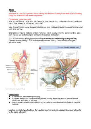
Anatomy
Abdominal wall:
Superficial to deep layers :
the skin, the Camper and Scarpa fascia, the external oblique aponeurosis, the internal oblique and
transversus muscles, the transversalis fascia, the preperitoneal fat, and the peritoneum
Inguinal canal
anterior wall: EOA and lateral 1/3 internal oblique muscle
posterior wall: transversalis fascia and medial 1/3 conjoint tendon
The floor: inguinal and pectineal ligaments
The roof: lower borders of the internal oblique and transversus muscles, forming the
conjoint muscle and tendon
Nerve :
1. Ilioinguinal nerve:
runs medially through the inguinal canal along with the cord structures traveling from the internal
ring to the external ring
innervates the upper and medial parts of the thigh, the anterior scrotum, and the base of the penis
Iliohypogastric nerve:
runs below the external oblique aponeurosis but cranial to the spermatic cord, then perforates the
external oblique cranial to the superficial ring
innervates the skin above the pubis
Genital branch of the genitofemoral nerve:
travels with the cremasteric vessels through the inguinal canal
innervates the cremaster muscle and provides sensory innervation to the scrotum
*NB:
impt of noting these nerves: avoid Causalgia syndromes: chronic groin pain when injury of these
nerves
Terminology
1. Indirect inguinal hernia: (finger pressure over deep ring will prevent formation >>This is the wat
to distinguish indirect and direct hernia>> but is often unreliable>>Age is reliable >> (age >50
usually direct inguinal hernia) (age <50 usually indirect inguinal hernia)
3. - protruding through the internal or deep inguinal ring
- lateral to the inferior epigastric artery
2. Direct inguinal hernia
- medial to the inferior epigastric artery
- through the Hesselbach triangle
Hesselbach triangle:
Inferior epigastric vessels laterally, lateral border of rectus sheath medially, and the
inguinal ligament inferiorly
Herniotomy (removal of the hernial sac only)
Herniorrhaphy (herniotomy plus repair of the
posterior wall of the inguinal canal)
Hernioplasty (reinforcement of the posterior
inguinal canal wall with a synthetic mesh)
Mechanism of Inguinal Hernia formation
Indirect inguinal hernia: Patent processus
vaginalis ( baby acutely crying and coughing may
ppt the process of indirect hernia formation)
Direct inguinal hernia: Old, weak muscle and
transversalis fascia , chronic cough, strain,
constipation, heavy lifting) ( Inguinal hernia
usually develop slowly)
(Neck of direct sac is board, in contrast to narrow neck of an indirect sac >> so indirect hernia
more likely to strangulate)
Direct + Indirect tgt : Pantaloon hernia
Strangulation: occur when hernia contents become constricted and by the neck of of the
sac and by twisting>> impair venous return >> swelling >> arterial obstruction >>
necrosis>> red + tender , irreducible >> bowel obstruction >> peritonitis)
Operation (Most inguinal hernia should be repaired ASAP to prevent risk of strangulation)
( Exception is some small, easily reducible and painless direct hernia n elderly man )
Direct hernia
>> if so medial
> maybe bladder so shd nt dissect the sac
Indirect hernia
if need widen of neck in deep inguinal ring
>> go lateral to avoid inferior epigastric vessels
if the sac is too wide in diameter
>>reduce radius of sac by circufenecial sticture
>>reduce pressure at sac by Laplace Law
Tension-free:
usu refer to Lichtenstein tension-free mesh-
based repair
but alternative tension free approach:
Herniorrhaphy Techniques
Shouldice repair
continue suture from pubic tubercle laterally &
then back to pubic tubercle
4. medial to lateral: inferolateral flap of transversalis fascia is sutured to the lateral edge of the rectus
sheath
lateral to medial: superior flap of transversalis fascia is sutured to the shelving portion of the
inguinal ligament
Bassini repair
Extraperitoneal approach: remove the peritoneal sac or educed it into the abdomen and suture the
approximate reflection of inguinal ligament (poupart’s) to the transversus abdominis aponeurosis/
conjoint tendon
Darn repair:
create a net along the conjoint tendon & inguinal ligament w/o tension apply
adv: weak point cn apply more stitch
Indications for laproscopic inguinal hernia repair
1. Bilateral inguinal hernia
2. Recurring hernia
3. Need to resume full activity asap
Complications of heniorrhaphy
1.chronic postherniorrhaphy pain
2. recurrence
3. seroma formation
4. bruising and hematoma
5. wound infection
6. Groin/ Scrotal swelling (haematoma)
7. testicular atrophy (damage to testicular artery, usually with diathermy or overtightening of the
deep ring)
5 Questions keep in mind
Lt vs Rt vs Both: cough & observe
scrotal mass vs hernia: cn get above?
femoral vs inguinal hernia: pubic tubercle
direct vs indirect hernia: occlusion test
reducible: ask patient to do so
Ddx
Groin mass
Skin & Subcutaneous lesions
Lipoma, Sebaceous cyst
Lymph nodes
Saphena varix
Inguinal canal
Inguinal hernia
Lipoma of spermatic cord
Encysted hydrocele of spermatic cord
Femoral hernia
rare in men & nulluparity
Scrotal mass
Skin lesions
Sebaceous cyst
Lymphedema of scrotum
Spermatic cord
Funiculitis
5. Varicocele
Inguinal-scrotal hernia
Epididymis
Epididymal cyst
epididymitis
Testis
Hydrocele
Testicular tumours
Orchitis
Torsion
Physical examination
Before exam
3C
privacy
exposure: abdomen to thigh
Position: legs apart
Inspection
swelling?
>> deviation of penis
scar?
in pain?
dilated vein?
instructed to look at the ceiling and cough:
cough impulse: 2 times for two side
extend to scrotum?
Palpation
groin vs scrotum
feel spermatic cord in relation with swelling
fet above the mass or not
inguinal vs femoral
palpate for pubic tubercle
palpate downward from umbilicus to pubic symphysis
palpate lateral to 1st bony prominent >> pubic tubercle
superior & medial >> inguinal
inferior & lateral >> femoral
ask pt to reduce it
direct vs indirect
occlusion test
locate deep inguinal ring: mid-pt btw ASIS & PT & 1 finger breath above it
if limit by occlusion & release upon standing
+ve >> indirect or narrow neck of direct
Tell the examiner
perform abdominal examination
Femoral hernia
3rd commonest hernia
20% of hernias in women and 5% in men
less common in nulliparity
most liable (40% of initial px) to become strangulated d/t narrowest neck & rigidity of femoral canal
>> OT asap
more in Rt side
Anatomy
Content:
6. fat, lymphatic vessels and the lymph node of Cloquet (femoral canal is narrow so cough impulse is
rarely detected and the hernia is usually irreducible . Thus small femoral hernia may be mistaken
as lipoma or cloquet’s lymph node. However a henia is deeply fixed while others tend to be mobile)
Borders of femoral hernia:
anterosuperiorly by the inguinal ligament
posteriorly by the pectineal ligament lying anterior to
the superior pubic ramus
medially by the lacunar ligament
laterally by the femoral vein
*NB:
widen femoral go medial avoid VAN
Strangulated femoral hernia : usually no local
symptom but may classically present as small bowel
obstruction (and usually a portion of bowel
circumference is trapped) (RICHTER’s HERNIA)
Lumbar hernia
2 weak pts ms folding
inferior lumbar hernia
triangle of petite by
lateral the external oblique muscle
inferior the crest of the ilium
medial: latissimus dorsi
superior lumbar hernia
triangle of Grynfelt by
superior: 12th rib
lateral: posterior border of IOM
medial: sacrospinalis
Spigelian hernia
Hernia through the linea semilunaris
occurring at the level of arcuate line
advances through that muscle & spreads out like a mushroom between EOM & IOM
but impalpable (because too many muscle covering)
soft, reducible mass lateral to rectus abdominalis below umbilicus
dx comfirmed by CT
7. Umbilical (hernia throught the umbilical ring, in adults associated with obesity
pregnancy and ascites)
congenital
failure of all or part of the midgut to return to coelom during early fetal life
failure of the round ligament
sac remains unruptured it is semitranslucent
symptomless but increase
RFs
ascites
hypothyroid
95% of hernias will disappear spontaneously at 2yo
persists at 2 years
>>low risk of strangulation
Paraumbilical
protrusion through the linea alba above or below umbilicus cicatrix instead of umbilicus
d/t increase abd pressure: obese
more in female
content: greater omentum +/- small intestine
narrow neck
high risk of strangulation
also px as IO
Epigasteic hernia
midline anywhere xiphoid process and the umbilicus, usually midway
protrusion of extraperitoneal fat through the linea alba
d/t abnormal decussation of the fibres of the aponeurosis
strong linea albu defect but protrude btw
two strong rectus abdominis >> small gap
extraperitoneal fat (but nt bowel)
>> pain as with nerve ending OR
>> fatty content being nipped sufficiently to produce partial strangulation
Related factors: obese
Divarication of rectus
nt hernia but fat
usu multiparous elderly
PE
strain >> gap btw rectus abdominalis
if abdomin relax >> finger cn insert btw them
no tx require
Obteratorhernia
Anatomy
Obturator canal
superior to obturator membrane in obturator foramen
formed by pectineal, external obturator & long adductor muscle
Px
elderly women with multiparity
aging lose fat
Atypical posture
pain refieved in semi-flexed position
worsen in hip extension adduction & internal rotation
usu no swelling d/t covered by the pectineus
8. 50% strangulated obturator hernia, pain refered along obturator nerve to knee
AXR
loop of gas at obturator foramen
Diaphragmatic hernia
Anatomy
components
central nonmuscular portion: central tendon
peripheral muscular rim
right and left diaphragmatic cura originated from L1-L3 and L1-L2
motor innervated by C3,4,5
sensory innervation
central nonmuscular portion: phrenic nerve
>> refer pain to neck
peripheral muscular part of diaphragmatic cura: intercostal nerve (T5-11), subcostal nerve (T12)
most case Lt posteriolateral defect
foramen of Bochdalek
dome of diaphragm posteriorly
the foramen of Morgagni: rare
anterior part with defect btw sternal and costal attachement
content: usu transverse colon
Prognosis
Severity of pulmonary HTN: earlier px >> worse prognosis
30% die even with respiratory support d/t lung hypoplasia
NB:
aplasia lung & small peritoneal cavity
acquired: traumatic close/open
Eventration of the diaphragm
diaphragmatic muscle is replaced by fibroelastic tissue
weakened hemidiaphragm: displaced into the thorax
d/t
congenital: inadequate development of phrenic nerve
acquired: trauma
Px
respiratory distress.
Prophylactic antibiotics in surgery
Indication
Prevention wound infection
Procedure involving prothestic insertion
Operation in patient with pre-inplanted prothesis: eg heart valve
Choice of Antibiotics:
use narrow spectrum: usu 1st G cephalosporin, cefazolin
CRC surgery & appendectomy
cefoxitin or cefotetan: coverage of anaserobes
alternative: cefuroxime + metronidazole
avoid routine use of vancomycin
>> promote VRE
>> indication of usage
serious allergy to penicillin & cephalosporin
high risk of post-OP MRSA
Timing
IV administrated at induction of GA
2nd dose
> 4hr OP
massive bleeding
NO indication of post-OP antibiotics
9. BUT some surgeons(Dr Au) still keep it
wound infection
clear: thyroid, breast, hernia
clean contaminated: 1st cephalosporin, metronidazole but depend on surgery
contaminated:
dirty: theurapeutic full course