Call Girls Hsr Layout Just Call 7001305949 Top Class Call Girl Service Available
Abc of burns
1. ABC of burns
Introduction
Shehan Hettiaratchy, Peter Dziewulski
Burns are one of the most devastating conditions encountered
in medicine. The injury represents an assault on all aspects of
the patient, from the physical to the psychological. It affects all
ages, from babies to elderly people, and is a problem in both
the developed and developing world. All of us have experienced
the severe pain that even a small burn can bring. However the
pain and distress caused by a large burn are not limited to the
immediate event. The visible physical and the invisible
psychological scars are long lasting and often lead to chronic
disability. Burn injuries represent a diverse and varied challenge
to medical and paramedical staff. Correct management requires
a skilled multidisciplinary approach that addresses all the
problems facing a burn patient.
This series provides an overview of the most important
aspects of burn injuries for hospital and non-hospital
healthcare workers.
How common are burns?
In the United Kingdom about 250 000 people are burnt each
year. Of these, 175 000 attend accident and emergency
departments, and 13 000 of these are admitted to hospital.
Some 1000 patients have severe enough burns to warrant
formal fluid resuscitation; half of these are children aged under
12 years. In an average year 300 burn deaths occur. These UK
figures are representative of most of the developed world
countries, although some, such as the United States, have a
higher incidence.
Burns are also a major problem in the developing world.
Over two million burn injuries are thought to occur each year
in India (population 500 million), but this may be a substantial
underestimate. Mortality in the developing world is much
higher than in the developed world. For example, Nepal has
about 1700 burn deaths a year for a population of 20 million,
giving a death rate 17 times that of Britain.
What are the causes of burns?
Most burns are due to flame injuries. Burns due to scalds are
the next most common. The most infrequent burns are those
caused by electrocution and chemical injuries. The type of
burns suffered is related to the type of patient injured. It is
therefore useful to break down burn aetiology by patient
groups as this reveals the varying causes of injury. In most
groups there is a male predominance. The only exception is in
elderly people, among whom more women are injured because
of the preponderance of women in that population.
Who gets burnt?
Young children—Children aged up to 4 years comprise 20% of
all patients with burn injuries. Most injuries (70%) are scalds
due to children spilling hot liquids or being exposed to hot
bathing water. These mechanisms can lead to large area burns.
Because of changes in the design and material of night
clothing, flame burns are less common than they were. Boys are
more likely to be injured, a reflection of the behavioural
differences between boys and girls.
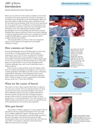
Top: Child with 70% full
thickness burns, which
required resuscitation,
intensive care support,
and extensive
debridement and skin
grafting. Left: The same
child one year later at a
burns camp, having made
a good recovery. A
reasonable outcome is
possible even with severe
burn injuries
Incidence of burns by ageCauses of burns
20%
10%
10%
55%
Flame
1-4 years old
5-14
years
old
15-64 years old
>65 years
old
Scalds
Chemical and
electrical
5%
40%
60%
Causes of burns (left) and incidence of burns by age (right)
Scald in young child caused by
spilling hot liquid. Most of the
burn is superficial, except for the
patch on the shoulder, which
required a skin graft and which
healed well
This is the first in a series of 12 articles
Clinical review
1366 BMJ VOLUME 328 5 JUNE 2004 bmj.com
on 30 September 2006bmj.comDownloaded from
2. Older children and adolescents—10% of burns happen to
children between the ages of 5 and 14. Teenagers are often
injured from illicit activities involving accelerants, such as petrol,
or electrocution.
Working age—Most burns ( > 60%) occur in patients aged
15-64. These are mainly due to flame burns, and up to a third
are due to work related incidents.
Elderly people—Some 10% of burns occur in people aged
over 65. Various effects of ageing (such as immobility, slowed
reactions, and decreased dexterity) mean elderly people are at
risk from scalds, contact burns, and flame burns.
Compromising factors—Burn victims’ health is often
compromised by some other factor, such as alcoholism,
epilepsy, or chronic psychiatric or medical illness. All such
problems need to be addressed when managing patients in
order to speed recovery and prevent repetition of injury.
Care of a major burn injury
The main aims of burn care are to restore form, function, and
feeling, and burn management can be divided up into seven
phases—rescue, resuscitate, retrieve, resurface, rehabilitate,
reconstruct, and review.
Rescue—The aim is to get the individual away from the
source of the injury and provide first aid. This is often done by
non-professionals—friends, relatives, bystanders, etc.
Resuscitate—Immediate support must be provided for any
failing organ system. This usually involves administering fluid to
maintain the circulatory system but may also involve supporting
the cardiac, renal, and respiratory systems.
Retrieve—After initial evacuation to an accident and
emergency department, patients with serious burns may need
transfer to a specialist burns unit for further care.
Resurface—The skin and tissues that have been damaged by
the burn must be repaired. This can be achieved by various
means, from simple dressings to aggressive surgical
debridement and skin grafting.
Rehabilitate—This begins on the day a patient enters hospital
and continues for years after he or she has left. The aim is to
return patients, as far as is possible, to their pre-injury level of
physical, emotional, and psychological wellbeing.
Reconstruct—The scarring that results from burns often leads
to functional impairment that must be addressed. The
operations needed to do this are often complex and may need
repeating as a patient grows or the scars re-form.
Review—Burn patients, especially children, require regular
review for many years so that problems can be identified early
and solutions provided.
The complexity of the injury and the chronic nature of the
sequelae of burns require an integrated multidisciplinary
approach with long follow up. Only such management can lead
to the best outcomes for burn patients.
Prognostication in major burns
Determining whether someone will survive a severe burn injury
is not simple but is important. Aggressive treatment for
someone with a non-survivable injury is inhumane, and it is
inappropriate not to treat a patient who has a severe but
potentially survivable injury. Unfortunately, there is no exact
way to predict who will survive a burn injury. Several formulae
have been devised to estimate the risk of death after burn injury.
None has been evaluated prospectively in large trials, however,
and so they should be used only for audit purposes. It is also
inappropriate to apply generic formulae to individuals. Each
patient should be considered individually.
Burn incurred by an adolescent boy while inhaling butane gas.
There was full thickness damage to the lower lip, which required
debridement and extensive reconstruction
Aims of burn care
Restore form—Return the damaged area to as close
to normality as is possible
Restore function—Maximise patient’s ability to
perform pre-injury activities
Restore feeling—Enable psychological and emotional
recovery
Bitumen burns to face in work related incident
Contact burns in an elderly patient after a collapse and
prolonged contact with a radiator. Treatment required excision
and split skin grafting as well as investigation into the cause of
the collapse
Clinical review
1367BMJ VOLUME 328 5 JUNE 2004 bmj.com
on 30 September 2006bmj.comDownloaded from
3. Certain factors increase the risk of death. The most
important are increasing age, increasing burn size, and the
presence of an inhalational injury. Exactly how these factors
interrelate is not clear. Evidence suggests that a patient
aged over 60 with a burn covering more than 40% of body
surface area and an inhalational injury has a > 90% chance of
dying.
As well as assessing the injury, it is also important to make
some estimation of the patient’s quality of life before the burn.
This can be obtained from relatives, carers, or the patient.
Deciding not to resuscitate a patient is difficult. It is often useful
to get a consensus opinion from the whole burn team.
Burn prevention and fire safety
The fact that 90% of burn injuries are preventable has led to
many attempts to decrease their incidence. These attempts fall
into two main categories—education and legislation. Education
is an “active” process that requires a change in an individual’s
behaviour. Legislation is “passive” and is independent of a
person’s actions. Both have advantages and disadvantages.
Education—The most successful campaigns have targeted
specific burn aetiologies or populations. A good example of this
is the campaign to reduce chip pan fires in Britain during the
late 1970s. This led to a 30% reduction in the incidence of
burns due to chip pan fires. The main problem with educational
prevention is that it relies on changing the way individuals
behave. This means the message must be repeated regularly, as
shown by the UK government launching a second chip pan fire
campaign in 1999. However, a successful educational campaign
has an instantaneous and widespread impact.
Legislation—Legislation (such as the compulsory fitting of
sprinklers in commercial buildings) has led to substantial
decreases in burn injury. The main problem with legislation is
that it takes time to pass and to have an effect. Compliance must
also be obtained and maintained. However, as it does not rely on
a change in individuals’ actions, legislation can be effective.
Effective prevention requires both passive and active
elements. The basis for all prevention is good epidemiological
data to reveal specific causes of burns and at risk populations,
both of which can be targeted. The UK government is currently
running the “Fire kills” campaign, which covers all aspects of
domestic fire prevention and safety. The related website,
www.firekills.gov.uk, is an excellent source of information.
Some risk factors for burns are not easy to change.
Overcrowding, poor housing, and the other attributes of
poverty are major contributors to the risk of burn injuries.
Shehan Hettiaratchy is specialist registrar in plastic and reconstructive
surgery, Pan-Thames Training Scheme, London; Peter Dziewulski is
consultant burns and plastic surgeon, St Andrews Centre for Plastic
Surgery and Burns, Broomfield Hospital, Chelmsford.
The ABC of burns is edited by Shehan Hettiaratchy; Remo Papini,
consultant and clinical lead in burns, West Midlands Regional Burns
Unit, Selly Oak University Hospital, Birmingham; and Peter
Dziewulski. The series will be published as a book in the autumn.
Competing interests: RP has been reimbursed by Johnson & Johnson,
manufacturer of Integra, and Smith & Nephew, manufacturer of Acticoat
and TransCyte, for attending symposiums on burn care.
Death
Increasing
age
Increasing
burn size
Inhalational
injury Factors that increase the risk
of death after a major burn
FIRE
KILLS
YOU CAN
PREVENT IT UK government’s “Fire kills”
campaign started in 2002
Information in UK government’s “Fire kills” campaign
“Top 10 safety tips”
How to make your house a safe home
x Fit a smoke alarm and check it regularly
x Make a fire action plan so that everyone in your house knows how
to escape in the event of fire
x Take care when cooking with hot oil and think about using
thermostatically controlled deep fat fryers
x Never leave lit candles unattended
x Ensure cigarettes are stubbed out and disposed of carefully
x Never smoke in bed
x Keep matches and lighters away from children
x Keep clothing away from heating appliances
x Take care in the kitchen. Accidents while cooking account for 59%
of fires in the home
x Take special care when you are tired or when you’ve been drinking.
Half of all deaths in domestic fires occur between 10 pm and 8 am
On finding a fire in the home
x Get out x Stay out x Call 999 (telephone number
for UK emergency services)
Key points
x Burns are a major cause of injury and death worldwide
x Flame burns are the most common type
x Young children, elderly people, and those who are mentally or
physically compromised are at particular risk
x Death is more likely with increasing age, increasing burn size, and
presence of inhalational injury
x 90% of burns are preventable
BMJ 2004;328:1366–8
Further reading
x Wilkinson E. The epidemiology of burns in secondary care, in a
population of 2.6 million people. Burns 1998;24:139-43
x Ryan CM, Schoenfeld DA, Thorpe WP, Sheridan RL, Cassem EH,
Tompkins RG. Objective estimates of the probability of death from
burn injuries. N Engl J Med 1998;338:362-6
x Fire kills. You can prevent it. www.firekills.gov.uk
x Herndon D. Total burn care. 2nd ed. London: WB Saunders, 2002
x National Community Fire Safety Centre Toolbox.
www.firesafetytoolbox.org.uk
x Liao C-C, Rossignol AM. Landmarks in burn prevention. Burns
2000;26:422-34
Clinical review
1368 BMJ VOLUME 328 5 JUNE 2004 bmj.com
on 30 September 2006bmj.comDownloaded from
4. ABC of burns
Pathophysiology and types of burns
Shehan Hettiaratchy, Peter Dziewulski
Understanding the pathophysiology of a burn injury is
important for effective management. In addition, different
causes lead to different injury patterns, which require different
management. It is therefore important to understand how a
burn was caused and what kind of physiological response it will
induce.
The body’s response to a burn
Burn injuries result in both local and systemic responses.
Local response
The three zones of a burn were described by Jackson in 1947.
Zone of coagulation—This occurs at the point of maximum
damage. In this zone there is irreversible tissue loss due to
coagulation of the constituent proteins.
Zone of stasis—The surrounding zone of stasis is
characterised by decreased tissue perfusion. The tissue in this
zone is potentially salvageable. The main aim of burns
resuscitation is to increase tissue perfusion here and prevent
any damage becoming irreversible. Additional insults—such as
prolonged hypotension, infection, or oedema—can convert this
zone into an area of complete tissue loss.
Zone of hyperaemia—In this outermost zone tissue perfusion is
increased. The tissue here will invariably recover unless there is
severe sepsis or prolonged hypoperfusion.
These three zones of a burn are three dimensional, and loss
of tissue in the zone of stasis will lead to the wound deepening
as well as widening.
Systemic response
The release of cytokines and other inflammatory mediators at
the site of injury has a systemic effect once the burn reaches
30% of total body surface area.
Cardiovascular changes—Capillary permeability is increased,
leading to loss of intravascular proteins and fluids into the
interstitial compartment. Peripheral and splanchnic
vasoconstriction occurs. Myocardial contractility is decreased,
possibly due to release of tumour necrosis factor . These
changes, coupled with fluid loss from the burn wound, result in
systemic hypotension and end organ hypoperfusion.
Respiratory changes—Inflammatory mediators cause
bronchoconstriction, and in severe burns adult respiratory
distress syndrome can occur.
Metabolic changes—The basal metabolic rate increases up to
three times its original rate. This, coupled with splanchnic
hypoperfusion, necessitates early and aggressive enteral feeding
to decrease catabolism and maintain gut integrity.
Immunological changes—Non-specific down regulation of the
immune response occurs, affecting both cell mediated and
humoral pathways.
Mechanisms of injury
Thermal injuries
Scalds—About 70% of burns in children are caused by scalds.
They also often occur in elderly people. The common
mechanisms are spilling hot drinks or liquids or being exposed
Clinical image of burn zones. There is central necrosis,
surrounded by the zones of stasis and of hyperaemia
Zone of
coagulation
Zone of
coagulation
Epidermis
Dermis
Zone of
stasis
Zone of
hyperaemia
Inadequate
resuscitation
Adequate
resuscitation
Zone of stasis preserved Zone of stasis lost
Jackson’s burns zones and the effects of adequate and inadequate
resuscitation
Cardiovascular
Reduced myocardial
contractility
Immunological
Reduced immune
response
Increased capillary
permeability
Peripheral and splanchnic
vasoconstriction
Respiratory
Bronchoconstriction
Metabolic
Basal metabolic rate
increased threefold
Adult respiratory
distress syndrome
Systemic changes that occur after a burn injury
This is the second in a series of 12 articles
Clinical review
1427BMJ VOLUME 328 12 JUNE 2004 bmj.com
on 30 September 2006bmj.comDownloaded from
5. to hot bathing water. Scalds tend to cause superficial to
superficial dermal burns (see later for burn depth).
Flame—Flame burns comprise 50% of adult burns. They are
often associated with inhalational injury and other concomitant
trauma. Flame burns tend to be deep dermal or full thickness.
Contact—In order to get a burn from direct contact, the
object touched must either have been extremely hot or the
contact was abnormally long. The latter is a more common
reason, and these types of burns are commonly seen in people
with epilepsy or those who misuse alcohol or drugs. They are
also seen in elderly people after a loss of consciousness; such a
presentation requires a full investigation as to the cause of the
blackout. Burns from brief contact with very hot substances are
usually due to industrial accidents. Contact burns tend to be
deep dermal or full thickness.
Electrical injuries
Some 3-4% of burn unit admissions are caused by electrocution
injuries. An electric current will travel through the body from
one point to another, creating “entry” and “exit” points. The
tissue between these two points can be damaged by the current.
The amount of heat generated, and hence the level of tissue
damage, is equal to 0.24×(voltage)2
×resistance. The voltage is
therefore the main determinant of the degree of tissue damage,
and it is logical to divide electrocution injuries into those
caused by low voltage, domestic current and those due to high
voltage currents. High voltage injuries can be further divided
into “true” high tension injuries, caused by high voltage current
passing through the body, and “flash” injuries, caused by
tangential exposure to a high voltage current arc where no
current actually flows through the body.
Domestic electricity—Low voltages tend to cause small, deep
contact burns at the exit and entry sites. The alternating nature
of domestic current can interfere with the cardiac cycle, giving
rise to arrhythmias.
“True” high tension injuries occur when the voltage is 1000 V
or greater. There is extensive tissue damage and often limb loss.
There is usually a large amount of soft and bony tissue necrosis.
Muscle damage gives rise to rhabdomyolysis, and renal failure
may occur with these injuries. This injury pattern needs more
aggressive resuscitation and debridement than other burns.
Contact with voltage greater than 70 000 V is invariably fatal.
“Flash” injury can occur when there has been an arc of
current from a high tension voltage source. The heat from this
arc can cause superficial flash burns to exposed body parts,
typically the face and hands. However, clothing can also be set
alight, giving rise to deeper burns. No current actually passes
through the victim’s body.
A particular concern after an electrical injury is the need for
cardiac monitoring. There is good evidence that if the patient’s
electrocardiogram on admission is normal and there is no
history of loss of consciousness, then cardiac monitoring is not
required. If there are electrocardiographic abnormalities or a
loss of consciousness, 24 hours of monitoring is advised.
Chemical injuries
Chemical injuries are usually as a result of industrial accidents
but may occur with household chemical products. These burns
tend to be deep, as the corrosive agent continues to cause
coagulative necrosis until completely removed. Alkalis tend to
penetrate deeper and cause worse burns than acids. Cement is a
common cause of alkali burns.
Certain industrial agents may require specific treatments in
addition to standard first aid. Hydrofluoric acid, widely used for
glass etching and in the manufacture of circuit boards, is one of
the more common culprits. It causes a continuing, penetrating
Examples of a scald burn (left) and a contact burn from a hot iron (right) in
young children
Flash injuryTrue high tension injury
Current arcs, causing flash
No current goes through patient
Current passes
through patient
Differences between true high tension burn and flash burn
Electrocardiogram after electrocution showing atrial
fibrillation
Chemical burn due to spillage of sulphuric acid
Clinical review
1428 BMJ VOLUME 328 12 JUNE 2004 bmj.com
on 30 September 2006bmj.comDownloaded from
6. injury and must be neutralised with calcium gluconate, either
applied topically in a gel or injected into the affected tissues.
The initial management of all chemical burns is the same
irrespective of the agent. All contaminated clothing must be
removed, and the area thoroughly irrigated. This is often best
achieved by showering the patient. This has been shown to limit
the depth of the burn. Litmus paper can be used to confirm
removal of alkali or acid. Eye injuries should be irrigated
copiously and referred to an ophthalmologist.
Non{accidental injury
An estimated 3-10% of paediatric burns are due to
non{accidental injury. Detecting these injuries is important as
up to 30% of children who are repeatedly abused die. Usually
young children ( < 3 years old) are affected. As with other
non{accidental injuries, the history and the pattern of injury
may arouse suspicion. A social history is also important. Abuse
is more common in poor households with single or young
parents. Such abuse is not limited to children: elderly and other
dependent adults are also at risk. A similar assessment can be
made in these scenarios.
It is natural for non{accidental injury to trigger anger
among healthcare workers. However, it is important that all
members of the team remain non-confrontational and try to
establish a relationship with the perpetrators. The time around
the burn injury is an excellent opportunity to try to break the
cycle of abuse. In addition, it is likely that the patient will
eventually be discharged back into the care of the individuals
who caused the injury. As well as treating the physical injury, the
burn team must try to prevent further abuse by changing the
relationship dynamics between victim and abuser(s).
Any suspicion of non{accidental injury should lead to
immediate admission of the child to hospital, irrespective of
how trivial the burn is, and the notification of social services.
The team should carry out the following:
x Examine for other signs of abuse
x Photograph all injuries
x Obtain a team opinion about parent-child interaction
x Obtain other medical information (from general practitioner,
health visitor, referring hospital)
x Interview family members separately about the incident
(check for inconsistencies) and together (observe interaction).
It should be remembered that the injury does not have to be
caused deliberately for social services to intervene; inadequate
supervision of children mandates their involvement.
Shehan Hettiaratchy is specialist registrar in plastic and reconstructive
surgery, Pan-Thames Training Scheme, London; Peter Dziewulski is
consultant burns and plastic surgeon, St Andrews Centre for Plastic
Surgery and Burns, Broomfield Hospital, Chelmsford.
The ABC of burns is edited by Shehan Hettiaratchy; Remo Papini,
consultant and clinical lead in burns, West Midlands Regional Burn
Unit, Selly Oak University Hospital, Birmingham; and Peter
Dziewulski. The series will be published as a book in the autumn.
Competing interests: See first article for series editors’ details.
Specific chemical burns and treatments
Chromic acid—Rinse with dilute sodium hyposulphite
Dichromate salts—Rinse with dilute sodium
hyposulphite
Hydrofluoric acid—10% calcium gluconate applied
topically as a gel or injected
Injury pattern of non{accidental burns
x Obvious pattern from cigarettes, lighters, irons
x Burns to soles, palms, genitalia, buttocks, perineum
x Symmetrical burns of uniform depth
x No splash marks in a scald injury. A child falling into a bath will
splash; one that is placed into it may not
x Restraint injuries on upper limbs
x Is there sparing of flexion creases—that is, was child in fetal position
(position of protection) when burnt? Does this correlate to a “tide
line” of scald—that is, if child is put into a fetal position, do the
burns line up?
x “Doughnut sign,” an area of spared skin surrounded by scald. If a
child is forcibly held down in a bath of hot water, the part in contact
with the bottom of the bath will not burn, but the tissue around will
x Other signs of physical abuse—bruises of varied age, poorly kempt,
lack of compliance with health care (such as no immunisations)
History of non-accidental burns
x Evasive or changing history
x Delayed presentation
x No explanation or an implausible mechanism
given for the burn
x Inconsistency between age of the burn and age
given by the history
x Inadequate supervision, such as child left in the
care of inappropriate person (older sibling)
x Lack of guilt about the incident
x Lack of concern about treatment or prognosis
“Doughnut sign” in a child with immersion scalds. An
area of spared skin is surrounded by burnt tissue. The
tissue has been spared as it was in direct contact with
the bath and protected from the water. This burn
pattern suggests non{accidental injury
Key points
x A burn results in three distinct zones—coagulation, stasis, and
hyperaemia
x The aim of burns resuscitation is to maintain perfusion of the zone
of stasis
x Systemic response occurs once a burn is greater than 30% of total
body surface area
x Different burn mechanisms lead to different injury patterns
x Identification of non{accidental burn injury is important
BMJ 2004;328:1427–9
Further reading
x Kao CC, Garner WL. Acute burns. Plast Reconstr Surg 2000;105:
2482{93
x Andronicus M, Oates RK, Peat J, Spalding S, Martin H.
Non-accidental burns in children. Burns 1998;24:552-8
x Herndon D. Total burn care. 2nd ed. London: WB Saunders, 2002
x Luce EA. Electrical burns. Clin Plast Surg 2000;27:133-43
x Kirkpatrick JJR, Enion DS, Burd DAR. Hydrofluoric acid burns; a
review. Burns 1995;21:483-93
x Burnsurgery.org. www.burnsurgery.org
Clinical review
1429BMJ VOLUME 328 12 JUNE 2004 bmj.com
on 30 September 2006bmj.comDownloaded from
7. ABC of burns
First aid and treatment of minor burns
Jackie Hudspith, Sukh Rayatt
Some 250 000 burns occur annually in the United Kingdom.
About 90% of these are minor and can be safely managed in
primary care. Most of these will heal regardless of treatment, but
the initial care can have a considerable influence on the
cosmetic outcome. All burns should be assessed by taking an
adequate history and examination.
First aid
The aims of first aid should be to stop the burning process, cool
the burn, provide pain relief, and cover the burn.
Stop the burning process—The heat source should be removed.
Flames should be doused with water or smothered with a
blanket or by rolling the victim on the ground. Rescuers should
take care to avoid burn injury to themselves. Clothing can retain
heat, even in a scald burn, and should be removed as soon as
possible. Adherent material, such as nylon clothing, should be
left on. Tar burns should be cooled with water, but the tar itself
should not be removed. In the case of electrical burns the victim
should be disconnected from the source of electricity before
first aid is attempted.
Cooling the burn—Active cooling removes heat and prevents
progression of the burn. This is effective if performed within
20 minutes of the injury. Immersion or irrigation with running
tepid water (15°C) should be continued for up to 20 minutes.
This also removes noxious agents and reduces pain, and may
reduce oedema by stabilising mast cells and histamine release.
Iced water should not be used as intense vasoconstriction can
cause burn progression. Cooling large areas of skin can lead to
hypothermia, especially in children. Chemical burns should be
irrigated with copious amounts of water.
Analgesia—Exposed nerve endings will cause pain. Cooling
and simply covering the exposed burn will reduce the pain.
Opioids may be required initially to control pain, but once first
aid measures have been effective non-steroidal
anti-inflammatory drugs such as ibuprofen or co-dydramol
taken orally will suffice.
Covering the burn—Dressings should cover the burn area
and keep the patient warm. Polyvinyl chloride film (cling film)
is an ideal first aid cover. The commercially available roll is
essentially sterile as long as the first few centimetres are
discarded. This dressing is pliable, non-adherent, impermeable,
acts as a barrier, and is transparent for inspection. It is
important to lay this on the wound rather than wrapping the
burn. This is especially important on limbs, as later swelling
may lead to constriction. A blanket laid over the top will keep
the patient warm. If cling film is not available then any clean
cotton sheet (preferably sterile) can be used. Hand burns can
be covered with a clear plastic bag so as not to restrict mobility.
Avoid using wet dressings, as heat loss during transfer to
hospital can be considerable.
Use of topical creams should be avoided at this stage as
these may interfere with subsequent assessment of the burn.
Cooling gels such as Burnshield are often used by paramedics.
These are useful in cooling the burn and relieving pain in the
initial stages.
A superficial scald suitable for management in primary care
Benefits of cooling burn injuries with water
x Stops burning process
x Minimises oedema
x Reduces pain
x Cleanses wound
Cling film for dressing burn wounds
x Essentially sterile
x Lay on wound—Do not wrap around
x Non-adherent
x Pliable
x Transparent for inspection
Burnshield is a cooling gel used to cover burn and
reduce pain
This is the third in a series of 12 articles
Clinical review
1487BMJ VOLUME 328 19 JUNE 2004 bmj.com
on 30 September 2006bmj.comDownloaded from
8. Management of minor burns
The cause of injury and depth and extent of burn should be
assessed in the same way as for more major burns and
recorded. Similarly, associated illness or injuries must be
considered (such as small burns as a result of fits, faints, or falls).
Burns suitable for outpatient management are usually small
and superficial and not affecting critical areas. Home
circumstances should be considered, as even small injuries to
the feet will progress if the legs are not elevated for at least
48 hours; this is rarely possible at home. Always consult a burns
unit if in doubt about management
Once the decision has been taken to treat a burn patient as
an outpatient, analgesia should be given and the wound
thoroughly cleaned and a dressing applied (except on the face).
Ensure that a follow up appointment is made.
There are a vast range of acceptable options in the
outpatient management of minor burns. The following should
be used as a guide
Cleaning the burn
It is important to realise that a new burn is essentially sterile,
and every attempt should be made to keep it so. The burn
wound should be thoroughly cleaned with soap and water or
mild antibacterial wash such as dilute chlorohexidine. Routine
use of antibiotics should be discouraged. There is some
controversy over management of blisters, but large ones should
probably be de-roofed, and dead skin removed with sterile
scissors or a hypodermic needle. Smaller blisters should be left
intact.
Dressings
Many different dressings are in use, with little or no data to
support any individual approach. We favour covering the clean
burn with a simple gauze dressing impregnated with paraffin
(Jelonet). Avoid using topical creams as these will interfere with
subsequent assessment of the burn. Apply a gauze pad over the
dressing, followed by several layers of absorbent cotton wool. A
firm crepe bandage applied in a figure of eight manner and
secured with plenty of adhesive tape (Elastoplast) will prevent
slippage of the dressing and shearing of the wound.
An elastic net dressing (Netelast) is useful for securing
awkward areas such as the head and neck and chest. Limb
burns should be elevated for the duration of treatment.
Dressing changes
The practice of subsequent dressing changes is varied. Ideally
the dressing should be checked at 24 hours. The burn wound
itself should be reassessed at 48 hours and the dressings
changed, as they are likely to be soaked through. At this stage
the depth of burn should be apparent, and topical agents such
as Flamazine can be used.
Depending on how healing is progressing, dressing changes
thereafter should be every three to five days. If the Jelonet
dressing has become adherent, it should be left in place to avoid
damage to delicate healing epithelium. If Flamazine is used it
should be changed on alternate days. The dressing should be
changed immediately if the wound becomes painful or smelly
or the dressing becomes soaked (“strike through”).
Any burn that has not healed within two weeks should be
seen by a burn surgeon.
Specialist dressings
Many specialist dressings are available, some developed for
specific cases, but most designed for their ease of use. The
following are among the more widely used.
Minor burns suitable for outpatient
management
x Partial thickness burns covering < 10% of total
body surface area in adults
x Partial thickness burns covering < 5% of body
surface area in children
x Full thickness burns covering < 1% of body
surface
x No comorbidity
Large blister on
thenar eminence
restricting movement
of hand (top). Blister
is de-roofed using
aseptic technique
(bottom)
Superficial scald burn on side of neck (top left) is cleaned and then a
layer of Jelonet applied over it (top right). Gauze square dressings on top
of the Jelonet (bottom left) are held in place with a Netelast type of
dressing (bottom right)
Dressing changes for burns
x Use aseptic technique
x First change after 48 hours, and every 3-5 days
thereafter
x Criteria for early dressing change:
Excessive “strike through” of fluid from wound
Smelly wound
Contaminated or soiled dressings
Slipped dressings
Signs of infection (such as fever)
Clinical review
1488 BMJ VOLUME 328 19 JUNE 2004 bmj.com
on 30 September 2006bmj.comDownloaded from
9. Flamazine is silver sulfadiazine cream and is applied topically
on the burn wound. It is effective against gram negative bacteria
including Pseudomonas. Infection with the latter will cause the
dressing to turn green with a distinctive smell. Apply the cream
in a 3-5 mm thick layer and cover with gauze. It should be
removed and reapplied every two days. There is a reported
3{5% incidence of reversible leucopenia.
Granulflex is a hydrocolloid dressing with a thin
polyurethane foam sheet bonded onto a semipermeable film.
The dressing is adhesive and waterproof and is therefore useful
in awkward areas or where normal dressings are not suitable. It
should be applied with a 2 cm border. By maintaining a moist
atmosphere over the wound, it creates an environment suitable
for healing. It usually needs to be changed every three or four
days, but it can be left for seven days. A thinner version
(Duoderm) is also available.
Mepitel is a flexible polyamide net coated with soft silicone
to give a Jelonet-type of dressing that is non adhesive. It is a
useful but expensive alternative to Jelonet when easy removal is
desirable, such as with children.
Facial burns
Facial burns should be referred to a specialist unit. However,
simple sunburn should be left exposed as dressings can be
awkward to retain on the face. The wound should be cleansed
twice daily with mild diluted chlorohexidine solution. The burn
should be covered with a bland ointment such as liquid paraffin.
This should be applied every 1-4 hours as necessary to
minimise crust formation. Men should shave daily to reduce
risk of infection. All patients should be advised to sleep
propped up on two pillows for the first 48 hours to minimise
facial oedema.
Follow up
Burns that fail to heal within three weeks should be referred to
a plastic surgery unit for review. Healed burns will be sensitive
and have dry scaly skin, which may develop pigmental changes.
Daily application of moisturiser cream should be encouraged.
Healed areas should be protected from the sun with sun block
for 6-12 months. Pruritis is a common problem.
Physiotherapy—Patients with minor burns of limbs may need
physiotherapy. It is important to identify these patients early
and start therapy. Hypertrophic scars may benefit from scar
therapy such as pressure garments or silicone. For these
reasons, all healed burns should be reviewed at two months for
referral to an occupational therapist if necessary.
Support and reassurance—Patients with burn injuries often
worry about disfigurement and ugliness, at least in the short
term, and parents of burnt children often have feelings of guilt.
It is important to address these issues with reassurance.
Jackie Hudspith is clinical nurse lead, Burns Centre, Chelsea and
Westminster Hospital, London. Sukh Rayatt is specialist registrar in
plastic and reconstructive surgery, West Midlands Training Scheme,
Birmingham.
The ABC of burns is edited by Shehan Hettiaratchy, specialist
registrar in plastic and reconstructive surgery, Pan-Thames Training
Scheme, London; Remo Papini, consultant and clinical lead in burns,
West Midlands Regional Burn Unit, Selly Oak University Hospital,
Birmingham; and Peter Dziewulski, consultant burns and plastic
surgeon, St Andrews Centre for Plastic Surgery and Burns,
Broomfield Hospital, Chelmsford. The series will be published as a
book in the autumn.
Competing interests: See first article for series editors’ details.
BMJ 2004;328:1487–9
Flamazine
x Silver sulfadiazine cream
x Covers gram negative bacteria including
Pseudomonas
x Needs to be changed every 24-48 hours
x Makes burn seem white and should be avoided if
burn needs reassessment
Management of facial burns
x Clean face twice a day with dilute chlorohexidine
solution
x Cover with cream such as liquid paraffin on
hourly basis
x Men should shave daily
x Sleep propped up on two pillows to minimise
oedema
Pruritis
x Common in healing and healed burn wounds
x Aggravated by heat, stress, and physical activity
x Worst after healing
x Massage with aqueous cream or aloe vera cream
x Use antihistamines (such as chlorphenamine)
and analgesics
Key points
x Initial first aid can influence final cosmetic outcome
x Cooling with tepid tap water is one of the most important first aid
measures
x Routine use of antibiotics should be discouraged
x Simple dressings suffice
x Aseptic technique should be used for dressing changes
x If in doubt, seek advice from regional burns unit or plastic surgery
department
Further reading
x Wilson G, French G. Plasticized polyvinylchloride as a temporary
dressing for burns. BMJ 1987;294:556-7
x Davies JWL. Prompt cooling of the burned area: a review of
benefits and the effector mechanisms. Burns 1982;9:1-6
x Slater RM, Hughes NC. A simplified method of treating burns of
the hands. Br J Plast Surg 1971;24:296-300
x Herndon D. Total burn care. 2nd ed. London: Harcourt, 2002
x Settle J, ed. Principles and practice of burns management. Edinburgh:
Churchill Livingstone, 1996
x National Burn Care Review. National burn injury referral
guidelines. In: Standards and strategy for burn care. London: NBCR,
2001: 68-9
Clinical review
1489BMJ VOLUME 328 19 JUNE 2004 bmj.com
on 30 September 2006bmj.comDownloaded from
10. ABC of burns
Initial management of a major burn: I—overview
Shehan Hettiaratchy, Remo Papini
A major burn is defined as a burn covering 25% or more of
total body surface area, but any injury over more than 10%
should be treated similarly. Rapid assessment is vital. The
general approach to a major burn can be extrapolated to
managing any burn. The most important points are to take an
accurate history and make a detailed examination of the patient
and the burn, to ensure that key information is not missed.
This article outlines the structure of the initial assessment.
The next article will cover the detailed assessment of burn
surface area and depth and how to calculate the fluid
resuscitation formula.
History taking
The history of a burn injury can give valuable information
about the nature and extent of the burn, the likelihood of
inhalational injury, the depth of burn, and probability of other
injuries. The exact mechanism of injury and any prehospital
treatment must be established.
A patient’s history must be obtained on admission, as this
may be the only time that a first hand history is obtainable.
Swelling may develop around the airway in the hours after
injury and require intubation, making it impossible for the
patient to give a verbal history. A brief medical history should
be taken, outlining previous medical problems, medications,
allergies, and vaccinations. Patients’ smoking habits should be
determined as these may affect blood gas analyses.
Primary survey
The initial management of a severely burnt patient is similar to
that of any trauma patient. A modified “advanced trauma life
support” primary survey is performed, with particular emphasis
on assessment of the airway and breathing. The burn injury
must not distract from this sequential assessment, otherwise
serious associated injuries may be missed.
A—Airway with cervical spine control
An assessment must be made as to whether the airway is
compromised or is at risk of compromise. The cervical spine
should be protected unless it is definitely not injured. Inhalation
of hot gases will result in a burn above the vocal cords. This
burn will become oedematous over the following hours,
especially after fluid resuscitation has begun. This means that an
airway that is patent on arrival at hospital may occlude after
admission. This can be a particular problem in small children.
Direct inspection of the oropharynx should be done by a
senior anaesthetist. If there is any concern about the patency of
the airway then intubation is the safest policy. However, an
unnecessary intubation and sedation could worsen a patient’s
condition, so the decision to intubate should be made carefully.
B—Breathing
All burn patients should receive 100% oxygen through a
humidified non-rebreathing mask on presentation. Breathing
problems are considered to be those that affect the respiratory
system below the vocal cords. There are several ways that a burn
injury can compromise respiration.
Initial assessment of a major burn
x Perform an ABCDEF primary survey
A—Airway with cervical spine control, B—Breathing,
C—Circulation, D—Neurological disability, E—Exposure with
environmental control, F—Fluid resuscitation
x Assess burn size and depth (see later article for detail)
x Establish good intravenous access and give fluids
x Give analgesia
x Catheterise patient or establish fluid balance monitoring
x Take baseline blood samples for investigation
x Dress wound
x Perform secondary survey, reassess, and exclude or treat associated
injuries
x Arrange safe transfer to specialist burns facility
Key points of a burn history
Exact mechanism
x Type of burn agent (scald,
flame, electrical, chemical)
x How did it come into contact
with patient?
x What first aid was performed?
x What treatment has been
started?
x Is there risk of concomitant
injuries (such as fall from height,
road traffic crash, explosion)?
x Is there risk of inhalational
injuries (did burn occur in an
enclosed space)?
Exact timings
x When did the injury occur?
x How long was patient exposed
to energy source?
x How long was cooling applied?
x When was fluid resuscitation
started?
Exact injury
Scalds
x What was the liquid? Was it
boiling or recently boiled?
x If tea or coffee, was milk in it?
x Was a solute in the liquid?
(Raises boiling temperature
and causes worse injury, such
as boiling rice)
Electrocution injuries
x What was the voltage (domestic
or industrial)?
x Was there a flash or arcing?
x Contact time
Chemical injuries
x What was the chemical?
Is there any suspicion of non-accidental injury?
x See previous article
Airway management
Signs of inhalational injury
x History of flame burns or burns in
an enclosed space
x Full thickness or deep dermal burns
to face, neck, or upper torso
x Singed nasal hair
x Carbonaceous sputum or carbon
particles in oropharynx
Indications for intubation
x Erythema or swelling of
oropharynx on direct
visualisation
x Change in voice, with
hoarseness or harsh cough
x Stridor, tachypnoea, or
dyspnoea
Carbonaceous particles
staining a patient’s face after
a burn in an enclosed space.
This suggests there is
inhalational injury
This is the fourth in a series of 12 articles
Clinical review
1555BMJ VOLUME 328 26 JUNE 2004 bmj.com
on 30 September 2006bmj.comDownloaded from
11. Mechanical restriction of breathing—Deep dermal or full
thickness circumferential burns of the chest can limit chest
excursion and prevent adequate ventilation. This may require
escharotomies (see next article).
Blast injury—If there has been an explosion, blast lung can
complicate ventilation. Penetrating injuries can cause tension
pneumothoraces, and the blast itself can cause lung contusions
and alveolar trauma and lead to adult respiratory distress
syndrome.
Smoke inhalation—The products of combustion, though
cooled by the time they reach the lungs, act as direct irritants to
the lungs, leading to bronchospasm, inflammation, and
bronchorrhoea. The ciliary action of pneumocytes is impaired,
exacerbating the situation. The inflammatory exudate created is
not cleared, and atelectasis or pneumonia follows. The situation
can be particularly severe in asthmatic patients. Non-invasive
management can be attempted, with nebulisers and positive
pressure ventilation with some positive end-expiratory pressure.
However, patients may need a period of ventilation, as this
allows adequate oxygenation and permits regular lung toileting.
Carboxyhaemoglobin—Carbon monoxide binds to
deoxyhaemoglobin with 40 times the affinity of oxygen. It also
binds to intracellular proteins, particularly the cytochrome
oxidase pathway. These two effects lead to intracellular and
extracellular hypoxia. Pulse oximetry cannot differentiate
between oxyhaemoglobin and carboxyhaemoglobin, and may
therefore give normal results. However, blood gas analysis will
reveal metabolic acidosis and raised carboxyhaemoglobin levels
but may not show hypoxia. Treatment is with 100% oxygen,
which displaces carbon monoxide from bound proteins six
times faster than does atmospheric oxygen. Patients with
carboxyhaemoglobin levels greater than 25-30% should be
ventilated. Hyperbaric therapy is rarely practical and has not
been proved to be advantageous. It takes longer to shift the
carbon monoxide from the cytochrome oxidase pathway than
from haemoglobin, so oxygen therapy should be continued
until the metabolic acidosis has cleared.
C—Circulation
Intravenous access should be established with two large bore
cannulas preferably placed through unburnt tissue. This is an
opportunity to take blood for checking full blood count, urea
and electrolytes, blood group, and clotting screen. Peripheral
circulation must be checked. Any deep or full thickness
circumferential extremity burn can act as a tourniquet,
especially once oedema develops after fluid resuscitation. This
may not occur until some hours after the burn. If there is any
suspicion of decreased perfusion due to circumferential burn,
the tissue must be released with escharotomies (see next article).
Profound hypovolaemia is not the normal initial response
to a burn. If a patient is hypotensive then it is may be due to
delayed presentation, cardiogenic dysfunction, or an occult
source of blood loss (chest, abdomen, or pelvis).
D—Neurological disability
All patients should be assessed for responsiveness with the
Glasgow coma scale; they may be confused because of hypoxia
or hypovolaemia.
E—Exposure with environment control
The whole of a patient should be examined (including the back)
to get an accurate estimate of the burn area (see later) and to
check for any concomitant injuries. Burn patients, especially
children, easily become hypothermic. This will lead to
hypoperfusion and deepening of burn wounds. Patients should
be covered and warmed as soon as possible.
Acute bronchoscopy being performed to assess amount of damage to the
bronchial tree. Patient has been covered in a blanket and a heat lamp placed
overhead to prevent excessive cooling
Signs of carboxyhaemoglobinaemia
COHb levels Symptoms
0-10% Minimal (normal level in heavy smokers)
10-20% Nausea, headache
20-30% Drowsiness, lethargy
30-40% Confusion, agitation
40-50% Coma, respiratory depression
> 50% Death
COHb = Carboxyhaemoglobin
Airway
Compromised or at
risk of compromise?
No
Yes
Yes
No
Yes
Yes
Circulation
Compromised perfusion
to an extremity?
Neurological disability
Impaired score on
Glasgow coma scale?
No
Breathing
Compromised?
Cause:
Mechanical
Carboxyhaemoglobin
Smoke inhalation
Blast injury
Consider:
Hypoxia (carboxyhaemoglobin level?)
Hypovolaemia
Escharotomies
Intubate and ventilate
Nebulisers
Non-invasive ventilation
Invasive ventilation
Invasive ventilation
Chest drains
Intubate
Escharotomies
Go back and re-evaluate
No
Exposure
Fully assess burn area and depth
Full examination for concomitant injuries
Keep warm
Fluids
Calculate resuscitation formula based on
surface area and time since burn
Algorithm for primary survey of a major burn injury
Clinical review
1556 BMJ VOLUME 328 26 JUNE 2004 bmj.com
on 30 September 2006bmj.comDownloaded from
12. F—Fluid resuscitation
The resuscitation regimen should be determined and begun.
This is based on the estimation of the burn area, and the
detailed calculation is covered in the next article. A urinary
catheter is mandatory in all adults with injuries covering > 20%
of total body surface area to monitor urine output. Children’s
urine output can be monitored with external catchment devices
or by weighing nappies provided the injury is < 20% of total
body area. In children the interosseous route can be used for
fluid administration if intravenous access cannot be obtained,
but should be replaced by intravenous lines as soon as possible.
Analgesia
Superficial burns can be extremely painful. All patients with
large burns should receive intravenous morphine at a dose
appropriate to body weight. This can be easily titrated against
pain and respiratory depression. The need for further doses
should be assessed within 30 minutes.
Investigations
The amount of investigations will vary with the type of burn.
Secondary survey
At the end of the primary survey and the start of emergency
management, a secondary survey should be performed. This is
a head to toe examination to look for any concomitant injuries.
Dressing the wound
Once the surface area and depth of a burn have been estimated,
the burn wound should be washed and any loose skin removed.
Blisters should be deroofed for ease of dressing, except for
palmar blisters (painful), unless these are large enough to
restrict movement. The burn should then be dressed.
For an acute burn which will be referred to a burn centre,
cling film is an ideal dressing as it protects the wound, reduces
heat and evaporative losses, and does not alter the wound
appearance. This will permit accurate evaluation by the burn
team later. Flamazine should not be used on a burn that is to be
referred immediately, since it makes assessment of depth more
difficult.
Referral to a burns unit
The National Burn Care Review has established referral
guidelines to specialist units. Burns are divided into complex
burns (those that require specialist intervention) and
non{complex burns (those that do not require immediate
admission to a specialist unit). Complex burns should be
referred automatically. If you are not sure whether a burn
should be referred, discuss the case with your local burns unit.
It is also important to discuss all burns that are not healed
within two weeks.
Shehan Hettiaratchy is specialist registrar in plastic and reconstructive
surgery, Pan-Thames Training Scheme, London; Remo Papini is
consultant and clinical lead in burns, West Midlands Regional Burn
Unit, Selly Oak University Hospital, Birmingham.
The ABC of burns is edited by Shehan Hettiaratchy; Remo Papini;
and Peter Dziewulski, consultant burns and plastic surgeon, St
Andrews Centre for Plastic Surgery and Burns, Broomfield Hospital,
Chelmsford. The series will be published as a book in the autumn.
Competing interests: RP has been reimbursed by Johnson & Johnson,
manufacturer of Integra, and Smith & Nephew, manufacturer of Acticoat
and TransCyte, for attending symposia on burn care.
BMJ 2004;328:1555–7
Investigations for major burns*
General
x Full blood count, packed cell volume, urea and electrolyte
concentration, clotting screen
x Blood group, and save or crossmatch serum
Electrical injuries
x 12 lead electrocardiography
x Cardiac enzymes (for high tension injuries)
Inhalational injuries
x Chest x ray
x Arterial blood gas analysis
Can be useful in any burn, as the base excess is predictive of the
amount of fluid resuscitation required
Helpful for determining success of fluid resuscitation and essential
with inhalational injuries or exposure to carbon monoxide
*Any concomitant trauma will have its own investigations
Indications for referral to a burns unit
All complex injuries should be referred
A burn injury is more likely to be complex if associated with:
x Extremes of age—under 5 or over 60 years
x Site of injury
Face, hands, or perineum
Feet (dermal or full thickness loss)
Any flexure, particularly the neck or axilla
Circumferential dermal or full thickness burn of limb, torso, or neck
x Inhalational injury
Any substantial injury, excluding pure carbon monoxide poisoning
x Mechanism of injury
Chemical injury > 5% of total body surface area
Exposure to ionising radiation
High pressure steam injury
High tension electrical injury
Hydrofluoric acid burn >1% of total body surface area
Suspicion of non-accidental injury
x Large size (dermal or full thickness loss)
Paediatric ( < 16 years old) > 5% of total body surface area
Adult ( ≥ 16 years) > 10% of total body surface area
x Coexisting conditions
Any serious medical conditions (cardiac dysfunction,
immunosuppression, pregnancy)
Any associated injuries (fractures, head injuries, crush injuries)
Key points
x Perform a systematic assessment as with any
trauma patient (don’t get distracted by the burn)
x Beware of airway compromise
x Provide adequate analgesia
x Exclude any concomitant injuries
x Discuss with a burns unit early
x If in doubt, reassess
Further reading
x Sheridan R. Burns. Crit Care Med 2002;30:S500-14
x British Burn Association. Emergency management of severe burns
course manual, UK version. Wythenshawe Hospital, Manchester,
1996
x Herndon D. Total burn care. 2nd ed. London: WB Saunders, 2002
x Kao CC, Garner WL. Acute burns. Plast Reconstr Surg 2000;105:
2482-93
x Burnsurgery.org. www.burnsurgery.org
Clinical review
1557BMJ VOLUME 328 26 JUNE 2004 bmj.com
on 30 September 2006bmj.comDownloaded from
13. ABC of burns
Initial management of a major burn: II—assessment and resuscitation
Shehan Hettiaratchy, Remo Papini
Assessment of burn area
Assessment of burn area tends to be done badly, even by those
who are expert at it. There are three commonly used methods
of estimating burn area, and each has a role in different
scenarios. When calculating burn area, erythema should not be
included. This may take a few hours to fade, so some
overestimation is inevitable if the burn is estimated acutely.
Palmar surface—The surface area of a patient’s palm (including
fingers) is roughly 0.8% of total body surface area. Palmar surface
are can be used to estimate relatively small burns ( < 15% of total
surface area) or very large burns ( > 85%, when unburnt skin is
counted). For medium sized burns, it is inaccurate.
Wallace rule of nines—This is a good, quick way of estimating
medium to large burns in adults. The body is divided into areas
of 9%, and the total burn area can be calculated. It is not
accurate in children.
Lund and Browder chart—This chart, if used correctly, is the
most accurate method. It compensates for the variation in body
shape with age and therefore can give an accurate assessment
of burns area in children.
It is important that all of the burn is exposed and assessed.
During assessment, the environment should be kept warm, and
small segments of skin exposed sequentially to reduce heat loss.
Pigmented skin can be difficult to assess, and in such cases it
may be necessary to remove all the loose epidermal layers to
calculate burn size.
Resuscitation regimens
Fluid losses from the injury must be replaced to maintain
homoeostasis. There is no ideal resuscitation regimen, and
many are in use. All the fluid formulas are only guidelines, and
their success relies on adjusting the amount of resuscitation
fluid against monitored physiological parameters. The main
aim of resuscitation is to maintain tissue perfusion to the zone
of stasis and so prevent the burn deepening. This is not easy, as
too little fluid will cause hypoperfusion whereas too much will
lead to oedema that will cause tissue hypoxia.
The greatest amount of fluid loss in burn patients is in the
first 24 hours after injury. For the first eight to 12 hours, there is
a general shift of fluid from the intravascular to interstitial fluid
compartments. This means that any fluid given during this time
will rapidly leave the intravascular compartment. Colloids have
no advantage over crystalloids in maintaining circulatory
volume. Fast fluid boluses probably have little benefit, as a rapid
rise in intravascular hydrostatic pressure will just drive more
fluid out of the circulation. However, much protein is lost
through the burn wound, so there is a need to replace this
oncotic loss. Some resuscitation regimens introduce colloid
after the first eight hours, when the loss of fluid from the
intravascular space is decreasing.
Burns covering more than 15% of total body surface area in
adults and more than 10% in children warrant formal
resuscitation. Again these are guidelines, and experienced staff
can exercise some discretion either way. The most commonly
used resuscitation formula is the Parkland formula, a pure
crystalloid formula. It has the advantage of being easy to
Adult Child
Back
= 18%
Back
= 18%
Head = 9%
(front and back)
Head = 18%
(front and back)
Right arm
= 9%
Perineum
= 1%
Perineum
= 1%
Left arm
= 9%
Left arm
= 9%
Right leg
= 18%
Right arm
= 9%
Right leg
= 13.5%
Left leg
= 18%
Left leg
= 13.5%
Chest = 18%
Chest = 18%
Wallace rule of nines
% Total Body Surface Area Burn
Be clear and accurate, and do not include erythema
(Lund and Browder)
REGION PTL
%
FTL
Head
Neck
Ant. trunk
Post. trunk
Right arm
Left arm
Buttocks
Genitalia
Right leg
Left leg
Total burn
AREA
A = 1
/2 OF HEAD
B = 1
/2 OF ONE THIGH
C = 1
/2 OF ONE LOWER LEG
Age 0
91
/2
23
/4
21
/2
1
81
/2
31
/4
21
/2
5
61
/2
4
23
/4
10
51
/2
41
/2
3
15
41
/2
41
/2
31
/4
Adult
31
/2
43
/4
31
/2
A
13
1
1
1
22
11
/2 11
/2
11
/2 11
/2
13
/413
/4
B B
C C
A
13
22
11
/2
21
/2 21
/2
11
/2
11
/2 11
/2
13
/413
/4
B B
C C
Lund and Browder chart
This is the fifth in a series of 12 articles
Clinical review
101BMJ VOLUME 329 10 JULY 2004 bmj.com
on 30 September 2006bmj.comDownloaded from
14. calculate and the rate is titrated against urine output. This
calculates the amount of fluid required in the first 24 hours.
Children require maintenance fluid in addition to this.
The starting point for resuscitation is the time of injury, not the
time of admission. Any fluid already given should be deducted
from the calculated requirement.
At the end of 24 hours, colloid infusion is begun at a rate of
0.5 ml×(total burn surface area (%))×(body weight (kg)), and
maintenance crystalloid (usually dextrose-saline) is continued at
a rate of 1.5 ml×(burn area)×(body weight). The end point to
aim for is a urine output of 0.5-1.0 ml/kg/hour in adults and
1.0-1.5 ml/kg/hour in children.
High tension electrical injuries require substantially more
fluid (up to 9 ml×(burn area)×(body weight) in the first 24
hours) and a higher urine output (1.5-2 ml/kg/hour).
Inhalational injuries also require more fluid.
In Britain Hartman’s solution (sodium chloride 0.6%,
sodium lactate 0.25%, potassium chloride 0.04%, calcium
chloride 0.027%) is the most commonly used crystalloid.
Colloid use is controversial: some units introduce colloid after
eight hours, as the capillary leak begins to shut down, whereas
others wait until 24 hours. Fresh frozen plasma is often used in
children, and albumin or synthetic high molecular weight
starches are used in adults.
The above regimens are merely guidelines to the probable
amount of fluid required. This should be continuously adjusted
according to urine output and other physiological parameters
(pulse, blood pressure, and respiratory rate). Investigations at
intervals of four to six hours are mandatory for monitoring a
patient’s resuscitation status. These include packed cell volume,
plasma sodium, base excess, and lactate.
Burns units use different resuscitation formulas, and it is
best to contact the local unit for advice.
Escharotomies
A circumferential deep dermal or full thickness burn is inelastic
and on an extremity will not stretch. Fluid resuscitation leads to
the development of burn wound oedema and swelling of the
tissue beneath this inelastic burnt tissue. Tissue pressures rise
and can impair peripheral circulation. Circumferential chest
burns can also cause problems by limiting chest excursion and
impairing ventilation. Both of these situations require
escharotomy, division of the burn eschar. Only the burnt tissue
is divided, not any underlying fascia, differentiating this
procedure from a fasciotomy.
Incisions are made along the midlateral or medial aspects of
the limbs, avoiding any underlying structures. For the chest,
longitudinal incisions are made down each mid-axillary line to
the subcostal region. The lines are joined up by a chevron
incision running parallel to the subcostal margin. This creates a
mobile breastplate that moves with ventilation. Escharotomies
are best done with electrocautery, as they tend to bleed. They
are then packed with Kaltostat alginate dressing and dressed
with the burn.
Although they are an urgent procedure, escharotomies are
best done in an operating theatre by experienced staff. They
should be discussed with the local burns unit, and performed
under instruction only when transfer is delayed by several
hours. Initially, at risk limbs should be elevated and observed.
Assessment of burn depth
The depth of burn is related to the amount of energy delivered
in the injury and to the relative thickness of the skin (the dermis
is thinner in very young and very old people).
Parkland formula for burns resuscitation
Total fluid requirement in 24 hours =
4 ml×(total burn surface area (%))×(body weight (kg))
50% given in first 8 hours
50% given in next 16 hours
Children receive maintenance fluid in addition, at hourly rate of
4 ml/kg for first 10 kg of body weight plus
2 ml/kg for second 10 kg of body weight plus
1 ml/kg for > 20 kg of body weight
End point
Urine output of 0.5-1.0 ml/kg/hour in adults
Urine output of 1.0-1.5 ml/kg/hour in children
Worked examples of burns resuscitation
Fluid resuscitation regimen for an adult
A 25 year old man weighing 70 kg with a 30% flame burn was
admitted at 4 pm. His burn occurred at 3 pm.
1) Total fluid requirement for first 24 hours
4 ml×(30% total burn surface area)×(70 kg) = 8400 ml in 24 hours
2) Half to be given in first 8 hours, half over the next 16 hours
Will receive 4200 ml during 0-8 hours and 4200 ml during 8-24 hours
3) Subtract any fluid already received from amount required for first
8 hours
Has already received 1000 ml from emergency services, and so needs
further 3200 ml in first 8 hours after injury
4) Calculate hourly infusion rate for first 8 hours
Divide amount of fluid calculated in (3) by time left until it is 8 hours
after burn
Burn occurred at 3 pm, so 8 hour point is 11 pm. It is now 4 pm, so
need 3200 ml over next 7 hours:
3200/7 = 457 ml/hour from 4 pm to 11 pm
5) Calculate hourly infusion rate for next 16 hours
Divide figure in (2) by 16 to give fluid infusion rate
Needs 4200 ml over 16 hours:
4200/16 = 262.5 ml/hour from 11 pm to 3 pm next day
Maintenance fluid required for a child
A 24 kg child with a resuscitation burn will need the following
maintenance fluid:
4 ml/kg/hour for first 10 kg of weight = 40 ml/hour plus
2 ml/kg/hour for next 10 kg of weight = 20 ml/hour plus
1 ml/kg/hour for next 4 kg of weight = 1×4 kg = 4 ml/hour
Total = 64 ml/hour
Escharotomy in a
leg with a
circumferential
deep dermal burn
Line of
escharotomies
Diagram of
escharotomies for
the chest
Clinical review
102 BMJ VOLUME 329 10 JULY 2004 bmj.com
on 30 September 2006bmj.comDownloaded from
15. Classification of burn depths
Burns are classified into two groups by the amount of skin loss.
Partial thickness burns do not extend through all skin layers,
whereas full thickness burns extend through all skin layers into
the subcutaneous tissues. Partial thickness burns can be further
divided into superficial, superficial dermal, and deep dermal:
x Superficial—The burn affects the epidermis but not the
dermis (such as sunburn). It is often called an epidermal burn
x Superficial dermal—The burn extends through the epidermis
into the upper layers of the dermis and is associated with
blistering
x Deep dermal—The burn extends through the epidermis into
the deeper layers of the dermis but not through the entire
dermis.
Estimation of burn depth
Assessing burn depth can be difficult. The patient’s history will
give clues to the expected depth: a flash burn is likely to be
superficial, whereas a burn from a flame that was not rapidly
extinguished will probably be deep. On direct examination,
there are four elements that should be assessed—bleeding on
needle prick, sensation, appearance, and blanching to pressure.
Bleeding—Test bleeding with a 21 gauge needle. Brisk
bleeding on superficial pricking indicates the burn is superficial
or superficial dermal. Delayed bleeding on a deeper prick
suggests a deep dermal burn, while no bleeding suggests a full
thickness burn.
Sensation—Test sensation with a needle also. Pain equates
with a superficial or superficial dermal burn, non-painful
sensation equates with deep dermal injury, while full thickness
injuries are insensate. However, this test is often inaccurate as
oedema also blunts sensation.
Appearance and blanching—Assessing burn depth by
appearance is often difficult as burns may be covered with soot
or dirt. Blisters should be de-roofed to assess the base. Capillary
refill should be assessed by pressing with a sterile cotton bud
(such as a bacteriology swab).
x A red, moist wound that obviously blanches and then rapidly
refills is superficial
x A pale, dry but blanching wound that regains its colour
slowly is superficial dermal
x Deep dermal injuries have a mottled cherry red colour that
does not blanch (fixed capillary staining). The blood is fixed
within damaged capillaries in the deep dermal plexus
x A dry, leathery or waxy, hard wound that does not blanch is
full thickness. With extensive burns, full thickness burns can
often be mistaken for unburnt skin in appearance.
Most burns are a mixture of different depths. Assessment of
depth is important for planning treatment, as more superficial
burns tend to heal spontaneously whereas deeper burns need
surgical intervention, but is not necessary for calculating
resuscitation formulas. Therefore, in acute situations lengthy
depth assessment is inappropriate. A burn is a dynamic wound,
and its depth will change depending on the effectiveness of
resuscitation. Initial estimates need to be reviewed later.
Shehan Hettiaratchy is specialist registrar in plastic and reconstructive
surgery, Pan-Thames Training Scheme, London; Remo Papini is
consultant and clinical lead in burns, West Midlands Regional Burn
Unit, Selly Oak University Hospital, Birmingham.
The ABC of burns is edited by Shehan Hettiaratchy; Remo Papini;
and Peter Dziewulski, consultant burns and plastic surgeon, St
Andrews Centre for Plastic Surgery and Burns, Broomfield Hospital,
Chelmsford. The series will be published as a book in the autumn.
Competing interests: RP has been reimbursed by Johnson & Johnson,
manufacturer of Integra, and Smith & Nephew, manufacturer of Acticoat
and TransCyte, for attending symposiums on burn care.
BMJ 2004;329:101–3
Epidermis
Superficial
Superficial
dermal
Deep
dermal
Full
thickness
Dermis
Subcutaneous
Diagram of the different burn depths
Full thickness burn in a black patient. In a white patient with extensive
burns, such full thickness burns can easily be mistaken for unburnt skin
Assessment of burn depth
Burn type
Superficial
Superficial
dermal
Deep
dermal
Full
thickness
Bleeding on
pin prick
Brisk Brisk Delayed None
Sensation Painful Painful Dull None
Appearance Red,
glistening
Dry, whiter Cherry red Dry, white,
leathery
Blanching to
pressure
Yes, brisk
return
Yes, slow
return
No No
Key points
x Accurate assessment of burn area is crucial to calculate
resuscitation formula
x Resuscitation formulas are only guidelines—monitor the patient
x Discuss resuscitation with a burns unit
x Be aware of the need for escharotomies
x Burn depth is difficult to estimate and changes with resuscitation
Further information
x Clarke J. Burns. Br Med Bull 1999;55:885-94
x Herndon D. Total burn care. 2nd ed. London: WB Saunders, 2002
x Kao CC, Garner WL. Acute burns. Plast Reconstr Surg 2000;105:
2482{93
x Yowler CJ, Fratianne RB. The current status of burn resuscitation.
Clin Plast Surg 2000;1:1-9
x Collis N, Smith G, Fenton OM. Accuracy of burn size estimation
and subsequent fluid resuscitation prior to arrival at the Yorkshire
Regional Burns Unit. A three year retrospective study. Burns 1999;
25: 345-51
x Burnsurgery.org (see www.burnsurgery.org)
Clinical review
103BMJ VOLUME 329 10 JULY 2004 bmj.com
on 30 September 2006bmj.comDownloaded from
16. ABC of burns
Management of burn injuries of various depths
Remo Papini
Accurate assessment of burn depth on admission is important
in making decisions about dressings and surgery. However, the
burn wound is a dynamic living environment that will alter
depending on both intrinsic factors (such as release of
inflammatory mediators, bacterial proliferation) and extrinsic
factors (such as dehydration, systemic hypotension, cooling). It
is therefore important to review the wound at regular intervals
until healing.
Optimum treatment of the wound reduces morbidity and, in
larger injuries, mortality. It also shortens the time for healing
and return to normal function and reduces the need for
secondary reconstruction.
When epithelialisation is delayed beyond three weeks, the
incidence of hypertrophic scarring rises. Hypertrophic scars
occur in 60% of burnt children aged under 5 years. Early
grafting of those burns that have not healed at three weeks has
been shown to improve the result, but because of delays in the
referral process, all injuries, which show no sign of healing by
10 days, should be referred for assessment.
Treatment
Epidermal burns
By definition these affect only the epidermis and are typified by
sunburn. Blistering may occur but is not common. Supportive
therapy is usually all that is required, with regular analgesia and
intravenous fluids for extensive injuries. Healing occurs rapidly,
within a week, by regeneration from undamaged keratinocytes
within skin adnexae.
Superficial partial thickness burns
These affect the upper dermis and the epidermis. Blistering is
common. The exposed superficial nerves make these injuries
painful.
Healing is expected within two weeks by regeneration of
epidermis from keratinocytes within sweat glands and hair
follicles. The rate of regeneration depends on the density of
these skin adnexae: thin hairless skin (inner arm, eyelids, etc)
heals more slowly than thick or hairy skin (back, scalp, and face).
Progression to a deeper burn is unlikely but can occur if the
wound dries out or becomes infected or the patient becomes
systemically unwell or hypotensive.
Treatment is aimed at preventing wound progression by the
use of antimicrobial creams and occlusive dressings, since
epithelialisation progresses faster in a moist environment.
Hypafix applied directly to superficial wounds can be useful to
preserve mobility and allow washing of the affected part with
the dressing intact. It must be soaked in oil (such as olive oil) for
an hour before removal, and should be changed at least weekly
until the burn has healed.
Alternatively, tulle gras dressing or Mepitel (a silicone
dressing) can be applied with or without silver sulfadiazine
cream, or Acticoat and gauze, and changed on alternate days.
Some burns units treat difficult wounds such as facial burns by
leaving them exposed and applying antimicrobial ointment.
If a burn has not healed by two weeks, the depth has
probably been assessed incorrectly and referral should be made
to a burns unit.
Deep dermal
Full thickness
Superficial
partial
thickness
Flame injury showing all burn depths
Hypertrophic scar in a child
Hypafix dressing applied to a burn wound allows
movement and washing
This is the sixth in a series of 12 articles
Clinical review
158 BMJ VOLUME 329 17 JULY 2004 bmj.com
on 30 September 2006bmj.comDownloaded from
17. Deep partial thickness
These injuries are the most difficult to assess and treat. They
may initially seem superficial, with blanching on pressure, but
have fixed capillary staining on re-examination after 48 hours.
The density of skin adnexae (and hence islands of regeneration)
is lower at this depth, and healing is slower and associated with
contraction. Therefore, if these injuries are extensive or in
functional or cosmetically sensitive areas, they are better excised
to a viable depth and then skin grafted to reduce morbidity and
to accelerate return to normal function.
Some deep partial thickness injuries will heal if the wound
environment is optimised to encourage endogenous healing.
This includes keeping it warm, moist, and free of infection.
Some of the newer tissue engineered dressings are designed to
encourage this by supplying exogenous cytokines. An example
is TransCyte, which contains allogeneic non-viable fibroblasts
that have produced wound healing factors during manufacture.
However, these dressings are highly expensive and need to be
applied by trained staff in theatre.
Full thickness injuries
All regenerative elements have been destroyed in these injuries,
and healing only occurs from the edges and is associated with
considerable contraction. All such injuries should therefore be
excised and grafted unless they are < 1 cm in diameter in an
area where function would not be compromised.
Timing of surgery
Ideally, all wounds should have epithelial cover within three
weeks to minimise scarring, but in practice the decision whether
to refer a patient must be made by day 10 to achieve this.
The burn eschar is shaved tangentially or excised to deep
fascia. From the surgical viewpoint, the best time to graft burns
is within five days of injury to minimise blood loss, and injuries
that are obviously deep at presentation must be referred early.
With major burns, treatment is skewed towards preservation
of life or limb, and large areas of deep burn must be excised
before the burnt tissue triggers multiple organ failure or
becomes infected. In such cases more superficial burns may be
treated with dressings until healing occurs late or fresh skin
donor sites become available.
The ideal covering is split skin autograft from unburnt areas.
Thickness is usually tailored to the depth of excision to obtain
good cosmesis, although thinner grafts are thought to contract
more. Donor sites should ideally be harvested adjacent to the
injury to improve colour match, and sheet graft is preferred to
improve the cosmetic result.
If donor sites are sparse, however, or the wound bed is likely
to bleed profusely (because excision is carried out late, for
instance) then the graft is perforated with a mesher to allow
expansion. Although this improves graft “take” where the
wound bed is bleeding after tangential excision, the mesh
pattern is permanent and unsightly. Unmeshed sheet graft is
used on hands and faces, and over any future site for
intravenous central lines and tracheostomies to obtain rapid
cover. Where unburnt split skin donor sites are in very short
supply, there are two possible solutions:
x Rotation of donor sites is practised, and unexcised burn
covered with antimicrobial creams
x The excised wound is resurfaced with a temporary covering
until donor sites have regenerated and can be re-harvested.
Examples of a temporary covering are cadaveric allograft
from an unrelated donor, xenograft (such as pigskin), synthetic
products, and cultured epithelial autograft. Development of
synthetic products (such as Integra dermal regeneration
Shave excision to healthy tissue
Thick and thin split skin grafts
Meshed graft
Persistent mesh pattern in patient whose extensive
burns were covered with meshed skin grafts
Clinical review
159BMJ VOLUME 329 17 JULY 2004 bmj.com
on 30 September 2006bmj.comDownloaded from
18. template) has allowed us to excise extremely large burns and
still achieve physiological closure, with potentially lower
mortality in these injuries. Cultured epithelial autografts also
permits us to extend the available donor sites. The cultured cells
can be applied as sheets (available after three weeks) or in
suspension (available within one week). A few burns units use
these cells for superficial skin loss or in combination with mesh
graft to improve the cosmetic result.
Major burns
These include injuries covering more than 20% of the total
body surface area, and represent a real challenge to burn
surgeons. Survival depends on accurate assessment and prompt
resuscitation initially, as well as on patients’ premorbid
conditions and associated injuries such as smoke inhalation.
Subsequently, constant attention to wound cleanliness and to
nutritional, respiratory, cardiovascular, and renal support is
necessary. Relentless but carefully timed removal of burnt tissue
and replacement with definitive wound cover is the key to survival
and return to function. Such injuries are best managed in large
centres where the necessary expertise is concentrated. Early
excision and grafting have been shown to reduce pain, shorten
hospital stay, and accelerate return to normal function in
moderate injuries. It is more difficult to show that this approach
improves survival in massive injuries because these are
uncommon and many factors other than surgery play a part.
Most major centres treating burns believe early aggressive
excision is the treatment of choice, and advances in intensive care
and the development of skin substitutes have facilitated this.
Summary
x Full thickness injuries have no regenerative elements left.
Unless they are very small they will take weeks to heal and
undergo severe contraction. They should be referred for
surgery as early as possible.
x Deep dermal injuries are unlikely to heal within three weeks.
The incidence of unsightly hypertrophic scarring rises from
33% to 78% if healing is delayed from three to six weeks.
Therefore these injuries should also be excised and grafted
within the first 5-10 days.
x Superficial wounds should heal by regeneration within two
weeks. They should be cleaned, dressed, and reviewed on
alternate days to optimise the wound healing environment. Any
burn not healed within two weeks should be referred for
assessment.
x Clean wounds can be dressed with a non-adherent primary
dressing such as tulle gras or Mepitel and an absorbent
secondary dressing such as gauze or Gamgee Tissue.
Antimicrobial agents are added where infection is likely
(perineum, feet) or heavy colonisation is evident on the
dressings or invasive infection is suspected.
Remo Papini is consultant and clinical lead in burns, West Midlands
Regional Burn Unit, Selly Oak University Hospital, Birmingham.
The ABC of burns is edited by Shehan Hettiaratchy, specialist
registrar in plastic and reconstructive surgery, Pan-Thames Training
Scheme, London; Remo Papini; and Peter Dziewulski, consultant
burns and plastic surgeon, St Andrews Centre for Plastic Surgery and
Burns, Broomfield Hospital, Chelmsford. The series will be published
as a book in the autumn.
Competing interests: RP has been reimbursed by Johnson & Johnson,
manufacturer of Integra, and Smith & Nephew, manufacturer of Acticoat
and TransCyte, for attending symposia on burn care.
BMJ 2004;329:158–60
Top: Deep dermal
injury from bath scald.
Bottom: Six weeks after
tangential excision and
grafting with 3:1 mesh
and cultured epithelial
autograft in suspension.
Note biopsy site for cell
culture on buttock
Major burn in elderly patient
Depth assessed (appearance, bleeding, capillary refill, sensation)
Superficial
(epidermal)
Dress with tulle
gras and gauze if
extensive until
healed (usually
within 1 week)
Superficial
partial thickness
Deep partial
thickness
Full
thickness
Dress with tulle
gras and gauze,
reassess at
48 hours
May be suitable for
Hypafix; wash
dressing daily and
take off with
oil in 1 week
Continue with tulle gras or Bactigras
and review every 2 days until healed
Contaminated
or signs
of infection?
Likely to heal
within 2-3 weeks?
Signs of improvement
or healing?
Dress with tulle
gras and gauze,
reassess at 48 hours
Apply antimicrobials (such as silver
sulfadiazine cream, antibiotics) Need to refer
Obvious deep
dermal injury?
Re-dress and review
every 2 days
Requires surgery -
refer to burns unit
Unhealed at
2 weeks?
No
No Yes
Yes
Yes No
Low exudate? High exudate?
Yes
No
Requires surgery,
preferably within
5 days, unless
<1 cm2 in area in a
non-essential area
Algorithm for assessing depth of burn wounds and suggested treatment
Clinical review
160 BMJ VOLUME 329 17 JULY 2004 bmj.com
on 30 September 2006bmj.comDownloaded from
19. ABC of burns
Intensive care management and control of infection
Mark Ansermino, Carolyn Hemsley
Intensive care management
The goal in management of an acute burn is to limit the extent
of the systemic insult. Intensive care management should not be
seen as rescue for failed initial treatment but as a preventive
measure in patients at high risk of organ failure. Intensive care
units have the resources for improved monitoring and expertise
in managing acute physiological changes. Intensive care
management should not, however, become an obstacle to early
aggressive surgical excision of the burn wound, which is
associated with improved outcome.
Airway burns
The term “inhalational injury” has been used to describe the
aspiration of toxic products of combustion, but also more
generally any pulmonary insult associated with a burn injury.
Patients with cutaneous burns are two to three times more likely
to die if they also have lower airway burns. Death may be a
direct result of lung injury but is usually due to the systemic
consequences of such injury. It may be impossible to distinguish
lung injury caused at the time of the burn directly to the lungs
by a burn from injury due to the systemic consequences of the
burn.
Diagnosis of lower airway burns is largely based on the
patient’s history and clinical examination. Clinicians should
have a high index of suspicion of airway burns in patients with
one or more of the warning signs. Special investigations will
support clinical suspicion. However, severity of injury or
prediction of outcome is not aided by additional tests.
The pathophysiology of airway burns is highly variable,
depending on the environment of the burn and the incomplete
products of combustion. The clinical manifestations are often
delayed for the first few hours but are usually apparent by 24
hours. Airway debris—including secretions, mucosal slough, and
smoke residue—can seriously compromise pulmonary function.
There is no specific treatment for airway burns other than
ensuring adequate oxygenation and minimising iatrogenic lung
insult. Prophylactic corticosteroids or antibiotics have no role in
treatment.
Control of the airway, by endotracheal intubation, is
essential before transporting any patient with suspected airway
burn. Rapid fluid administration, with inevitable formation of
oedema, may lead to life threatening airway compromise if
control of the airway is delayed. Endotracheal intubation before
oedema formation is far safer and simpler. Oxygen (100%)
should be given until the risk of carbon monoxide toxicity has
been excluded, since high concentrations of oxygen will clear
carbon monoxide from the body more rapidly than
atmospheric concentrations. Importantly, carbon monoxide
toxicity may result in a falsely elevated pulse oximetry
saturation.
Airway burns are associated with a substantially increased
requirement for fluid resuscitation. Reducing the fluid volume
administered, to avoid fluid accumulation in the lung, results in
a worse outcome. Invasive monitoring may be required to guide
fluid administration, especially with failure to respond to
increasing volumes of fluid. Adequate oxygen delivery to all the
tissues of the body is essential to prevent multi-organ failure.
Patient with burns in intensive care unit. Note the bilateral slings raising the
burnt hands, air fluidised mattress, warm air heater, haemofiltration, and
ventilator
Warning signs of airway burns
Suspect airway burn if:
x Burns occurred in an enclosed space
x Stridor, hoarseness, or cough
x Burns to face, lips, mouth, pharynx, or nasal mucosa
x Soot in sputum, nose, or mouth
x Dyspnoea, decreased level of consciousness, or confusion
x Hypoxaemia (low pulse oximetry saturation or arterial oxygen
tension) or increased carbon monoxide levels ( > 2%)
Onset of symptoms may be delayed
Mechanisms of pulmonary insult after lower airway burns
x Mucosal inflammation
x Mucosal burn
x Bronchorrhoea
x Bronchospasm
x Ciliary paralysis
x Reduced surfactant
x Obstruction by debris
x Systemic inflammatory response
Bronchoscopy image showing mucosal inflammation
This is the seventh in a series of 12 articles
Clinical review
220 BMJ VOLUME 329 24 JULY 2004 bmj.com
on 30 September 2006bmj.comDownloaded from
20. Aggressive airway toilet is essential. Diluted heparin and
acetyl cystine nebulisation may be helpful. Early surgical
debridement, enteral feeding, mobilisation of the patient, and
early extubation are desirable. Antibiotics should be reserved
for established infections and guided by regular microbiological
surveillance.
Several ventilatory strategies have been proposed to
improve outcome following airway burns. Adequate systemic
oxygenation and minimising further alveolar injury is the
primary clinical objective. Prolonging survival will permit
spontaneous lung recovery.
Intensive monitoring—The intensive care environment
facilitates rapid, graded response to physiological disturbance.
Frequent reassessment, based on a range of clinical and
monitored parameters, should guide treatment. Fluid
administration should not be guided by calculated fluid
requirements alone. Failure to respond to treatment should
trigger an escalation in the invasiveness of the monitoring.
Heart failure
Myocardial dysfunction is a potential consequence of major
burn injury. It has been attributed to a circulating myocardial
depressant factor, primarily causing myocardial diastolic
dysfunction. It may also be caused by myocardial oedema.
Administration of an inotropic agent is preferable to
overloading a failing myocardium with large volumes of fluid.
However, the inotropic drug can produce vasoconstriction in
the burn wound, reducing the viability of critically injured
tissue. Inotropic drugs should not be used until adequate fluid
resuscitation has been ensured (usually by invasive monitoring).
Inotropic drugs that do not produce vasoconstriction (such as
dopexamine or dobutamine) will preserve wound viability,
providing they do not produce unacceptable hypotension.
Kidney failure
Early renal failure after burn injury is usually due to delayed or
inadequate fluid resuscitation, but it may also result from
substantial muscle break down or haemolysis. Delayed renal
failure is usually the consequence of sepsis and is often
associated with other organ failure.
A reduced urine output, despite adequate fluid
administration, is usually the first sign of acute renal failure.
This will be followed by a rise in serum creatinine and urea
concentrations. Early renal support (haemodialysis or
haemodiafiltration) will control serum electrolytes and
accommodate the large volumes of nutritional supplementation
required in a major burn.
Cerebral failure
Hypoxic cerebral insults and closed head injuries are not
uncommonly associated with burn injuries. Fluid administration
for the burn injury will increase cerebral oedema and
intracranial pressure. Monitoring intracranial pressure may help
in minimising the adverse effects of trying to achieve two
contradictory treatment goals.
Nutrition
Burn injury is associated with a considerable hypermetabolic
response, mediated by the systemic response to the burn and
related to the extent of the burn injury. The hypermetabolism
may result in a resting energy expenditure increase in excesses
of 100% of basal metabolic rate. Even small burns can be
associated with hyperpyrexia directly due to hypermetabolism.
Only limited success has been achieved in reducing the
hypermetabolic state, which may persist for many months.
Close attention to nutritional needs is critical to prevent protein
breakdown, decreased wound healing, immune suppression,
and an increase in infective complications.
Airway burns—key clinical points
x Restricting fluids increases mortality
x If in doubt, intubate
x Give 100% oxygen until carbon monoxide
toxicity excluded
x Ventilatory strategies to avoid lung injury (low
volume or pressure)
x Aggressive airway toilet
x Early surgical debridement of wounds
x Early enteral feeding
Possible ventilatory strategies for patients
with airway burns
x Low volume ventilation
x Permissive hypercapnia
x High frequency percussive ventilation
x Nitric oxide
x Surfactant replacement
x Partial liquid ventilation (experimental)
x Extracorporeal membrane oxygenation (limited
application)
End points to guide fluid administration
x Vital signs (blood pressure, heart rate, capillary
refill)
x Urine output
x Peripheral perfusion (temperature gradient)
x Gastric mucosal pH
x Serum lactate or base deficit
x Central venous pressure or pulmonary capillary
wedge pressure
x Cardiac output—oxygen delivery and
consumption
Myoglobinuria in patient after receiving high tension
electrical burns
Management of the hypermetabolic response
x Reduce heat loss—environmental conditioning
x Excision and closure of burn wound
x Early enteral feeding
x Recognition and treatment of infection
Clinical review
221BMJ VOLUME 329 24 JULY 2004 bmj.com
on 30 September 2006bmj.comDownloaded from
21. Energy requirements are proportional to the size of the
burn and should be met by enteral nutrition, and this should be
established as soon as possible after the burn injury. Total
parenteral nutrition is associated with immunosuppression, an
increase in infective complications, and reduced survival.
Glutamine, arginine, and omega 3 fatty acid supplementation
may improve immunity and gut function.
Infection in burns patients
After the initial resuscitation, up to 75% of mortality in burns
patients is related to infection. Preventing infection, recognising
it when it occurs, and treating it successfully present
considerable challenges. Infective pulmonary complications are
now the commonest types of infection seen in burns patients,
but infection is common in many other sites. Several factors
contribute to the high frequency and severity of infection at
multiple sites in burns patients:
x Destruction of the skin or mucosal surface barrier allows
microbial access
x Presence of necrotic tissue and serosanguinous exudate
provides a medium to support growth of microorganisms
x Invasive monitoring provides portals for bacterial entry
x Impaired immune function allows microbial proliferation.
Deciding whether infection is present can be difficult. Burns
patients have an inflammatory state from the injury itself that
can mimic infection. Extensive microbial colonisation of
wounds makes interpretation of surface cultures difficult.
Patients may have open wounds and repeated episodes of
infection over weeks. Excessive use of antibiotics will encourage
the appearance of resistant colonising organisms. A sensible
approach is to limit antibiotic use to short courses of drugs with
as narrow a spectrum of activity as is feasible.
Pathogenesis
The burn injury destroys surface microbes except for Gram
positive organisms located in the depths of the sweat glands or
hair follicles. Without prophylactic use of topical antimicrobial
agents, the wound becomes colonised with large numbers of
Gram positive organisms within 48 hours. Gram negative
bacteria appear from three to 21 days after the injury. Invasive
fungal infection is seen later.
The microbiology reflects the hospital environment and
varies from centre to centre. In general there has been a change
in the main infective organisms over time from haemolytic
streptococci to resistant Gram negative organisms including
pseudomonas, resistant Gram positive organisms, and fungi.
Tube feeding in burns patients
x In all patients with burns covering more than
20% of total body surface area
x Established during initial resuscitation
x Early enteral feeding improves success in
establishing feeding
x Nasojejunal feeding will bypass gastric stasis
Risk factors for pneumonia
x Inhalational injury:
a) Destruction of respiratory
epithelial barrier
b) Loss of ciliary function and
impaired secretion clearance
c) Bronchospasm
d) Mucus and cellular plugging
x Intubation
x Circumferential, full thickness
chest wall burns
Decreased chest wall compliance
x Immobility
x Uncontrolled wound sepsis
Can lead to secondary
pneumonia from haematogenous
spread of organisms from wound
Eyes
Secondary infection
of corneal burns
Sinuses and middle ear
Complications of
nasogastric feeding
Supparative chondritis
Ear commonest site
Intra-abdominal
Rare
Infective endocarditis
Risk from transient
bacteraemias secondary
to manipulation of burn
wound and line placement
Septic thrombophlebitis
Complications of long
line placement
Pneumonia
Burn wound
Urinary tract infection
Common with urinary
catheters and for
burns to perineum
Sites of potential infection in a burns patient
Streptococcal cellulitis in a superficial burn wound
Pseudomonal colonisation of a grafted burn wound on
a thigh (note vivid green coloration of dressing)
Causative agents of wound infection
Bacteria
haemolytic streptococci—Such as Streptococcus pyogenes. Cause acute
cellulitis, and occasionally associated with toxic shock syndrome
Staphylococci—Such as methicillin resistant Staphylococcus aureus
(MRSA). Cause abscesses and subeschar pus
Gram negative bacteria—Such as Pseudomonas aeruginosa, Acinetobacter
baumanii, Proteus species. Mini epidemics seen in specialised
centres secondary to antibiotic pressure
Fungi
Candida—Most common fungal isolate, act as surface colonisers but
have low potential for invasion
Filamentous fungi—Such as Aspergillus, Fusarium, and phycomycetes.
Can be aggressive invaders of subcutaneous tissues. Treatment
must include debridement of infected tissue
Viruses
Herpes simplex—Characterised by vesicular lesions
Clinical review
222 BMJ VOLUME 329 24 JULY 2004 bmj.com
on 30 September 2006bmj.comDownloaded from