SlideShare a Scribd company logo
1 of 19
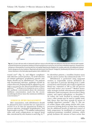
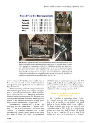
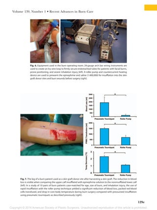
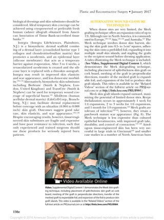
Download to read offline
Copyright © 2016 American Society of Plastic Surgeons. Unauthorized reproduction of this article is prohibited.
www.PRSJournal.com120e
T
hermal injury represents one of the most
severe, complex forms of traumatic injury.
Large burns require prolonged patient hos-
pitalizations, lead to enormous costs of care, and
impose significant physical and psychological bur-
dens on recovering victims and their families.
EPIDEMIOLOGY
The World Health Organization estimates that
over 265,000 deaths result annually from fire-related
burns,1
with over 95 percent occurring in low- and
middle-income countries.2
Survival after burn injury
with or without inhalation injury is a function of the
size of the second- and third-degree burn injury
(total body surface area burned)3
(Fig. 1).
In North America, over 30,000 burn patients
are admitted each year to specialized burn care
facilities.3
Most burn victims are male patients (68
percent) who suffer accidental, non–work-related
flame injuries (43 percent) at home (73 percent).
Children younger than 5 years account for almost
one-fifth of burn admissions, most commonly
caused by scald injuries.4
In the United States, the
number of fire-related deaths has decreased by
20.6 percent from 3380 deaths in 20025
to 2855
cases in 2012,4
with the highest death rates occur-
ring in blacks and Native Americans (Table  1).
Currently, the total body surface area burn that
represents 50 percent survival is approximately 70
percent.4
Importantly, delivery of care to these patients
involves burn quality improvement programs in
Disclosure: Dr. Tredget is a principal investigator
for Scar X Therapeutics, British Canadian BioSci-
ences Corp., and Klox Therapeutics. The remaining
authors have no financial interest to declare in rela-
tion to the content of this article.
Copyright © 2016 by the American Society of Plastic Surgeons
DOI: 10.1097/PRS.0000000000002908
Kevin J. Zuo, M.D.
Abelardo Medina, M.D.,
Ph.D.
Edward E. Tredget, M.D.,
M.Sc.
Toronto, Ontario, and Edmonton,
Alberta, Canada
Learning Objectives: After studying this article, the participant should be able
to: 1. Explain the epidemiology of severe burn injury in the context of socio-
economic status, gender, age, and burn cause. 2. Describe challenges with burn
depth evaluation and novel methods of adjunctive assessment. 3. Summarize
the survival and functional outcomes of severe burn injury. 4. State strategies
of fluid resuscitation, endpoints to guide fluid titration, and sequelae of over-
resuscitation. 5. Recognize preventative measures of sepsis. 6. Explain intraop-
erative strategies to improve patient outcomes, including hemostasis, restrictive
transfusion, temperature regulation, skin substitutes, and Meek skin grafting.
7. Translate updates in the pathophysiology of hypertrophic scarring into novel
methods of clinical management. 8. Discuss the potential role of free tissue
transfer in primary and secondary burn reconstruction.
Summary: Management of burn-injured patients is a challenging and unique
field for plastic surgeons. Significant advances over the past decade have oc-
curred in resuscitation, burn wound management, sepsis, and reconstruction
that have improved outcomes and quality of life after thermal injury. However,
as patients with larger burns are resuscitated, an increased risk of nosocomial
infections, sepsis, compartment syndromes, and venous thromboembolic phe-
nomena have required adjustments in care to maintain quality of life after
injury. This article outlines a number of recent developments in burn care that
illustrate the evolution of the field to assist plastic surgeons involved in burn
care.  (Plast. Reconstr. Surg. 139: 120e, 2017.)
From the Division of Plastic and Reconstructive Surgery,
Department of Surgery, University of Toronto; and the Di-
visions of Plastic and Reconstructive Surgery and Critical
Care, Department of Surgery, University of Alberta.
Received for publication October 28, 2015; accepted April
11, 2016.
Important Developments in Burn Care
Related Video content is available for this
article. The videos can be found under the
“Related Videos” section of the full-text article,
or, for Ovid users, using the URL citations pub-
lished in the article.
CME
Copyright © 2016 American Society of Plastic Surgeons. Unauthorized reproduction of this article is prohibited.
Volume 139, Number 1 • Recent Advances in Burn Care
121e
verified burn centers6
developed jointly by the
American College of Surgeons and the Ameri-
can Burn Association that have established stan-
dards of burn care delivery, infection control,
rehabilitation, and disaster management.7
There
are currently 67 American Burn Association–­
verified burn centers in the United States, three
in Canada, and one in Australia.8
BURN SEVERITY ASSESSMENT
The severity of burn injury guides the initial
resuscitation strategy and decision to transfer the
patient to a specialized burn center (Table 2). Man-
ual methods such as the rule of nines, the patient’s
palm method, or the Lund-Browder chart tend to
overestimate total body surface area burned,9–12
even among experienced clinicians.13
This leads to
excessive fluid resuscitation volumes,14
especially for
obese patients.15,16
As a result, digital assessment pro-
grams are becoming more prevalent in practice.17–20
Fig. 1. Survival after burn injury as a function of age and total body surface area burned for patients without inhalation injury
(left) and with inhalation injury (right). Numerically, survival can be calculated using the following formula: p = eu
/1 + eu
, where
u = 4.99 − 4.26 × 10⁻²
(TBSA) − 1.30 (I) − 8.65 × 10⁻⁶
(A)3
− 2.12 × 10⁻⁴
(TBSA) (A), where p is the probability of survival, e is the
base of the natural logarithm, I is 1 with inhalation injury and 0 if no inhalation injury is present, TBSA is the total body surface
area burned, and A is the age of the patient. (See American Burn Association. National Burn Repository Annual Report 2014.
Available at: http://www.ameriburn.org/2014NBRAnnualReport.pdf. Accessed December 20, 2014.)
Table 1.  Burn Mortality Rates by Race in the United
States, 1999 to 2013
Race
Age-Adjusted* Death Rate per
100,000
White 1.03
Black 2.39
American Indian 1.62
Asian 0.45
Other 0.65
All Races 1.17
*Age-adjusted to standard year 2000. Data obtained from Centers
for Disease Control and Prevention Fatal Injury Reports (U.S. Fire
Administration. Fire statistics. Available at: http://webappa.cdc.gov/
sasweb/ncipc/mortrate10_us.html. Accessed December 20, 2014).
Table 2.  American Burn Association Burn Center Referral Criteria*
1. Partial-thickness burns greater than 10% total body surface area in patients <10 yr or >50 yr
2. Partial-thickness burns greater than 20% total body surface area in all patients
3. Burns that involve the face, hands, feet, genitalia, perineum, or major joints
4. Third-degree burns in any age group
5. Electrical burns, including lightning injury
6. Chemical burns
7. Inhalation injury
8. Burn injury in patients with preexisting medical disorders that could complicate management, prolong recovery, or affect
mortality
9. Any patient with burns and concomitant trauma (such as fractures) in which the burn injury poses the greatest risk of
morbidity or mortality
10. Burned children in hospitals without qualified personnel or equipment for the care of children
11. Burn injury in patients who will require special social, emotional, or rehabilitative intervention
*At the time of writing of this article, 71 burn centers have been verified by the American Burn Association (http://www.ameriburn.org/verifi-
cation_verifiedcenters.php), including 67 centers in the United States, three centers in Canada, and one in Australia. Criteria from the Ameri-
can College of Surgeons Committee on Trauma. Guidelines for Trauma Centers Caring for Burn Patients. Chicago: American College of Surgeons;
2014. Available at: http://www.ameriburn.org/ACS%20Resources%20Burn%20Chapter%2014.pdf. Accessed August 8, 2016.
Copyright © 2016 American Society of Plastic Surgeons. Unauthorized reproduction of this article is prohibited.
122e
Plastic and Reconstructive Surgery • January 2017
In pediatric burns, the possibility of child
abuse should be considered when a history of the
injury is inconsistent with physical examination
findings such as uniform burn depth with sharp
borders; symmetrical isolated lower limb and but-
tock injury; skin fold sparing; absence of splash
marks; associated unrelated injuries; and a pas-
sive, introverted, fearful child.21,22
Therefore, all
pediatric burns are also screened by local dedi-
cated child abuse teams.
BURN DEPTH ASSESSMENT
Timely, accurate burn depth assessment is
critical to management strategy. Surgical interven-
tion is indicated for burn wounds not expected
to reepithelialize within 14 to 21 days,23
because
deep dermal wounds heal from unique activated
fibroblasts in the reticular regions of the dermis
and are prone to severe hypertrophic scarring.24
Clinical evaluation can differentiate very
superficial burns, which may be managed conser-
vatively, from full-thickness burns, which require
early eschar excision and skin grafting to facilitate
wound healing, decrease the risk of hypertrophic
scarring, prevent infection, and reduce mortal-
ity25,26
(Table  3). The major challenge in burn
depth assessment is in partial-thickness wounds
where clinical evaluation by experienced clini-
cians is often inaccurate, in part because of the
evolving inflammation that progresses in deep
dermal wounds in the zone of stasis.27,28
Thus, new
instruments to evaluate burn depth are becoming
useful tools and include the laser Doppler imag-
ing system, which evaluates microvascular dermal
perfusion (Figs. 2 and 3).29,30
Laser Doppler imag-
ing is performed between 48 hours and 5 days
after burn and has an accuracy ranging from 90
to 97 percent, compared with 52.5 to 71.4 percent
with clinical evaluation.31–35
Laser Doppler imag-
ing has a positive predictive value for burns that
will not heal within 14 to 21 days of 85.1 to 98
percent36–38
and is accurate and noninvasive; how-
ever, sedation is often required for burns in young
children, where it likely has its greatest applicabil-
ity.36
Commercial videos illustrating laser Doppler
imaging application are available online.39,40
Other modalities to distinguish burn depth at
early time points have been investigated, including
thermography,41
ultrasonography,42
nuclear mag-
netic resonance,43
near infrared spectroscopy, and
confocal microscopy44
; to date, however, they have
gained only modest application in clinical prac-
tice. These various approaches require expensive
equipment, standardized training, and controlled
environmental conditions during assessment.
DECISION TO TREAT
Numerous models have been developed to
predict mortality in major burn patients.45,46
The
revised Baux score is the sum of age, total body
surface area, and inhalation injury (+17), and has
a point-of-futility score of 160 and 50 percent pre-
dicted mortality score of 11047–49
; however, these
values do not reflect advances in clinical care since
Table 3.  Clinical Features of Burn Wounds*
Degree Depth Layers Involved Features
Healing
Mechanism
Healing
Time Management
First Superficial Epidermis only Pink, red, brisk ­
capillary refill, painful
Dermal
­appendages,
contact
­inhibition
<7 days Symptomatic
Second Superficial
partial-­
thickness
Epidermis, ­papillary
(upper) dermis
Pink, red, moist,
­edematous, brisk
capillary refill, very
painful
Dermal
­appendages,
contact
­inhibition
7–10 days Daily wound
care, debride
sloughed skin
Deep partial-
thickness
Epidermis, reticular
(lower) dermis
White, pink, red,
dry, ­nonblanching,
reduced
sensation
Contact
­inhibition,
wound
­contraction
Variable,
10–28 days
Daily wound care,
surgical excision
and resurfacing
Third Full thickness Epidermis, entire
dermis
White, brown, dry,
leathery, nonblanch-
ing, insensate
Contact
­inhibition,
wound
­contraction
>21 days Surgical excision
and resurfacing
Fourth Full thickness Epidermis, entire
dermis, fat, fascia,
muscle, bone
Exposed deep tissue N/A >21 days Amputation,
­complex
­reconstruction
N/A, not applicable.
*Superficial (first-degree) burns are not included in the calculation of total body surface area burned. Although full-thickness burns are
insensate, there may be areas of mixed burn depth resulting in an inconsistent sensory examination. Dermal appendages include hair follicles,
sebaceous glands, and sweat glands.
Copyright © 2016 American Society of Plastic Surgeons. Unauthorized reproduction of this article is prohibited.
Volume 139, Number 1 • Recent Advances in Burn Care
123e
the 1960s, when total mortality was ascribed to a
Baux score of 100.50
The expected quality of life
after a severe burn injury is currently the major
consideration in the decision to resuscitate.51–54
In
patients with greater than 70 percent total body
surface area burned receiving modern care, stan-
dardized outcome scales such as the Short Form-
36 have demonstrated high health-related quality
of life relative to healthy, nonburned individu-
als from the same populations, and greater than
patients receiving solid organ transplants in five
of six domains.51
Currently, standardized databases are main-
tained by modern burn centers and their out-
comes are shared with the National Burn
Repository, facilitating comparison of local out-
comes with national standards. Based on the age
of the patient, size and depth of burn, and pres-
ence of inhalation injury, local burn centers are
able to establish their own survival outcomes. For
adults with thermal injury where the probability
of recovery is poor, palliative care may be appro-
priate54,55
; however, in modern burn care, all pedi-
atric burns are considered nonfutile and should
be actively resuscitated.49
FLUID RESUSCITATION AND
MONITORING
Major burns exceeding 30 percent total body
surface area in children, 20 percent total body
surface area in adults, and 15 percent total body
surface area in elderly patients (older than 65
years) trigger a systemic inflammatory response
that results in nitric oxide–induced endothelial
relaxation, increased capillary permeability, and
interstitial fluid translocation that is not localized
to the burn wound alone, requiring extensive fluid
resuscitation to prevent hypovolemic burn shock.56
Intravenous fluid resuscitation should be com-
menced in adult burns exceeding 15 to 20 per-
cent total body surface area and pediatric burns
exceeding 10 percent total body surface area. The
most commonly used formula for calculating fluid
requirements in the first 24 hours is the Parkland
formula (2 to 4 ml/kg/percent total body surface
area burned of lactated Ringer solution), in which
Fig. 2. The Moor scanning laser Doppler instrument.
Fig. 3. A diagrammatic illustration of the Moor scanning laser Doppler device depicting the components of the equipment and the
principle of laser beam deflection by blood flowing in the viable tissues in the skin.
Copyright © 2016 American Society of Plastic Surgeons. Unauthorized reproduction of this article is prohibited.
124e
Plastic and Reconstructive Surgery • January 2017
half of the total calculated volume is administered
in the first 8 hours and the other half in the next
16 hours. Patients with inhalation injury, delayed
resuscitation, high-voltage electrical injuries, and
extensive deep burns will require higher volumes
than predicted.57
Children should also receive a
weight-appropriate maintenance fluid infusion
of 5% dextrose in half-normal saline to support
their limited glycogen stores58
(Table 4). A useful,
self-directed review course of existing knowledge
and guidelines for burn assessment, fluid manage-
ment, rule of nines, and guidelines for treatment
or transfer to a regional burn unit is available
online through the American Burn Association
Advanced Burn Life Support Now course.59
The fluid infusion rate should be rigorously
monitored and titrated according to hourly urine
output, base deficit, serum lactate, central venous
pressure, and bladder pressure (Table 5).60,61
It is
very important to avoid overresuscitation; there-
fore, the least amount of fluid should be infused
to maintain urine output at 30 to 50 ml/hour in
adults, or 0.5 to 1.0 ml/kg/hour in children weigh-
ing less than 30 kg.58
In general, invasive hemo-
dynamic monitoring with Swan-Ganz catheters is
not recommended, as this leads to excessive fluid
administration without improved outcomes.62
FLUID CREEP AND COMPARTMENT
HYPERTENSION
Fluidcreepreferstothephenomenonofincreas-
ingly larger volumes of fluid being administered to
Table 4.  Burn Resuscitation Formulas*†
Formula Fluid Infusion Volume in First 24 Hr Rate of Administration
Adult
  Parkland Lactated Ringer solution 2–4 ml/kg/% TBSA burn First half over 8 hr,
second half over 16 hr
  Modified Brooke Lactated Ringer solution 2 ml/kg/% TBSA burn First half over 8 hr,
second half over 16 hr
Pediatric
  Parkland Lactated Ringer solution 2–4 ml/kg/% TBSA burn
plus maintenance fluids
First half over 8 hr,
second half over 16 hr
  Shriners-Cincinnati Lactated Ringer solution plus
50 mg sodium bicarbonate
4 ml/kg/% TBSA burn
+
First 8 hr
Lactated Ringer solution 1500 ml/m2
BSA Second 8 hr
Lactated Ringer solution plus
12.5 g albumin 5%
Third 8 hr
  Shriners-Galveston Lactated Ringer solution 5000 ml/m2
TBSA burn
plus 2000 ml/m2
BSA
First half over 8 hr,
second half over 16 hr
TBSA, total body surface area; BSA, bovine serum albumin.
*Adapted with permission of Elsevier Ltd. from Warden GD. Fluid resuscitation and early management. In: Herndon DN, ed. Total Burn Care.
4th ed. New York: Saunders Elsevier; 2012:115–124.
†Intravenous fluid resuscitation should be commenced in adult burns exceeding 20 percent total body surface area and pediatric burns
exceeding 10 percent total body surface area. Pediatric patients following the Parkland formula should also receive maintenance fluids with
dextrose 5% in half-normal saline at 4 ml/hr for the first 10 kg of body mass, 2 ml/hr for the second 10 kg of body mass, and 1 ml/hr for the
remaining kilograms of body mass. Maintenance fluid requirements for the first 24 hr are already factored into the Shriners-Cincinnati and
Shriners-Galveston formulas.
Table 5.  Markers of Fluid Resuscitation
Index of Response Normal (Target) Range
Vital signs
  HR, beats/min <140
  BP, mmHg >90/60
  Sao2
, % >90
Urine output, ml/kg/hr
  Adults 0.5–1.0 (or 30–50 ml/hr)
  Children 1.0
Base deficit, mM
  Normal −3 to 0
  Target  >−6*
Serum lactate, mM
  Normal 0.5–2.2
  Target ≤4†
Central venous pressure, mmHg
  Normal 2–6
  Target 8–12‡
Mean arterial pressure, mmHg ≥65‡
Bladder pressure, mmHg
  Normal 0–5
  IAH >12§
  ACS >20§
Intrathoracic blood volume
index, ml/m2
>800
Cardiac index, liters/min/m2
>3.5
HR, heart rate; BP, blood pressure; Sao2
, oxygen saturation; IAH,
intraabdominal hypertension; ACS, abdominal compartment
syndrome.
*Cartotto R, Choi J, Gomez M, Cooper A. A prospective study on the
implications of a base deficit during fluid resuscitation. J Burn Care
Rehabil. 2003;24:75–84.
†Casserly B, Phillips GS, Schorr C, et al. Lactate measurements in
sepsis-induced tissue hypoperfusion: Results from the Surviving Sep-
sis Campaign database. Crit Care Med. 2015;43:567–573.
‡Dellinger RP, Levy MM, Rhodes A, et al. Surviving Sepsis Campaign:
International guidelines for management of severe sepsis and septic
shock, 2012. Intensive Care Med. 2013;39:165–228.
§Malbrain ML, Cheatham ML, Kirkpatrick A, et al. Results from the
International Conference of Experts on Intra-abdominal Hyperten-
sion and Abdominal Compartment Syndrome: I. Definitions. Inten-
sive Care Med. 2006;32:1722–1732.
Copyright © 2016 American Society of Plastic Surgeons. Unauthorized reproduction of this article is prohibited.
Volume 139, Number 1 • Recent Advances in Burn Care
125e
burn patients than predicted by the Parkland for-
mula,63–65
with infusion rates averaging as high as
8.0 ml/kg/percent total body surface area in the
first 24 hours after injury.66
Fluid creep can lead to
abdominal, orbital, and extremity fascial compart-
ment syndromes; acute respiratory distress syn-
drome; multiorgan failure; nosocomial infection;
and increased mortality.67
It is associated with exces-
sive and continuous opioid use (opioid creep),68
larger severe burns,57
persistent capillary perme-
ability, obese patients assessed on actual rather than
ideal or adjusted body weight,57
and a desire to “play
it safe” by exceeding the target urine output rate.
Burn providers must recognize signs of a failed
resuscitation, including serial low urine output val-
ues despite increasing fluid infusion rates, repeated
episodes of hypotension or need for vasopressors,
worsening base deficit, or fluid infusion in excess of
200 to 250 ml/kg in the first 24 hours.67,69
Modern burn care monitors abdominal, ocu-
lar,70,71
and extremity fascial compartments for
hypertension (Table 6). Abdominal compartment
syndrome is defined as sustained intraabdomi-
nal pressure exceeding 20 mmHg with new-onset
organ failure,72
such as oliguria or decreased pul-
monary compliance.69
Intraabdominal hyperten-
sion (intraabdominal pressure >12 mmHg), can be
managed with escharotomy, percutaneous drain-
age, nasogastric tube decompression, or sedation,73
but abdominal compartment syndrome requires
emergent decompression laparotomy. However,
this procedure is associated with mortality rates of
44 to 100 percent73
; thus, prevention of abdominal
compartment syndrome by avoiding excessive fluid
and narcotic or sedative administration is critical.
The role of colloid solutions such as plasma and
albumin, to limit fluid requirements is unclear.74–76
Historically, Baxter recommended colloid
supplementation with 0.3 to 0.5 ml/kg/percent total
body surface area burned of plasma during the sec-
ond 24-hour period to reexpand intravascular vol-
ume; however, colloids were later excluded because
of concerns about persistent capillary permeability
leading to pulmonary edema.77
Contemporary strat-
egies may include albumin during early resuscita-
tion as a volume expander78
or, more commonly, as
a rescue fluid following the initial 12 to 24 hours57,79,80
when capillary integrity is thought to be restored58
to
assist in the fluid-overloaded, failed resuscitation.
Hypertonic saline may also be beneficial in
limiting fluid volumes,81
but careful monitoring is
needed, as hypernatremia is associated with acute
renal failure.58
Most recently, computerized fluid
resuscitation software has shown promising results
in precise standardized fluid titration.82
VENOUS THROMBOEMBOLISM
PROPHYLAXIS
Although this was not appreciated in the
past, burn patients are in a hypercoagulable state
and should be prophylactically anticoagulated to
reduce the risk of venous thromboembolism,83
including adults and adolescents. Compared to
unfractionated heparin, enoxaparin has a lower
incidence of venous thromboembolism and hep-
arin-induced thrombocytopenia84
but requires a
higher initial dosing, good renal function, and
routine monitoring of anti–factor Xa because of
altered pharmacokinetics in burn patients.85
TOPICAL BURN WOUND MANAGEMENT
A plethora of burn dressing materials are avail-
able (Table 7).60
Silver sulfadiazine is the most com-
mon topical antimicrobial but is associated with
resistantnosocomialpathogenssuchasPseudomonas
Table 6.  Methods of Monitoring Compartment Hypertension*
Compartment Method of Monitoring Dangerous Signs Sequelae Management
Ocular Tonometry IOP >30 mmHg Ischemic optic neuropathy,
blindness
Lateral canthotomy
Abdominal IAP; typically obtained
by intrabladder
­pressure
IAP >12 mmHg
(­intraabdominal
­hypertension)
Intracranial hypertension,
reduced cardiac output,
respiratory failure, gastro-
intestinal ischemia, acute
kidney failure, multisystem
organ failure
Escharotomy, percutaneous
drainage, nasogastric tube
decompression, sedation
IAP >20 mmHg
(­abdominal
­compartment
syndrome)
Decompression laparotomy
Extremity Clinical examination Circumferential burns,
pain on passive stretch,
decreased pulses, cool,
paresthesias
Ischemic myonecrosis, res-
piratory failure, rhabdomy-
olysis, acute renal failure,
Volkmann contracture
Urgent bedside escharotomy
and/or fasciotomy with
sedation and analgesia
IOP, intraocular pressure; IAP, intra-abdominal pressure.
*Cumulative resuscitative volumes exceeding 250 ml/kg are associated with a higher risk of compartment hypertension.
Copyright © 2016 American Society of Plastic Surgeons. Unauthorized reproduction of this article is prohibited.
126e
Plastic and Reconstructive Surgery • January 2017
species86
and poorer healing outcomes than newer
silver dressings.87
Some newer silver dressings such
as Acticoat (Smith & Nephew, Montreal, Quebec,
Canada) contain unique nanocrystalline silver with
sustained release, leading to enhanced antibacte-
rial effects and reducing the frequency of dressing
changes, infection risk, and patient discomfort.
They can be effective against methicillin-resistant
Staphylococcus aureus, have relatively low mamma-
lian cell toxicity, reduce pain and pruritus, and may
accelerate healing and thus decrease costs.86,88,89
The role of negative-pressure wound therapy in
partial-thickness burns is unclear.90
SEPSIS
Infection leading to sepsis and multiorgan fail-
ure is a major cause of burn mortality. Pneumonia,
central venous lines, and burn wounds are the most
common sources of bloodstream infections, which
typically occur within 5 to 7 days of injury.91,92
The
most common pathogens in the first 7 days after
burn are Staphylococcal species; thereafter, bactere-
mia is more commonly with Gram-negative organ-
isms.92–94
Bloodstream infections are associated with
significantly higher mortality, hospital length of stay,
and number of ventilator days, and 10 times higher
cost.93
Nosocomial Pseudomonas infection is particu-
larly aggressive, increasing length of stay, number of
ventilator days, blood transfusions, surgical proce-
dures, and mortality from 8 to 33 percent95
(Fig. 4).
Preventative measures against infection are
critical for survival of the burn patient and include
early excision of burn eschar to improve local per-
fusion and prevent microbial colonization, pru-
dent use of invasive devices, appropriate choice
of antimicrobial burn dressings, elimination of
potential water-borne sources of bacteria during
Table 7.  Burn Wound Dressings and Topical Antimicrobials*
Dressing Type Features Example
Topical Antimi-
crobial Advantages Disadvantages
Paraffin gauze Nonadherent moist
coverage
Adaptic† Polymyxin B
(Polysporin†)
Action against MDR
Pseudomonas and
Enterobacter species
Nephrotoxic,
­neurotoxicity,
hypersensitivity,
limited Gram-
positive activity
Hydrocolloid Forms gel on contact
with exudate
Comfeel‡,
DuoDERM§
0.5% silver
nitrate
Broad spectrum
against bacte-
ria and fungi,
­antiinflammatory
properties
Stains surfaces,
requires frequent
application (every
2 hr) due to
­inactivation
Polyurethane Permeable to water
vapor and oxygen
but not liquid or
bacteria
OpSite‖,
Tegaderm¶
1% silver
sulfadiazine
Broad spectrum
against bacteria
and fungi, sooth-
ing, antiinflamma-
tory properties
Poor eschar
­penetration, forms
pseudoeschar,
requires twice
daily applica-
tion because of
­inactivation
Hydrogel High fluid-absorbing
capacity
IntraSite‖,
SoluGel†
Mafenide
(Sulfamylon**)
Broad spectrum
against bacteria
and fungi, good
eschar penetration
Painful, metabolic
acidosis
Silicone-coated
nylon
Nonadherent,
exudate drainage
Mepitel Silicone#
Antimicrobial Contains silver or
iodine
Acticoat, Iodosorb‖,
Aquacel Ag§
Biosynthetic
skin substitute
Supports
reepithelialization
BioBrane‖,
TransCyte‖,
Integra††
Foam Easy to change,
absorbent
Mepilex Ag#
MDR, multidrug resistant.
*Some data used from Wasiak J, Cleland H, Campbell F, Spinks A. Dressings for superficial and partial thickness burns. Cochrane Database Syst
Rev. 2013;3:CD002106.
†Johnson & Johnson, New Brunswick, N.J.
‡Coloplast, Humlebæk, Denmark.
§ConvaTec, Greensboro, N.C.
‖Smith & Nephew.
¶3M, St. Paul, Minn.
#Mölnlycke Health Care, Gothenburg, Sweden.
**UDL Laboratories, Inc., Rockford, Ill.
††Integra Life Sciences, Plainsboro, N.J.
Copyright © 2016 American Society of Plastic Surgeons. Unauthorized reproduction of this article is prohibited.
Volume 139, Number 1 • Recent Advances in Burn Care
127e
wound care96
(Fig.  5), and diligent compliance
with infection control practices. To avoid selection
of resistant pathogens, prophylactic systemic anti-
biotics should not be administered.97
For patients
with documented infection, antibiotics should be
culture-directed. Dosing should be adjusted accord-
ingly to account for the altered metabolism of burn
patients,98–100
as 60 percent of patients never achieve
free antibiotic concentrations above the minimum
inhibitory concentration, much less the recom-
mended goal of four times the minimum inhibitory
concentration.101
SURGICAL BURN MANAGEMENT
After resuscitation, early débridement should
be planned for burn wounds that are expected to
exceed 14 to 21 days for spontaneous healing to
prevent infection, prolonged hospitalization, and
hypertrophic scarring.25,26
To stabilize endotracheal
tubes and avoid complications on injured facial
skin, interdental Ivy wire fixation is used (Fig. 6).102
In edentulous patients, a maxillary fixation screw
may be used to anchor the endotracheal tube.103,104
Débridement is performed using tangential
excision to sequentially remove devitalized tis-
sue until there is punctate bleeding from a viable
wound bed; unfortunately, this results in significant
blood loss, estimated at 190 to 270  ml/percent
total body surface area excised.105
Modern hemo-
static strategies include subcutaneous epinephrine
infiltration, limb tourniquets, electrocautery, fibrin
sealant, and topical epinephrine or thrombin.106
To reduce the work involved in manual tumes-
cence, roller pumps may be used to rapidly insuf-
flate donor sites and burn wounds by means of
multiple large-bore cannulas107
(Fig.  7). The use
of cardiac bypass roller pump systems with coun-
tercurrent heating devices in large burn excisions
has the additional benefit of improved control of
body temperature by insufflation of warmed epi-
nephrine-containing crystalloid solutions, which
greatly facilitates the amount of burn eschar that
Fig. 4. A 37-year-old man with an industrial scald burn injury to the left thigh and abdomen that became infected with hospital-
acquired Pseudomonasaeruginosa, leading to Ecthymagangrenosum and tissue necrosis from embolized organisms. Pseudomonas
aeruginosa is illustrated by a scanning electron micrograph depicting the flagellum for motility, polar pili, and additional virulence
factors dangerous for burn patients. (Reprinted with permission from Elsevier:Tredget EE, Shankowsky HA, Rennie R, et al. Pseudo-
monas infections in the thermally injured patient. Burns 2004;30:3–26.)
Copyright © 2016 American Society of Plastic Surgeons. Unauthorized reproduction of this article is prohibited.
128e
Plastic and Reconstructive Surgery • January 2017
may be removed. Up to 50 percent total body sur-
face area can be safely excised and resurfaced in
one operation, with significantly less blood loss and
hypothermia.108
Blood-conserving protocols using a combination
of these techniques during burn surgery are impor-
tant to avoid immunosuppressive effects and infec-
tious complications.105,109–112
Ongoing multicenter
randomized studies will elucidate the threshold level
ofhemoglobinforbloodtransfusioninburnpatients,
similar to the restrictive transfusion strategy found
efficacious in other intensive care patients.113–116
Intraoperative hypothermia (<36.0°C) sig-
nificantly increases blood loss,117,118
wound infec-
tion,119,120
and acute lung injury121
during surgery.
Strategies to maintain normothermia include
increasing the ambient room temperature, infus-
ing warmed fluids, and using forced-warm-air
inflatable blanket technologies, such as the Bair
hugger (3M, St. Paul, Minn), although caution is
necessary.122–125
Novel closed-loop thermoregulat-
ing strategies include thermal water mattresses126,127
and intravascular warming catheters.128–130
BURN WOUND CLOSURE: SKIN
SUBSTITUTES
After excision of devitalized tissue, defini-
tive wound coverage with split-thickness sheet
skin grafts is preferred, particularly for hand
and facial burns, smaller injuries, and children.
For larger injuries, skin grafts are meshed with
expansion ratios of 1:1.5 to 1:3 to permit greater
surface area coverage and drainage of wound
fluid. In burns exceeding 60 percent total body
surface area where donor sites are very limited,
Fig. 5. Pulsed field gel electrophoresis of Pseudomonas aeruginosa isolated from four burn
patients tracked to contaminated sinks of defective design in the burn unit (above, left) that
were subsequently replaced by newly designed sinks (above, right) that have splash pan-
els, polished stainless steel surfaces, deep contoured bowls with U-traps situated at least
6 inches below the bottom of the sink (below, left), and an ultraviolet irradiation cell that
treats the effluent and creates the blue/green color in the drain (below, right). (Reprinted
with permission from Elsevier: Tredget EE, Shankowsky HA, Rennie R, et al. Pseudomonas
infections in the thermally injured patient. Burns 2004;30:3–26.)
Copyright © 2016 American Society of Plastic Surgeons. Unauthorized reproduction of this article is prohibited.
Volume 139, Number 1 • Recent Advances in Burn Care
129e
Fig. 6. Equipment used in the burn operating room; 24-gauge arch bar wiring instruments are
used to create an Ivy wire loop to firmly secure endotracheal tubes for patients with facial burns,
prone positioning, and severe inhalation injury (left). A roller pump and countercurrent heating
device are used to prewarm the epinephrine and saline (1:400,000) for insufflation into the skin
graft donor sites and burn wounds before surgery (right).
Fig. 7. The leg of a burn patient used as a skin graft donor site after harvesting a skin graft. The reduction in blood
loss is visible when comparing the upper calf insufflated with epinephrine solution to the noninsufflated lower calf
(left). In a study of 10 pairs of burn patients case-matched for age, size of burn, and inhalation injury, the use of
rapid insufflation with the roller pump technique yielded a significant reduction of blood loss, packed red blood
cells transfused, and drop in core body temperature during burn surgery compared with pressurized insufflation
using pneumatic tourniquets as described previously (right).
Copyright © 2016 American Society of Plastic Surgeons. Unauthorized reproduction of this article is prohibited.
130e
Plastic and Reconstructive Surgery • January 2017
biological dressings and skin substitutes should be
considered. Ideal temporary skin coverage can be
achieved using cryopreserved or preferably fresh
human cadaver allograft obtained from Ameri-
can Association of Tissue Bank–accredited tissue
banks.131
Integra (Integra LifeSciences, Plainsboro,
N.J.) is a biosynthetic dermal scaffold consist-
ing of a dermal layer (cross-linked bovine type 1
collagen and chondroitin-6-sulfate matrix) that
promotes a neodermis, and an epidermal layer
(silicone membrane) that acts as a temporary
barrier against evaporation. After 3 to 4 weeks, a
revascularized neodermis is created and the sili-
cone layer is replaced with a thin-skin autograft.
Integra may result in improved skin elasticity
and scar appearance, and less donor-site morbid-
ity.132–134
Alternatively, biosynthetic skin substitutes
including Biobrane (Smith & Nephew, Lon-
don, United Kingdom) and TransCyte (Smith &
Nephew) can be used for temporary wound cov-
erage of superficial burns.135
AlloDerm (human
acellular dermal matrix) (LifeCell Corp., Branch-
burg, N.J.) may facilitate dermal replacement
before coverage with an ultrathin (0.004 to 0.008
inch) skin graft. Outcomes suggest good take
rate, skin elasticity, and scar appearance.136–138
Despite encouraging results, however, tissue-engi-
neered skin substitutes are fragile and expensive
and have poor resistance to infection, such that
only experienced and trained surgeons should
use these products for seriously injured burn
patients.
ALTERNATIVE WOUND CLOSURE
TECHNIQUES
When donor sites are very limited, the Meek
grafting technique offers an expansion ratio of up to
1:9. Although rare in North America, it is commonly
used in Europe,139–142
Asia,143–146
and Australia.147
The
Meek technique involves donor skin harvest, slic-
ing the skin graft into 0.5- to 1-cm2
squares, adher-
ing the skin onto a prefolded foil, expanding it into
multiple small skin islands, and stapling the grafts
to the recipient wound before dressing application.
A video illustrating the Meek technique is included.
(See Video, Supplemental Digital Content 1, which
demonstrates the Meek skin-grafting technique,
including placement of split-thickness skin graft on
cork board, meshing of the graft in perpendicular
directions, transfer of the meshed graft to expand-
able foil, and expansion of the foil to produce skin
graft islands. This video is available in the “Related
Videos” section of the full-text article on PRSJour-
nal.com or at http://links.lww.com/PRS/B984.)
Meek skin graft islands expand outward, maxi-
mizing the potential of limited donor sites. Reepi-
thelialization occurs in approximately 1 week for
1:4 expansions, 2 to 3 weeks for 1:6 expansions,
and 1 month for 1:9 expansions.145
Meek grafts are
tolerant of infection139,143
; however, they retain a
patchwork mature scar appearance (Fig. 8). The
Meek technique is less expensive than cultured
epithelial keratinocytes, with improved graft take,
durability, and control of contraction.147–149
Autol-
ogous tissue-engineered skin has been very suc-
cessful in large trials in Cincinnati150
and smaller
case studies in a number of North American burn
Video.SupplementalDigitalContent1demonstratestheMeekskin-graft-
ing technique, including placement of split-thickness skin graft on cork
board, meshing of the graft in perpendicular directions, transfer of the
meshed graft to expandable foil, and expansion of the foil to produce skin
graft islands. This video is available in the “Related Videos” section of the
full-text article on PRSJournal.com or at http://links.lww.com/PRS/B984.
Copyright © 2016 American Society of Plastic Surgeons. Unauthorized reproduction of this article is prohibited.
Volume 139, Number 1 • Recent Advances in Burn Care
131e
centers151
but is currently in further U.S. Food and
Drug Administration–regulated clinical trials.
BURN WOUND CLOSURE: FACIAL
TRANSPLANTATION
Vascularized composite allotransplantation of
the face represents a new reconstructive avenue
for patients with disfiguring full-face injuries, with
10 cases reported involving major facial burns.152
Rigorous patient screening is essential and further
research is ongoing to establish whether the long-
term effects of immunosuppression are justifiable
and what reconstructive options are available if
there is graft failure.153
HYPERTROPHIC SCARRING AFTER
BURN INJURY
Despite early wound resurfacing, joint splint-
ing, compression garments, and physiotherapy,
complications may inevitably develop and confer
significant distress to patients. Hypertrophic scar-
ring is a common complication of burn injuries
involving the deep dermis associated with pain,
pruritus, disfiguration, and functional restric-
tion with joint contractures. At the cellular level,
unique features of hypertrophic scar fibroblasts
compared with site-matched cells from normal
skin include increased synthesis of collagen types
I and III, high-molecular-weight proteoglycans
including versican, and the fibrogenic transform-
ing growth factor (TGF)-β.154
More importantly,
hypertrophic scar fibroblasts consistently synthe-
size less collagenase or matrix metalloprotein-
ase-1, which normally facilitates remodeling of
the extracellular matrix, and less decorin, a small
leucine-rich proteoglycan important for the fibril-
logenesis of small, tightly packed collagen fibers
and fiber bundles typical of the morphology of
normal skin.155
These features of hypertrophic scar fibro-
blasts are characteristic of fibroblasts located in
the deeper layers of the skin or reticular dermis
compared with superficial papillary fibroblasts.
Recently, two different groups have reaffirmed
distinct lineages of skin fibroblasts that possess
intrinsic fibrogenic potential154
and determine the
ultimate dermal architecture after wound heal-
ing.156
The systemic immunologic response typical
of recovering burn patients with severe hypertro-
phic scarring includes a polarized T-helper cell 2
environment157
that also promotes the differen-
tiation of blood-borne fibrocytes,156
which secrete
extracellular matrix proteins, proteases, and
fibrotic cytokines, including TGF-β. This response
to burn injury persists for up to 1 year after burn
injury. Thus, reconstruction of patients with large
burns and limited skin donor sites is best delayed
where possible, until resolution of the systemic
inflammatory response.
Established management strategies for imma-
ture hypertrophic burn scars include massage,
topical emollients, pressure garments, silicone
Fig. 8. The Meek skin graft meshing approach to skin graft
expansion using 4:1 expansion illustrating the nylon mesh car-
rying the micrografts to the patient’s arm and back at 1 week
(above), 1 month (center), and 3 months postoperatively (below).
Copyright © 2016 American Society of Plastic Surgeons. Unauthorized reproduction of this article is prohibited.
132e
Plastic and Reconstructive Surgery • January 2017
sheeting, steroid injections, and surgical exci-
sion.158–161
An experimental treatment for hyper-
trophic scarring is interferon-α2b, an antifibrotic
T-helper cell 1 cytokine that significantly improves
scar remodeling and normalizes TGF-β.162–164
Other approaches include topical imiquimod,
calcium channel blockers, tacrolimus, 5-fluoro-
uracil, and bleomycin, but newer experimental
approaches such as interleukin-10, microinhibi-
tory RNA to TGF-β, and peptide inhibitors of
CXCR4 offer potential future therapies.155,165
Pulsed-dye laser and fractional carbon dioxide
laser have shown promise as an adjunct to estab-
lished treatments for burn scar treatment. Pulsed
dye laser therapy selectively targets hemoglobin
in the 585-nm wavelength, making it effective in
hypervascular immature burn scars to reduce ery-
thema. Using pulsed-dye and fractional carbon
dioxide lasers, Hultman et al. demonstrated sig-
nificant improvements in before-and-after burn
scar scale scores and patient-reported outcomes.166
Ablative lasers such as the neodymium:yttrium-
aluminum-garnet laser have been effective in con-
tact mode, where 102 scar patients treated every
3 to 4 weeks for 1 year demonstrated significant
improvements overall. Unfortunately, scar recur-
rence developed in the upper chest, arm, and
back areas, particularly if residual erythema and
induration persisted following therapy.167
Thus,
althoughlasertreatmentofpostburnhypertrophic
scar is offering a new, potentially transformative
approach to difficult scar challenges, further
objective controlled trials are required.
An important complication of massive burn
injury is heterotopic ossification, the formation
of mature lamellar bone in extraskeletal tissue.
With an incidence of 0.2 to 4 percent, heterotopic
ossification occurs most commonly at the elbow
of burns exceeding 20 percent total body surface
area, and it is associated with skin breakdown,
soft-tissue deformity, nerve palsy, chronic pain,
and limitation of joint excursion.168
Risk factors
include prolonged immobilization, burn wound
infection, delayed wound closure, and repeated
forceful passive mobilization. Postoperative radio-
therapy has been shown to be slightly more effec-
tive than nonsteroidal antiinflammatory drugs in
preventing heterotopic ossification169
; however,
surgical excision is the procedure of choice for
restoration of range of motion.170,171
For both
hypertrophic scarring and heterotopic ossifica-
tion, radiation-induced Marjolin ulcer can occur,
limiting radiotherapy treatment to severe prob-
lems and patients older than 16 years.172
BURN RECONSTRUCTIVE SURGERY
Reconstructive surgery to improve the aes-
thetic and functional outcomes of burn patients
with severe contractures, disfiguring scars, or
exposed vital structures is ideally reserved until
scar maturation.155
Prevention of burn scarring
involves the understanding that, beyond a critical
Fig. 9. (Left) Keloid scarring in an African patient after minor injury as a child, multiple unsuc-
cessful attempts at excision, and local scar modification treatment. (Right) Appearance following
excision of the keloid and resurfacing with a left anterolateral thigh free flap, postoperative radio-
therapy, and one defatting procedure.
Copyright © 2016 American Society of Plastic Surgeons. Unauthorized reproduction of this article is prohibited.
Volume 139, Number 1 • Recent Advances in Burn Care
133e
depth, activated deep dermal fibroblasts of spe-
cific lineage with fibrogenic potential will lead
to hypertrophic scarring. Therefore, accurate
determination of burn depth with serial exami-
nation aided by objective instruments31
will avoid
unnecessary surgery. Despite the creation of a
new wound and possible scar at the donor site,
skin graft resurfacing is indicated for deep dermal
burns to avoid hypertrophic scarring, particularly
in critical cosmetic regions such as the face.173
Burn reconstruction may be accomplished with
contracture release; scar excision and resurfacing;
local transposition, rotation, and advancement
flaps; tissue expansion; or distant axial flaps.174
Plastic surgeons offer significant reconstruction
advantages for burn patients through the use of
microsurgery. Acutely, free flaps may be used for
limb salvage or defect coverage, permitting pres-
ervation of exposed vital structures such as nerves,
tendons, vessels, or bone, often in high-voltage
electrical burns, to avoid limb amputation.175
Microvascular free flaps may also be used in sec-
ondary burn reconstruction for joint contractures
and hypertrophic scars when injured or deficient
regional tissue precludes local flaps, skin grafts, or
tissue expansion (Fig. 9). Success rates for free flap
transfer in burn reconstruction range from 78176
to
96 percent.175
Excessive free flap bulk is averted by
the use of thinner fasciocutaneous flaps such as the
anterolateral thigh177
or parascapular178
flaps in the
head and neck region179
and thin fascial flaps such
as the temporoparietal fascial or serratus fascial
flaps in the dorsum of the hand,175,180,181
which offer
better color, thickness, and texture match (Table 8).
When donor vessels are too distant or deficient
at the recipient site, arteriovenous loops are an
innovative strategy to ensure a robust blood sup-
ply from large patent proximal vessels182–184
to dif-
ficult regions such as in high cranial vault and distal
extremity injuries. As with all free flaps, vascular
thrombosis is a threat to flap viability requiring care-
ful monitoring, particular in the first 72 hours.185
CONCLUSIONS
Modern advancements in burn care have
greatly increased the survival of major burn
patients. Plastic surgeons are well-positioned to
improve the functional reintegration of these
patients into society using novel approaches to
burn care.
Edward E. Tredget, M.D., M.Sc.
2D2.28 WMC, 8440-112 Street
University of Alberta
Edmonton, Alberta T6G 2B7, Canada
etredget@ualberta.ca
ACKNOWLEDGMENTS
This work was supported by the Firefighters’ Burn
Trust Fund of the University of Alberta Hospital, the
Canadian Institutes for Health Research, and the
Alberta Heritage Trust Fund for Medical Research.
PATIENT CONSENT
The patient provided written consent for the use of
his images.
REFERENCES
	 1.	 World Health Organization. Burns: Fact sheet. Available at:
http://www.who.int/mediacentre/factsheets/fs365/en/.
Accessed December 30, 2014.
Table 8.  Types of Free Flaps Used in Patients with Burn Injuries*
Type of Free Flap Indications Example Defects
Example Free
Flaps Vascular Supply
Muscle Primary coverage of exposed vital
structures or secondary recon-
struction of complex three-
dimensional defects
Limb salvage or limb
reconstruction
Latissimus dorsi Thoracodorsal artery
Gracilis Medial femoral
­circumflex artery
Fasciocutaneous Secondary reconstruction in shal-
lower defects with provision of
gliding surface and minimization
of donor-site morbidity
Exposed bone without
periosteum, joint con-
tracture release
Anterolateral
thigh
Descending branch of
lateral femoral circum-
flex artery
Parascapular Descending branch of
circumflex scapular
artery
Radial forearm Radial artery
Fascial Coverage of areas with thin
­overlying protective soft tissue
Dorsum of the hand or
digits, head and neck
Serratus fascia Serratus branch of thora-
codorsal artery
Temporoparietal
fascia
Superficial temporal
artery
*Both primary and secondary reconstruction may be performed with microvascular free flap transfer.
Copyright © 2016 American Society of Plastic Surgeons. Unauthorized reproduction of this article is prohibited.
134e
Plastic and Reconstructive Surgery • January 2017
	 2.	 Peck MD. Epidemiology of burns throughout the world: Part
I. Distribution and risk factors. Burns 2011;37:1087–1100.
	 3.	 American Burn Association Burn. Burn incidence and
treatment in the United States: 2016. Available at: http://
www.ameriburn.org/resources_factsheet.php. Accessed
December 20, 2014.
	 4.	 American Burn Association. 2014 national burn
repository. Available at: http://www.ameriburn.
org/2014NBRAnnualReport.pdf. Accessed December 20,
2014.
	 5.	 U.S. Fire Administration. Fire statistics. Available at: http://
webappa.cdc.gov/sasweb/ncipc/mortrate10_us.html.
Accessed December 20, 2014.
	 6.	 American Burn Association. Consultation/verification pro-
gram for burn centers. Available at: http://www.ameriburn.
org/verification_about.php. Accessed December 20, 2014.
	 7.	 American Burn Association. Advanced burn life support.
Available at: www.ameriburn.org/ablscoursedescriptions.
php. Accessed June 10, 2015.
	 8.	 American Burn Association. Burn center verification.
Available at: www.ameriburn.org/verification_verifiedcen-
ters.php. Accessed June 10, 2015.
	 9.	 Chan QE, Barzi F, Cheney L, Harvey JG, Holland AJ. Burn
size estimation in children: Still a problem. Emerg Med
Australas. 2012;24:181–186.
	10.	 Baartmans MG, van Baar ME, Boxma H, Dokter J, Tibboel
D, Nieuwenhuis MK. Accuracy of burn size assessment prior
to arrival in Dutch burn centres and its consequences in chil-
dren: A nationwide evaluation. Injury 2012;43:1451–1456.
	11.	 Harish V, Raymond AP, Issler AC, et al. Accuracy of burn size
estimationinpatientstransferredtoadultBurnUnitsinSydney,
Australia: An audit of 698 patients. Burns 2015;41:91–99.
	12.	 Hammond JS, Ward CG. Transfers from emergency room
to burn center: Errors in burn size estimate. J Trauma
1987;27:1161–1165.
	13.	 Giretzlehner M, Dirnberger J, Owen R, Haller HL, Lumenta
DB, Kamolz LP. The determination of total burn surface
area: How much difference? Burns 2013;39:1107–1113.
	14.	 Parvizi D, Kamolz LP, Giretzlehner M, et al. The potential
impact of wrong TBSA estimations on fluid resuscitation in
patients suffering from burns: Things to keep in mind. Burns
2014;40:241–245.
	15.	 Rae L, Pham TN, Carrougher G, et al. Differences in resus-
citation in morbidly obese burn patients may contribute to
high mortality. J Burn Care Res. 2013;34:507–514.
	16.	 Neaman KC, Andres LA, McClure AM, Burton ME,
Kemmeter PR, Ford RD. A new method for estimation of
involved BSAs for obese and normal-weight patients with
burn injury. J Burn Care Res. 2011;32:421–428.
	17.	 Prieto MF, Acha B, Gómez-Cía T, Fondón I, Serrano C. A sys-
tem for 3D representation of burns and calculation of burnt
skin area. Burns 2011;37:1233–1240.
	18.	 Berry MG, Goodwin TI, Misra RR, Dunn KW. Digitisation of
the total burn surface area. Burns 2006;32:684–688.
	19.	 Godwin Z, Tan J, Bockhold J, Ma J, Tran NK. Development
and evaluation of a novel smart device-based application for
burn assessment and management. Burns 2015;41:754–760.
	20.	 Williams JF, King BT, Aden JK, et al. Comparison of tradi-
tional burn wound mapping with a computerized program.
J Burn Care Res. 2013;34:e29–e35.
	21.	 Maguire S, Moynihan S, Mann M, Potokar T, Kemp AM. A
systematic review of the features that indicate intentional
scalds in children. Burns 2008;34:1072–1081.
	22.	 Toon MH, Maybauer DM, Arceneaux LL, et al. Children with
burn injuries: Assessment of trauma, neglect, violence and
abuse. J Inj Violence Res. 2011;3:98–110.
	23.	 Monstrey S, Hoeksema H, Verbelen J, Pirayesh A, Blondeel
P. Assessment of burn depth and burn wound healing poten-
tial. Burns 2008;34:761–769.
	24.	 Ladak A, Tredget EE. Pathophysiology and management of
the burn scar. Clin Plast Surg. 2009;36:661–674.
	25.	 Herndon DN, Barrow RE, Rutan RL, et al. A comparison
of conservative versus early excision: Therapies in severely
burned patients. Ann Surg. 1989;209:547–552; discussion
552–553.
	26.	 Barret JP, Herndon DN. Effects of burn wound excision
on bacterial colonization and invasion. Plast Reconstr Surg.
2003;111:744–750; discussion 751–752.
	27.	 Jackson DM. The treatment of burns: An exercise in emer-
gency surgery. Ann R Coll Surg Engl. 1953;13:236–257.
	28.	 Singh V, Devgan L, Bhat S, Milner SM. The pathogenesis of
burn wound conversion. Ann Plast Surg. 2007;59:109–115.
	29.	 Jaskille AD, Shupp JW, Jordan MH, Jeng JC. Critical review of
burn depth assessment techniques: Part I. Historical review.
J Burn Care Res. 2009;30:937–947.
	30.	 Jaskille AD, Ramella-Roman JC, Shupp JW, Jordan MH, Jeng
JC. Critical review of burn depth assessment techniques:
Part II. Review of laser Doppler technology. J Burn Care Res.
2010;31:151–157.
	31.	 Stewart TL, Ball B, Schembri PJ, et al.; Wound Healing
Research Group. The use of laser Doppler imaging as a pre-
dictor of burn depth and hypertrophic scar postburn injury.
J Burn Care Res. 2012;33:764–771.
	32.	 Pape SA, Skouras CA, Byrne PO. An audit of the use of laser
Doppler imaging (LDI) in the assessment of burns of inter-
mediate depth. Burns 2001;27:233–239.
	33.	 Hoeksema H, Baker RD, Holland AJ, et al. A new, fast LDI
for assessment of burns: A multi-centre clinical evaluation.
Burns 2014;40:1274–1282.
	34.	 Niazi ZB, Essex TJ, Papini R, Scott D, McLean NR, Black MJ.
New laser Doppler scanner, a valuable adjunct in burn depth
assessment. Burns 1993;19:485–489.
	35.	 Brown RF, Rice P, Bennett NJ. The use of laser Doppler imag-
ing as an aid in clinical management decision making in the
treatment of vesicant burns. Burns 1998;24:692–698.
	36.	 La Hei ER, Holland AJ, Martin HC. Laser Doppler imaging
of paediatric burns: Burn wound outcome can be predicted
independent of clinical examination. Burns 2006;32:550–553.
	37.	 Jeng JC, Bridgeman A, Shivnan L, et al. Laser Doppler imag-
ing determines need for excision and grafting in advance
of clinical judgment: A prospective blinded trial. Burns
2003;29:665–670.
	38.	 Park YS, Choi YH, Lee HS, et al. The impact of laser Doppler
imaging on the early decision-making process for surgi-
cal intervention in adults with indeterminate burns. Burns
2013;39:655–661.
	39.	 Moor Instruments, Inc. moorLDI2 Laser Doppler Imager
2014 promotional video. Available at: https://www.youtube.
com/watch?v=t-egppJ9kEU. Accessed November 30, 2015.
	40.	 Moor Instruments, Inc. Early and accurate assessment of
burns. Available at: http://us.moor.co.uk/product/burn-
assessment-burn-assessment/286/o/41/video-channel.
Accessed November 30, 2015.
	41.	 Mladick R, Georgiade N, Thorne F. A clinical evaluation of
the use of thermography in determining degree of burn
injury. Plast Reconstr Surg. 1966;38:512–518.
	42.	 Iraniha S, Cinat ME, VanderKam VM, et al. Determination
of burn depth with noncontact ultrasonography. J Burn Care
Rehabil. 2000;21:333–338.
	43.	 Koruda MJ, Zimbler A, Settle RG, et al. Assessing burn
wound depth using in vitro nuclear magnetic resonance
(NMR). J Surg Res. 1986;40:475–481.
Copyright © 2016 American Society of Plastic Surgeons. Unauthorized reproduction of this article is prohibited.
Volume 139, Number 1 • Recent Advances in Burn Care
135e
	44.	 Altintas MA, Altintas AA, Knobloch K, Guggenheim M,
Zweifel CJ, Vogt PM. Differentiation of superficial-partial vs.
deep-partial thickness burn injuries in vivo by confocal-laser-
scanning microscopy. Burns 2009;35:80–86.
	45.	 Gravante G, Delogu D, Esposito G, Montone A. Analysis
of prognostic indexes and other parameters to predict the
length of hospitalization in thermally burned patients. Burns
2007;33:312–315.
	46.	 Brusselaers N, Agbenorku P, Hoyte-Williams PE. Assessment
of mortality prediction models in a Ghanaian burn popula-
tion. Burns 2013;39:997–1003.
	47.	 Osler T, Glance LG, Hosmer DW. Simplified estimates of
the probability of death after burn injuries: Extending and
updating the Baux score. J Trauma 2010;68:690–697.
	48.	 Dokter J, Meijs J, Oen IM, van Baar ME, van der Vlies CH,
Boxma H. External validation of the revised Baux score for
the prediction of mortality in patients with acute burn injury.
J Trauma Acute Care Surg. 2014;76:840–845.
	49.	 Roberts G, Lloyd M, Parker M, et al. The Baux score is dead:
Long live the Baux score. A 27-year retrospective cohort
study of mortality at a regional burns service. J Trauma Acute
Care Surg. 2012;72:251–256.
	50.	 Baux S. Contribution a l’Etude du traitement local des brulures
thermigues etendues. Paris: These; 1961.
	51.	 Anzarut A, Chen M, Shankowsky H, Tredget EE. Quality-of-
life and outcome predictors following massive burn injury.
Plast Reconstr Surg. 2005;116:791–797.
	52.	 Cleland H. Death and the burn patient: Who, how and when.
Burns 2014;40:786–787.
	53.	 Stavrou D, Weissman O, Tessone A, et al. Health related qual-
ity of life in burn patients: A review of the literature. Burns
2014;40:788–796.
	54.	 Pham TN, Otto A, Young SR, et al. Early withdrawal of life sup-
port in severe burn injury. J Burn Care Res. 2012;33:130–135.
	55.	 Hemington-Gorse SJ, Clover AJ, Macdonald C, et al. Comfort
care in burns: The Burn Modified Liverpool Care Pathway
(BM-LCP). Burns 2011;37:981–985.
	56.	 Jeschke MG, Herndon DN. Burns in children: Standard and
new treatments. Lancet 2014;383:1168–1178.
	57.	 Cartotto R. Fluid resuscitation of the thermally injured
patient. Clin Plast Surg. 2009;36:569–581.
	58.	 Pham TN, Cancio LC, Gibran NS; American Burn
Association. American Burn Association practice guidelines
burn shock resuscitation. J Burn Care Res. 2008;29:257–266.
	59.	 American Burn Association. Advanced burn life support:
ABLS now. A self-directed, Web-based learning program pro-
duced by the American Burn Association (online course).
Available at: http://www.ameriburn.org/ablsnow.php.
Accessed November 30, 2015.
	60.	 Cancio LC. Initial assessment and fluid resuscitation of burn
patients. Surg Clin North Am. 2014;94:741–754.
	61.	 Paratz JD, Stockton K, Paratz ED, et al. Burn resuscitation:
Hourly urine output versus alternative endpoints. A system-
atic review. Shock 2014;42:295–306.
	62.	 Holm C, Mayr M, Tegeler J, et al. A clinical randomized study
on the effects of invasive monitoring on burn shock resusci-
tation. Burns 2004;30:798–807.
	63.	 Pruitt BA Jr. Protection from excessive resuscitation:
“Pushing the pendulum back”. J Trauma 2000;49:567–568.
	64.	 Engrav LH, Colescott PL, Kemalyan N, et al. A biopsy of the
use of the Baxter formula to resuscitate burns or do we do it
like Charlie did it? J Burn Care Rehabil. 2000;21:91–95.
	65.	 Cartotto RC, Innes M, Musgrave MA, Gomez M, Cooper
AB. How well does the Parkland formula estimate
actual fluid resuscitation volumes? J Burn Care Rehabil.
2002;23:258–265.
	66.	 Friedrich JB, Sullivan SR, Engrav LH, et al. Is supra-Baxter
resuscitation in burn patients a new phenomenon? Burns
2004;30:464–466.
	67.	 Klein MB, Hayden D, Elson C, et al. The association between
fluid administration and outcome following major burn: A
multicenter study. Ann Surg. 2007;245:622–628.
	68.	 Sullivan SR, Friedrich JB, Engrav LH, et al. “Opioid
creep” is real and may be the cause of “fluid creep”. Burns
2004;30:583–590.
	69.	 Ivy ME, Atweh NA, Palmer J, Possenti PP, Pineau M, D’Aiuto
M. Intra-abdominal hypertension and abdominal compart-
ment syndrome in burn patients. J Trauma 2000;49:387–391.
	70.	 Singh CN, Klein MB, Sullivan SR, et al. Orbital compart-
ment syndrome in burn patients. Ophthal Plast Reconstr Surg.
2008;24:102–106.
	71.	 Sullivan SR, Ahmadi AJ, Singh CN, et al. Elevated orbital
pressure: Another untoward effect of massive resuscitation
after burn injury. J Trauma 2006;60:72–76.
	72.	 MalbrainML,CheathamML,KirkpatrickA,etal.Resultsfrom
the International Conference of Experts on Intra-abdominal
Hypertension and Abdominal Compartment Syndrome: I.
Definitions. Intensive Care Med. 2006;32:1722–1732.
	73.	 Strang SG, Van Lieshout EM, Breederveld RS, Van Waes OJ.
A systematic review on intra-abdominal pressure in severely
burned patients. Burns 2014;40:9–16.
	74.	 Perel P, Roberts I, Ker K. Colloids versus crystalloids for fluid
resuscitation in critically ill patients. Cochrane Database Syst
Rev. 2013;2:CD000567.
	75.	 Annane D, Siami S, Jaber S, et al.; CRISTAL Investigators.
Effects of fluid resuscitation with colloids vs crystalloids
on mortality in critically ill patients presenting with hypo-
volemic shock: The CRISTAL randomized trial. JAMA
2013;310:1809–1817.
	76.	 Melinyshyn A, Callum J, Jeschke MC, Cartotto R. Albumin
supplementation for hypoalbuminemia following burns:
Unnecessary and costly! J Burn Care Res. 2013;34:8–17.
	77.	 Goodwin CW, Dorethy J, Lam V, Pruitt BA Jr. Randomized
trial of efficacy of crystalloid and colloid resuscitation on
hemodynamic response and lung water following thermal
injury. Ann Surg. 1983;197:520–531.
	78.	 O’Mara MS, Slater H, Goldfarb IW, Caushaj PF. A prospec-
tive, randomized evaluation of intra-abdominal pressures
with crystalloid and colloid resuscitation in burn patients.
J Trauma 2005;58:1011–1018.
	79.	 Yowler CJ, Fratianne RB. Current status of burn resuscita-
tion. Clin Plast Surg. 2000;27:1–10.
	80.	 Cochran A, Morris SE, Edelman LS, Saffle JR. Burn patient
characteristics and outcomes following resuscitation with
albumin. Burns 2007;33:25–30.
	81.	 Saffle JI. The phenomenon of “fluid creep” in acute burn
resuscitation. J Burn Care Res. 2007;28:382–395.
	82.	 Salinas J, Chung KK, Mann EA, et al. Computerized
decision support system improves fluid resuscitation fol-
lowing severe burns: An original study. Crit Care Med.
2011;39:2031–2038.
	83.	 VanHarenRM,ThorsonCM,ValleEJ,etal.Hypercoagulability
after burn injury. J Trauma Acute Care Surg. 2013;75:37–43;
discussion 43.
	84.	 Busche MN, Herold C, Krämer R, Knobloch K, Vogt PM,
Rennekampff HO. Evaluation of prophylactic anticoagula-
tion, deep venous thrombosis, and heparin-induced throm-
bocytopenia in 21 burn centers in Germany, Austria, and
Switzerland. Ann Plast Surg. 2011;67:17–24.
	85.	 Lin H, Faraklas I, Cochran A, Saffle J. Enoxaparin and
antifactor Xa levels in acute burn patients. J Burn Care Res.
2011;32:1–5.
Copyright © 2016 American Society of Plastic Surgeons. Unauthorized reproduction of this article is prohibited.
136e
Plastic and Reconstructive Surgery • January 2017
	 86.	 Tredget EE, Shankowsky HA, Groeneveld A, Burrell R. A
matched-pair, randomized study evaluating the efficacy and
safety of Acticoat silver-coated dressing for the treatment of
burn wounds. J Burn Care Rehabil. 1998;19:531–537.
	 87.	 Wasiak J, Cleland H, Campbell F, Spinks A. Dressings for
superficial and partial thickness burns. Cochrane Database
Syst Rev. 2013;3:CD002106.
	 88.	 Strand O, San Miguel L, Rowan S, Sahlqvist A. Retrospective
comparison of two years in a paediatric burns unit, with
and without Acticoat as a standard dressing. Ann Burns Fire
Disasters 2010;23:182–185.
	 89.	 Khundkar R, Malic C, Burge T. Use of Acticoat dressings in
burns: What is the evidence? Burns 2010;36:751–758.
	 90.	 Dumville JC, Munson C, Christie J. Negative pressure wound
therapy for partial-thickness burns. Cochrane Database Syst
Rev. 2014;12:CD006215.
	 91.	 Raz-Pasteur A, Hussein K, Finkelstein R, Ullmann Y, Egozi
D. Blood stream infections (BSI) in severe burn patients:
Early and late BSI. A 9-year study. Burns 2013;39:636–642.
	 92.	 Patel BM, Paratz JD, Mallet A, et al. Characteristics of blood-
stream infections in burn patients: An 11-year retrospective
study. Burns 2012;38:685–690.
	 93.	 Shupp JW, Pavlovich AR, Jeng JC, et al. Epidemiology
of bloodstream infections in burn-injured patients: A
review of the national burn repository. J Burn Care Res.
2010;31:521–528.
	 94.	 Erol S, Altoparlak U, Akcay MN, Celebi F, Parlak M.
Changes of microbial flora and wound colonization in
burned patients. Burns 2004;30:357–361.
	 95.	 Armour AD, Shankowsky HA, Swanson T, Lee J, Tredget
EE. The impact of nosocomially-acquired resistant
Pseudomonas aeruginosa infection in a burn unit. J Trauma
2007;63:164–171.
	 96.	 Hota S, Hirji Z, Stockton K, et al. Outbreak of multidrug-
resistant Pseudomonas aeruginosa colonization and infection
secondary to imperfect intensive care unit room design.
Infect Control Hosp Epidemiol. 2009;30:25–33.
	 97.	 Barajas-Nava LA, Lopez-Alcalde J, Roque i Figuls M, Sola
I, Bonfill Cosp X. Antibiotic prophylaxis for prevent-
ing burn wound infection. Cochrane Database Syst Rev.
2013;6:CD008738.
	 98.	 Elligsen M, Walker SA, Walker SE, Simor A. Optimizing
initial vancomycin dosing in burn patients. Burns
2011;37:406–414.
	 99.	 Dolton M, Xu H, Cheong E, et al. Vancomycin pharma-
cokinetics in patients with severe burn injuries. Burns
2010;36:469–476.
	100.	 Doh K, Woo H, Hur J, et al. Population pharmacokinet-
ics of meropenem in burn patients. J Antimicrob Chemother.
2010;65:2428–2435.
	101.	 Patel BM, Paratz J, See NC, et al. Therapeutic drug monitor-
ing of beta-lactam antibiotics in burns patients: A one-year
prospective study. Ther Drug Monit. 2012;34:160–164.
	102.	 Rooney KD, Poolacherla R. Use of the nasal bridle to secure
fixation of an endotracheal tube in a child with facial blis-
tering secondary to toxic epidermal necrolysis. Burns
2010;36:e143–e144.
	103.	 Davis C. Endotracheal tube fixation to the maxilla in patients
with facial burns. Plast Reconstr Surg. 2004;113:982–984.
	104.	 Fleissig Y, Rushinek H, Regev E. Intermaxillary fixa-
tion screw for endotracheal tube fixation in the edentu-
lous patient with facial burns. Int J Oral Maxillofac Surg.
2014;43:1257–1258.
	105.	 Cartotto R, Musgrave MA, Beveridge M, Fish J, Gomez
M. Minimizing blood loss in burn surgery. J Trauma
2000;49:1034–1039.
	106.	 Sterling JP, Heimbach DM. Hemostasis in burn surgery: A
review. Burns 2011;37:559–565.
	107.	 Fraulin FO, Tredget EE. Subcutaneous instillation of donor
sites in burn patients. Br J Plast Surg. 1993;46:324–326.
	108.	 Mowbrey K, Shankowsky HA, Tredget EE. Rapid insuffla-
tion using the roller pump technique: Achieving better
intra-operative hemostasis and body temperatures in burn
patients. J Burn Care Rehab. Submitted for publication.
	109.	 Losee JE, Fox I, Hua LB, Cladis FP, Serletti JM. Transfusion-
free pediatric burn surgery: Techniques and strategies. Ann
Plast Surg. 2005;54:165–171.
	110.	 O’Mara MS, Hayetian F, Slater H, Goldfarb IW, Tolchin E,
Caushaj PF. Results of a protocol of transfusion threshold
and surgical technique on transfusion requirements in
burn patients. Burns 2005;31:558–561.
	111.	 Sheridan RL, Szyfelbein SK. Trends in blood conservation
in burn care. Burns 2001;27:272–276.
	112.	 Gomez M, Logsetty S, Fish JS. Reduced blood loss during
burn surgery. J Burn Care Rehabil. 2001;22:111–117.
	113.	 Jeschke MG, Chinkes DL, Finnerty CC, Przkora R, Pereira
CT, Herndon DN. Blood transfusions are associated with
increased risk for development of sepsis in severely burned
pediatric patients. Crit Care Med. 2007;35:579–583.
	114.	 Palmieri TL, Caruso DM, Foster KN, et al.; American Burn
Association Burn Multicenter Trials Group. Effect of blood
transfusion on outcome after major burn injury: A multi-
center study. Crit Care Med. 2006;34:1602–1607.
	115.	 Palmieri TL, Lee T, O’Mara MS, Greenhalgh DG. Effects of
a restrictive blood transfusion policy on outcomes in chil-
dren with burn injury. J Burn Care Res. 2007;28:65–70.
	116.	 Kwan P, Gomez M, Cartotto R. Safe and successful restric-
tion of transfusion in burn patients. J Burn Care Res.
2006;27:826–834.
	117.	 Rajagopalan S, Mascha E, Na J, Sessler DI. The effects of
mild perioperative hypothermia on blood loss and transfu-
sion requirement. Anesthesiology 2008;108:71–77.
	118.	 Watts DD, Trask A, Soeken K, Perdue P, Dols S, Kaufmann
C. Hypothermic coagulopathy in trauma: Effect of varying
levels of hypothermia on enzyme speed, platelet function,
and fibrinolytic activity. J Trauma 1998;44:846–854.
	119.	 Kurz A, Sessler DI, Lenhardt R. Perioperative normother-
mia to reduce the incidence of surgical-wound infection
and shorten hospitalization. Study of Wound Infection and
Temperature Group. N Engl J Med. 1996;334:1209–1215.
	120.	 Melling AC, Ali B, Scott EM, Leaper DJ. Effects of pre-
operative warming on the incidence of wound infection
after clean surgery: A randomised controlled trial. Lancet
2001;358:876–880.
	121.	 Oda J, Kasai K, Noborio M, Ueyama M, Yukioka T.
Hypothermia during burn surgery and postoperative
acute lung injury in extensively burned patients. J Trauma
2009;66:1525–1529; discussion 1529–1530.
	122.	 Truell KD, Bakerman PR, Teodori MF, Maze A. Third-
degree burns due to intraoperative use of a Bair Hugger
warming device. Ann Thorac Surg. 2000;69:1933–1934.
	123.	 Siddik-Sayyid SM, Abdallah FW, Dahrouj GB. Thermal
burns in three neonates associated with intraoperative
use of Bair Hugger warming devices. Paediatr Anaesth.
2008;18:337–339.
	124.	 Azzam FJ, Krock JL. Thermal burns in two infants associated
with a forced air warming system. Anesth Analg. 1995;81:661.
	125.	 Dewar DJ, Fraser JF, Choo KL, Kimble RM. Thermal injuries
in three children caused by an electrical warming mattress.
Br J Anaesth. 2004;93:586–589.
	126.	 Kjellman BM, Fredrikson M, Glad-Mattsson G, Sjöberg
F, Huss FR. Comparing ambient, air-convection, and
Copyright © 2016 American Society of Plastic Surgeons. Unauthorized reproduction of this article is prohibited.
Volume 139, Number 1 • Recent Advances in Burn Care
137e
fluid-convection heating techniques in treating hypo-
thermic burn patients, a clinical RCT. Ann Surg Innov Res.
2011;5:4.
	127.	 Nesher N, Wolf T, Kushnir I, et al. Novel thermoregulation
system for enhancing cardiac function and hemodynam-
ics during coronary artery bypass graft surgery. Ann Thorac
Surg. 2001;72:S1069–S1076.
	128.	 Davis JS, Rodriguez LI, Quintana OD, et al. Use of a warm-
ing catheter to achieve normothermia in large burns. J Burn
Care Res. 2013;34:191–195.
	129.	 Prunet B, Asencio Y, Lacroix G, et al. Maintenance of
normothermia during burn surgery with an intravascular
temperature control system: A non-randomised controlled
trial. Injury 2012;43:648–652.
	130.	 Corallo JP, King B, Pizano LR, Namias N, Schulman CI.
Core warming of a burn patient during excision to prevent
hypothermia. Burns 2008;34:418–420.
	131.	 American Association of Tissue Bank Accredited Tissue
Banks. Available at: http://www.aatb.org/Accredited-Bank-
Search. Accessed June 10, 2015.
	132.	 BranskiLK,HerndonDN,PereiraC,etal.Longitudinalassess-
ment of Integra in primary burn management: A random-
ized pediatric clinical trial. Crit Care Med. 2007;35:2615–2623.
	133.	 Danin A, Georgesco G, Touze AL, Penaud A, Quignon
R, Zakine G. Assessment of burned hands reconstructed
with Integra by ultrasonography and elastometry. Burns
2012;38:998–1004.
	134.	 Heimbach D, Luterman A, Burke J, et al. Artificial dermis
for major burns: A multi-center randomized clinical trial.
Ann Surg. 1988;208:313–320.
	135.	 Purdue GF, Hunt JL, Still JM Jr, et al. A multicenter clini-
cal trial of a biosynthetic skin replacement, Dermagraft-TC,
compared with cryopreserved human cadaver skin for
temporary coverage of excised burn wounds. J Burn Care
Rehabil. 1997;18:52–57.
	136.	 Li X, Meng X, Wang X, et al. Human acellular dermal
matrix allograft: A randomized, controlled human trial for
the long-term evaluation of patients with extensive burns.
Burns 2015;41:689–699.
	137.	 Yim H, Cho YS, Seo CH, et al. The use of AlloDerm on
major burn patients: AlloDerm prevents post-burn joint
contracture. Burns 2010;36:322–328.
	138.	 Wainwright D, Madden M, Luterman A, et al. Clinical evalu-
ation of an acellular allograft dermal matrix in full-thick-
ness burns. J Burn Care Rehabil. 1996;17:124–136.
	139.	 Chang LY, Yang JY. Clinical experience of postage stamp
autograft with porcine skin onlay dressing in extensive
burns. Burns 1998;24:264–269.
	140.	Lumenta DB, Kamolz LP, Frey M. Adult burn patients
with more than 60% TBSA involved-Meek and other tech-
niques to overcome restricted skin harvest availability: The
Viennese concept. J Burn Care Res. 2009;30:231–242.
	141.	 Papp A, Härmä M. A collagen based dermal substitute and
the modified Meek technique in extensive burns: Report of
three cases. Burns 2003;29:167–171.
	142.	 Zermani RG, Zarabini A, Trivisonno A. Micrografting in the
treatmentofseverelyburnedpatients.Burns1997;23:604–607.
	143.	 Lari AR, Gang RK. Expansion technique for skin grafts
(Meek technique) in the treatment of severely burned
patients. Burns 2001;27:61–66.
	144.	 Lee SS, Lin TM, Chen YH, Lin SD, Lai CS. “Flypaper tech-
nique” a modified expansion method for preparation of
postage stamp autografts. Burns 2005;31:753–757.
	145.	 Hsieh CS, Schuong JY, Huang WS, Huang TT. Five years’
experience of the modified Meek technique in the manage-
ment of extensive burns. Burns 2008;34:350–354.
	146.	 Xu Q, Cai C, Yu Y, et al. Meek technique skin graft for treat-
ing exceptionally large area burns (in Chinese). Zhongguo
Xiu Fu Chong Jian Wai Ke Za Zhi 2010;24:650–652.
	147.	 Menon S, Li Z, Harvey JG, Holland AJ. The use of the Meek
technique in conjunction with cultured epithelial auto-
graft in the management of major paediatric burns. Burns
2013;39:674–679.
	148.	 Raff T, Hartmann B, Wagner H, Germann G. Experience with
themodifiedMeektechnique.ActaChirPlast.1996;38:142–146.
	149.	 Medina A, Nystad D, Tredget EE. The use of the ­modified
Meek technique for major burn injury. J Burn Care Res
2016;37:305–315.
	150.	 Boyce ST, Kagan RJ, Greenhalgh DG, et al. Cultured skin
substitutes reduce requirements for harvesting of skin auto-
graft for closure of excised, full-thickness burns. J Trauma
2006;60:821–829.
	151.	 Medina A, Tredget EE. Strategies to increase flap survival in
nasal reconstruction in patients with deep panfacial burns.
J Burn Care Res. 2013;34:e42–e47.
	152.	 Smeets R, Rendenbach C, Birkelbach M, et al. Face trans-
plantation: On the verge of becoming clinical routine?
Biomed Res Int. 2014;2014:907272.
	153.	 Gordon CR, Siemionow M, Coffman K, et al. The Cleveland
Clinic FACES Score: A preliminary assessment tool for iden-
tifying the optimal face transplant candidate. J Craniofac
Surg. 2009;20:1969–1974.
	154.	 Rinkevich Y, Walmsley GG, Hu MS, et al. Skin fibrosis:
Identification and isolation of a dermal lineage with intrin-
sic fibrogenic potential. Science 2015;348:aaa2151.
	155.	 Tredget EE, Levi B, Donelan MB. Biology and principles of
scar management and burn reconstruction. Surg Clin North
Am. 2014;94:793–815.
	156.	 Driskell RR, Lichtenberger BM, Hoste E, et al. Distinct
fibroblast lineages determine dermal architecture in skin
development and repair. Nature 2013;504:277–281.
	157.	 Tredget EE, Yang L, Delehanty M, Shankowsky H, Scott PG.
Polarized Th2 cytokine production in patients with hyper-
trophic scar following thermal injury. J Interferon Cytokine
Res. 2006;26:179–189.
	158.	Cho YS, Jeon JH, Hong A, et al. The effect of burn
rehabilitation massage therapy on hypertrophic scar
after burn: A randomized controlled trial. Burns
2014;40:1513–1520.
	159.	 Arno AI, Gauglitz GG, Barret JP, Jeschke MG. Up-to-date
approach to manage keloids and hypertrophic scars: A use-
ful guide. Burns 2014;40:1255–1266.
	160.	 O’Brien L, Jones DJ. Silicone gel sheeting for prevent-
ing and treating hypertrophic and keloid scars. Cochrane
Database Syst Rev. 2013;9:CD003826.
	161.	 Friedstat JS, Hultman CS. Hypertrophic burn scar man-
agement: What does the evidence show? A systematic
review of randomized controlled trials. Ann Plast Surg.
2014;72:S198–S201.
	162.	 Tredget EE, Shankowsky HA, Pannu R, et al. Transforming
growth factor-beta in thermally injured patients with hyper-
trophic scars: Effects of interferon alpha-2b. Plast Reconstr
Surg. 1998;102:1317–1328; discussion 1329–1330.
	163.	 Wang J, Chen H, Shankowsky HA, Scott PG, Tredget EE.
Improved scar in postburn patients following interferon-
alpha2b treatment is associated with decreased angiogen-
esis mediated by vascular endothelial cell growth factor.
J Interferon Cytokine Res. 2008;28:423–434.
	164.	 Wang J, Jiao H, Stewart TL, Shankowsky HA, Scott PG,
Tredget EE. Improvement in postburn hypertrophic scar
aftertreatmentwithIFN-alpha2bisassociatedwithdecreased
fibrocytes. J Interferon Cytokine Res. 2007;27:921–930.
Copyright © 2016 American Society of Plastic Surgeons. Unauthorized reproduction of this article is prohibited.
138e
Plastic and Reconstructive Surgery • January 2017
	165.	 Ding J, Ma Z, Liu H, et al. The therapeutic potential of
a C-X-C chemokine receptor type 4 (CXCR-4) antago-
nist on hypertrophic scarring in vivo. Wound Repair Regen.
2014;22:622–630.
	166.	 Hultman CS, Friedstat JS, Edkins RE, Cairns BA, Meyer AA.
Laser resurfacing and remodeling of hypertrophic burn
scars: The results of a large, prospective, before-after cohort
study, with long-term follow-up. Ann Surg. 2014;260:519–
529; discussion 529–532.
	167.	 Koike S, Akaishi S, Nagashima Y, Dohi T, Hyakusoku H,
Ogawa R. Nd:YAG laser treatment for keloids and hypertro-
phic scars: An analysis of 102 cases. Plast Reconstr Surg Glob
Open 2014;2:e272.
	168.	 Medina A, Shankowsky H, Savaryn B, Shukalak B, Tredget
EE. Characterization of heterotopic ossification in burn
patients. J Burn Care Res. 2014;35:251–256.
	169.	 Pakos EE, Ioannidis JP. Radiotherapy vs. nonsteroidal
anti-inflammatory drugs for the prevention of hetero-
topic ossification after major hip procedures: A meta-anal-
ysis of randomized trials. Int J Radiat Oncol Biol Phys.
2004;60:888–895.
	170.	 Chen HC, Yang JY, Chuang SS, Huang CY, Yang SY.
Heterotopic ossification in burns: Our experience and lit-
erature reviews. Burns 2009;35:857–862.
	171.	 Maender C, Sahajpal D, Wright TW. Treatment of het-
erotopic ossification of the elbow following burn injury:
Recommendations for surgical excision and perioperative
prophylaxis using radiation therapy. J Shoulder Elbow Surg.
2010;19:1269–1275.
	172.	 Zuo KJ, Tredget EE. Multiple Marjolin’s ulcers arising from
irradiated post-burn hypertrophic scars: A case report.
Burns 2014;40:e21–e25.
	173.	 Fraulin FO, Illmayer SJ, Tredget EE. Assessment of cosmetic
and functional results of conservative versus surgical man-
agement of facial burns. J Burn Care Rehabil. 1996;17:19–29.
	174.	 Orgill DP, Ogawa R. Current methods of burn reconstruc-
tion. Plast Reconstr Surg. 2013;131:827e–836e.
	175.	 Sauerbier M, Ofer N, Germann G, Baumeister S.
Microvascular reconstruction in burn and electrical burn
injuries of the severely traumatized upper extremity. Plast
Reconstr Surg. 2007;119:605–615.
	176.	 Platt AJ, McKiernan MV, McLean NR. Free tissue transfer in
the management of burns. Burns 1996;22:474–476.
	177.	 Yang JY, Tsai FC, Chana JS, Chuang SS, Chang SY, Huang
WC. Use of free thin anterolateral thigh flaps combined
with cervicoplasty for reconstruction of postburn anterior
cervical contractures. Plast Reconstr Surg. 2002;110:39–46.
	178.	 Angrigiani C. Aesthetic microsurgical reconstruction
of anterior neck burn deformities. Plast Reconstr Surg.
1994;93:507–518.
	179.	 Parrett BM, Pomahac B, Orgill DP, Pribaz JJ. The role of
free-tissue transfer for head and neck burn reconstruction.
Plast Reconstr Surg. 2007;120:1871–1878.
	180.	 Baumeister S, Köller M, Dragu A, Germann G, Sauerbier
M. Principles of microvascular reconstruction in burn and
electrical burn injuries. Burns 2005;31:92–98.
	181.	 De Lorenzi F, van der Hulst R, Boeckx W. Free flaps in burn
reconstruction. Burns 2001;27:603–612.
	182.	 Oswald TM, Stover SA, Gerzenstein J, et al. Immediate and
delayed use of arteriovenous fistulae in microsurgical flap
procedures: A clinical series and review of published cases.
Ann Plast Surg. 2007;58:61–63.
	183.	 Brüner S, Jester A, Sauerbier M, Germann G. Use of a cross-
over arteriovenous fistula for simultaneous microsurgical
tissue transfer and restoration of blood flow to the lower
extremity. Microsurgery 2004;24:114–117.
	184.	 Reichenberger MA, Harenberg PS, Pelzer M, et al.
Arteriovenous loops in microsurgical free tissue transfer in
reconstruction of central sternal defects. J Thorac Cardiovasc
Surg. 2010;140:1283–1287.
	185.	 Chen KT, Mardini S, Chuang DC, et al. Timing of presenta-
tion of the first signs of vascular compromise dictates the
salvage outcome of free flap transfers. Plast Reconstr Surg.
2007;120:187–195.

More Related Content

Similar to Important developments in_burn_care.41

Chemical disasters
Chemical disastersChemical disasters
Chemical disastersGoshisohail
 
WOUND_COVER_OPTIONS_IN_BURNS_PATIENTS_ppt_[1.pptx
WOUND_COVER_OPTIONS_IN_BURNS_PATIENTS_ppt_[1.pptxWOUND_COVER_OPTIONS_IN_BURNS_PATIENTS_ppt_[1.pptx
WOUND_COVER_OPTIONS_IN_BURNS_PATIENTS_ppt_[1.pptxharshal1994
 
Benefits of extracorporeal shockwave in the treatment of skin ulcers: a liter...
Benefits of extracorporeal shockwave in the treatment of skin ulcers: a liter...Benefits of extracorporeal shockwave in the treatment of skin ulcers: a liter...
Benefits of extracorporeal shockwave in the treatment of skin ulcers: a liter...Clínica de Acupuntura Dr. Hong Jin Pai
 
epidemiology of burns in Nicaragua
epidemiology of burns in Nicaraguaepidemiology of burns in Nicaragua
epidemiology of burns in NicaraguaRichard Sieller
 
Acs0714 Management Of The Patient With Thermal Injuries
Acs0714 Management Of The Patient With Thermal InjuriesAcs0714 Management Of The Patient With Thermal Injuries
Acs0714 Management Of The Patient With Thermal Injuriesmedbookonline
 
Traumagram winter 2016 content only
Traumagram winter 2016 content onlyTraumagram winter 2016 content only
Traumagram winter 2016 content onlybahlinnm
 
Traumagram winter 2016 final
Traumagram winter 2016 finalTraumagram winter 2016 final
Traumagram winter 2016 finalbahlinnm
 
Physical Dysfunction: Burn Rehabilitation
Physical Dysfunction: Burn RehabilitationPhysical Dysfunction: Burn Rehabilitation
Physical Dysfunction: Burn RehabilitationYeasir Arafat Alve
 
Hyperbaric oxygen therapy for wound management
Hyperbaric oxygen therapy for wound managementHyperbaric oxygen therapy for wound management
Hyperbaric oxygen therapy for wound managementPratikJugnake1
 
Woundontology Consortium
Woundontology ConsortiumWoundontology Consortium
Woundontology ConsortiumSven Van Poucke
 
Hyperbaric oxygen treatment for University of Texas grade.pptx
Hyperbaric oxygen treatment for University of Texas grade.pptxHyperbaric oxygen treatment for University of Texas grade.pptx
Hyperbaric oxygen treatment for University of Texas grade.pptxPratik Jugnake
 

Similar to Important developments in_burn_care.41 (20)

Chemical disasters
Chemical disastersChemical disasters
Chemical disasters
 
WOUND_COVER_OPTIONS_IN_BURNS_PATIENTS_ppt_[1.pptx
WOUND_COVER_OPTIONS_IN_BURNS_PATIENTS_ppt_[1.pptxWOUND_COVER_OPTIONS_IN_BURNS_PATIENTS_ppt_[1.pptx
WOUND_COVER_OPTIONS_IN_BURNS_PATIENTS_ppt_[1.pptx
 
Benefits of extracorporeal shockwave in the treatment of skin ulcers: a liter...
Benefits of extracorporeal shockwave in the treatment of skin ulcers: a liter...Benefits of extracorporeal shockwave in the treatment of skin ulcers: a liter...
Benefits of extracorporeal shockwave in the treatment of skin ulcers: a liter...
 
73 groah et al int wound journal
73 groah et al int wound journal73 groah et al int wound journal
73 groah et al int wound journal
 
Thermal burns
Thermal burnsThermal burns
Thermal burns
 
epidemiology of burns in Nicaragua
epidemiology of burns in Nicaraguaepidemiology of burns in Nicaragua
epidemiology of burns in Nicaragua
 
Acs0714 Management Of The Patient With Thermal Injuries
Acs0714 Management Of The Patient With Thermal InjuriesAcs0714 Management Of The Patient With Thermal Injuries
Acs0714 Management Of The Patient With Thermal Injuries
 
70570065 (3)
70570065 (3)70570065 (3)
70570065 (3)
 
Early excision and grafting for deep burns
Early excision and grafting for deep burnsEarly excision and grafting for deep burns
Early excision and grafting for deep burns
 
The Burnt child
The Burnt childThe Burnt child
The Burnt child
 
Traumagram winter 2016 content only
Traumagram winter 2016 content onlyTraumagram winter 2016 content only
Traumagram winter 2016 content only
 
Traumagram winter 2016 final
Traumagram winter 2016 finalTraumagram winter 2016 final
Traumagram winter 2016 final
 
Physical Dysfunction: Burn Rehabilitation
Physical Dysfunction: Burn RehabilitationPhysical Dysfunction: Burn Rehabilitation
Physical Dysfunction: Burn Rehabilitation
 
Hyperbaric oxygen therapy for wound management
Hyperbaric oxygen therapy for wound managementHyperbaric oxygen therapy for wound management
Hyperbaric oxygen therapy for wound management
 
05 n141 16396
05 n141 1639605 n141 16396
05 n141 16396
 
Woundontology Consortium
Woundontology ConsortiumWoundontology Consortium
Woundontology Consortium
 
Hyperbaric oxygen treatment for University of Texas grade.pptx
Hyperbaric oxygen treatment for University of Texas grade.pptxHyperbaric oxygen treatment for University of Texas grade.pptx
Hyperbaric oxygen treatment for University of Texas grade.pptx
 
The Toughest triage
The Toughest triageThe Toughest triage
The Toughest triage
 
Clinical Study of Causative Factors, Precautionary Measures and the Treatment...
Clinical Study of Causative Factors, Precautionary Measures and the Treatment...Clinical Study of Causative Factors, Precautionary Measures and the Treatment...
Clinical Study of Causative Factors, Precautionary Measures and the Treatment...
 
Burn SlideShare
Burn SlideShare Burn SlideShare
Burn SlideShare
 

Recently uploaded

Russian Call Girls in Pune Tanvi 9907093804 Short 1500 Night 6000 Best call g...
Russian Call Girls in Pune Tanvi 9907093804 Short 1500 Night 6000 Best call g...Russian Call Girls in Pune Tanvi 9907093804 Short 1500 Night 6000 Best call g...
Russian Call Girls in Pune Tanvi 9907093804 Short 1500 Night 6000 Best call g...Miss joya
 
Call Girl Service Bidadi - For 7001305949 Cheap & Best with original Photos
Call Girl Service Bidadi - For 7001305949 Cheap & Best with original PhotosCall Girl Service Bidadi - For 7001305949 Cheap & Best with original Photos
Call Girl Service Bidadi - For 7001305949 Cheap & Best with original Photosnarwatsonia7
 
Call Girls Colaba Mumbai ❤️ 9920874524 👈 Cash on Delivery
Call Girls Colaba Mumbai ❤️ 9920874524 👈 Cash on DeliveryCall Girls Colaba Mumbai ❤️ 9920874524 👈 Cash on Delivery
Call Girls Colaba Mumbai ❤️ 9920874524 👈 Cash on Deliverynehamumbai
 
College Call Girls Pune Mira 9907093804 Short 1500 Night 6000 Best call girls...
College Call Girls Pune Mira 9907093804 Short 1500 Night 6000 Best call girls...College Call Girls Pune Mira 9907093804 Short 1500 Night 6000 Best call girls...
College Call Girls Pune Mira 9907093804 Short 1500 Night 6000 Best call girls...Miss joya
 
Russian Call Girls Chickpet - 7001305949 Booking and charges genuine rate for...
Russian Call Girls Chickpet - 7001305949 Booking and charges genuine rate for...Russian Call Girls Chickpet - 7001305949 Booking and charges genuine rate for...
Russian Call Girls Chickpet - 7001305949 Booking and charges genuine rate for...narwatsonia7
 
Bangalore Call Girls Marathahalli 📞 9907093804 High Profile Service 100% Safe
Bangalore Call Girls Marathahalli 📞 9907093804 High Profile Service 100% SafeBangalore Call Girls Marathahalli 📞 9907093804 High Profile Service 100% Safe
Bangalore Call Girls Marathahalli 📞 9907093804 High Profile Service 100% Safenarwatsonia7
 
Bangalore Call Girls Majestic 📞 9907093804 High Profile Service 100% Safe
Bangalore Call Girls Majestic 📞 9907093804 High Profile Service 100% SafeBangalore Call Girls Majestic 📞 9907093804 High Profile Service 100% Safe
Bangalore Call Girls Majestic 📞 9907093804 High Profile Service 100% Safenarwatsonia7
 
Call Girls Service Chennai Jiya 7001305949 Independent Escort Service Chennai
Call Girls Service Chennai Jiya 7001305949 Independent Escort Service ChennaiCall Girls Service Chennai Jiya 7001305949 Independent Escort Service Chennai
Call Girls Service Chennai Jiya 7001305949 Independent Escort Service ChennaiNehru place Escorts
 
Call Girl Chennai Indira 9907093804 Independent Call Girls Service Chennai
Call Girl Chennai Indira 9907093804 Independent Call Girls Service ChennaiCall Girl Chennai Indira 9907093804 Independent Call Girls Service Chennai
Call Girl Chennai Indira 9907093804 Independent Call Girls Service ChennaiNehru place Escorts
 
Russian Call Girls Chennai Madhuri 9907093804 Independent Call Girls Service ...
Russian Call Girls Chennai Madhuri 9907093804 Independent Call Girls Service ...Russian Call Girls Chennai Madhuri 9907093804 Independent Call Girls Service ...
Russian Call Girls Chennai Madhuri 9907093804 Independent Call Girls Service ...Nehru place Escorts
 
Low Rate Call Girls Pune Esha 9907093804 Short 1500 Night 6000 Best call girl...
Low Rate Call Girls Pune Esha 9907093804 Short 1500 Night 6000 Best call girl...Low Rate Call Girls Pune Esha 9907093804 Short 1500 Night 6000 Best call girl...
Low Rate Call Girls Pune Esha 9907093804 Short 1500 Night 6000 Best call girl...Miss joya
 
Kesar Bagh Call Girl Price 9548273370 , Lucknow Call Girls Service
Kesar Bagh Call Girl Price 9548273370 , Lucknow Call Girls ServiceKesar Bagh Call Girl Price 9548273370 , Lucknow Call Girls Service
Kesar Bagh Call Girl Price 9548273370 , Lucknow Call Girls Servicemakika9823
 
Call Girls Service in Bommanahalli - 7001305949 with real photos and phone nu...
Call Girls Service in Bommanahalli - 7001305949 with real photos and phone nu...Call Girls Service in Bommanahalli - 7001305949 with real photos and phone nu...
Call Girls Service in Bommanahalli - 7001305949 with real photos and phone nu...narwatsonia7
 
VIP Call Girls Tirunelveli Aaradhya 8250192130 Independent Escort Service Tir...
VIP Call Girls Tirunelveli Aaradhya 8250192130 Independent Escort Service Tir...VIP Call Girls Tirunelveli Aaradhya 8250192130 Independent Escort Service Tir...
VIP Call Girls Tirunelveli Aaradhya 8250192130 Independent Escort Service Tir...narwatsonia7
 
Artifacts in Nuclear Medicine with Identifying and resolving artifacts.
Artifacts in Nuclear Medicine with Identifying and resolving artifacts.Artifacts in Nuclear Medicine with Identifying and resolving artifacts.
Artifacts in Nuclear Medicine with Identifying and resolving artifacts.MiadAlsulami
 
Vip Call Girls Anna Salai Chennai 👉 8250192130 ❣️💯 Top Class Girls Available
Vip Call Girls Anna Salai Chennai 👉 8250192130 ❣️💯 Top Class Girls AvailableVip Call Girls Anna Salai Chennai 👉 8250192130 ❣️💯 Top Class Girls Available
Vip Call Girls Anna Salai Chennai 👉 8250192130 ❣️💯 Top Class Girls AvailableNehru place Escorts
 
Call Girls Doddaballapur Road Just Call 7001305949 Top Class Call Girl Servic...
Call Girls Doddaballapur Road Just Call 7001305949 Top Class Call Girl Servic...Call Girls Doddaballapur Road Just Call 7001305949 Top Class Call Girl Servic...
Call Girls Doddaballapur Road Just Call 7001305949 Top Class Call Girl Servic...narwatsonia7
 
Low Rate Call Girls Ambattur Anika 8250192130 Independent Escort Service Amba...
Low Rate Call Girls Ambattur Anika 8250192130 Independent Escort Service Amba...Low Rate Call Girls Ambattur Anika 8250192130 Independent Escort Service Amba...
Low Rate Call Girls Ambattur Anika 8250192130 Independent Escort Service Amba...narwatsonia7
 
Call Girl Coimbatore Prisha☎️ 8250192130 Independent Escort Service Coimbatore
Call Girl Coimbatore Prisha☎️  8250192130 Independent Escort Service CoimbatoreCall Girl Coimbatore Prisha☎️  8250192130 Independent Escort Service Coimbatore
Call Girl Coimbatore Prisha☎️ 8250192130 Independent Escort Service Coimbatorenarwatsonia7
 

Recently uploaded (20)

Russian Call Girls in Pune Tanvi 9907093804 Short 1500 Night 6000 Best call g...
Russian Call Girls in Pune Tanvi 9907093804 Short 1500 Night 6000 Best call g...Russian Call Girls in Pune Tanvi 9907093804 Short 1500 Night 6000 Best call g...
Russian Call Girls in Pune Tanvi 9907093804 Short 1500 Night 6000 Best call g...
 
Call Girl Service Bidadi - For 7001305949 Cheap & Best with original Photos
Call Girl Service Bidadi - For 7001305949 Cheap & Best with original PhotosCall Girl Service Bidadi - For 7001305949 Cheap & Best with original Photos
Call Girl Service Bidadi - For 7001305949 Cheap & Best with original Photos
 
Call Girls Colaba Mumbai ❤️ 9920874524 👈 Cash on Delivery
Call Girls Colaba Mumbai ❤️ 9920874524 👈 Cash on DeliveryCall Girls Colaba Mumbai ❤️ 9920874524 👈 Cash on Delivery
Call Girls Colaba Mumbai ❤️ 9920874524 👈 Cash on Delivery
 
College Call Girls Pune Mira 9907093804 Short 1500 Night 6000 Best call girls...
College Call Girls Pune Mira 9907093804 Short 1500 Night 6000 Best call girls...College Call Girls Pune Mira 9907093804 Short 1500 Night 6000 Best call girls...
College Call Girls Pune Mira 9907093804 Short 1500 Night 6000 Best call girls...
 
Russian Call Girls Chickpet - 7001305949 Booking and charges genuine rate for...
Russian Call Girls Chickpet - 7001305949 Booking and charges genuine rate for...Russian Call Girls Chickpet - 7001305949 Booking and charges genuine rate for...
Russian Call Girls Chickpet - 7001305949 Booking and charges genuine rate for...
 
Bangalore Call Girls Marathahalli 📞 9907093804 High Profile Service 100% Safe
Bangalore Call Girls Marathahalli 📞 9907093804 High Profile Service 100% SafeBangalore Call Girls Marathahalli 📞 9907093804 High Profile Service 100% Safe
Bangalore Call Girls Marathahalli 📞 9907093804 High Profile Service 100% Safe
 
sauth delhi call girls in Bhajanpura 🔝 9953056974 🔝 escort Service
sauth delhi call girls in Bhajanpura 🔝 9953056974 🔝 escort Servicesauth delhi call girls in Bhajanpura 🔝 9953056974 🔝 escort Service
sauth delhi call girls in Bhajanpura 🔝 9953056974 🔝 escort Service
 
Bangalore Call Girls Majestic 📞 9907093804 High Profile Service 100% Safe
Bangalore Call Girls Majestic 📞 9907093804 High Profile Service 100% SafeBangalore Call Girls Majestic 📞 9907093804 High Profile Service 100% Safe
Bangalore Call Girls Majestic 📞 9907093804 High Profile Service 100% Safe
 
Call Girls Service Chennai Jiya 7001305949 Independent Escort Service Chennai
Call Girls Service Chennai Jiya 7001305949 Independent Escort Service ChennaiCall Girls Service Chennai Jiya 7001305949 Independent Escort Service Chennai
Call Girls Service Chennai Jiya 7001305949 Independent Escort Service Chennai
 
Call Girl Chennai Indira 9907093804 Independent Call Girls Service Chennai
Call Girl Chennai Indira 9907093804 Independent Call Girls Service ChennaiCall Girl Chennai Indira 9907093804 Independent Call Girls Service Chennai
Call Girl Chennai Indira 9907093804 Independent Call Girls Service Chennai
 
Russian Call Girls Chennai Madhuri 9907093804 Independent Call Girls Service ...
Russian Call Girls Chennai Madhuri 9907093804 Independent Call Girls Service ...Russian Call Girls Chennai Madhuri 9907093804 Independent Call Girls Service ...
Russian Call Girls Chennai Madhuri 9907093804 Independent Call Girls Service ...
 
Low Rate Call Girls Pune Esha 9907093804 Short 1500 Night 6000 Best call girl...
Low Rate Call Girls Pune Esha 9907093804 Short 1500 Night 6000 Best call girl...Low Rate Call Girls Pune Esha 9907093804 Short 1500 Night 6000 Best call girl...
Low Rate Call Girls Pune Esha 9907093804 Short 1500 Night 6000 Best call girl...
 
Kesar Bagh Call Girl Price 9548273370 , Lucknow Call Girls Service
Kesar Bagh Call Girl Price 9548273370 , Lucknow Call Girls ServiceKesar Bagh Call Girl Price 9548273370 , Lucknow Call Girls Service
Kesar Bagh Call Girl Price 9548273370 , Lucknow Call Girls Service
 
Call Girls Service in Bommanahalli - 7001305949 with real photos and phone nu...
Call Girls Service in Bommanahalli - 7001305949 with real photos and phone nu...Call Girls Service in Bommanahalli - 7001305949 with real photos and phone nu...
Call Girls Service in Bommanahalli - 7001305949 with real photos and phone nu...
 
VIP Call Girls Tirunelveli Aaradhya 8250192130 Independent Escort Service Tir...
VIP Call Girls Tirunelveli Aaradhya 8250192130 Independent Escort Service Tir...VIP Call Girls Tirunelveli Aaradhya 8250192130 Independent Escort Service Tir...
VIP Call Girls Tirunelveli Aaradhya 8250192130 Independent Escort Service Tir...
 
Artifacts in Nuclear Medicine with Identifying and resolving artifacts.
Artifacts in Nuclear Medicine with Identifying and resolving artifacts.Artifacts in Nuclear Medicine with Identifying and resolving artifacts.
Artifacts in Nuclear Medicine with Identifying and resolving artifacts.
 
Vip Call Girls Anna Salai Chennai 👉 8250192130 ❣️💯 Top Class Girls Available
Vip Call Girls Anna Salai Chennai 👉 8250192130 ❣️💯 Top Class Girls AvailableVip Call Girls Anna Salai Chennai 👉 8250192130 ❣️💯 Top Class Girls Available
Vip Call Girls Anna Salai Chennai 👉 8250192130 ❣️💯 Top Class Girls Available
 
Call Girls Doddaballapur Road Just Call 7001305949 Top Class Call Girl Servic...
Call Girls Doddaballapur Road Just Call 7001305949 Top Class Call Girl Servic...Call Girls Doddaballapur Road Just Call 7001305949 Top Class Call Girl Servic...
Call Girls Doddaballapur Road Just Call 7001305949 Top Class Call Girl Servic...
 
Low Rate Call Girls Ambattur Anika 8250192130 Independent Escort Service Amba...
Low Rate Call Girls Ambattur Anika 8250192130 Independent Escort Service Amba...Low Rate Call Girls Ambattur Anika 8250192130 Independent Escort Service Amba...
Low Rate Call Girls Ambattur Anika 8250192130 Independent Escort Service Amba...
 
Call Girl Coimbatore Prisha☎️ 8250192130 Independent Escort Service Coimbatore
Call Girl Coimbatore Prisha☎️  8250192130 Independent Escort Service CoimbatoreCall Girl Coimbatore Prisha☎️  8250192130 Independent Escort Service Coimbatore
Call Girl Coimbatore Prisha☎️ 8250192130 Independent Escort Service Coimbatore
 

Important developments in_burn_care.41

  • 1. Copyright © 2016 American Society of Plastic Surgeons. Unauthorized reproduction of this article is prohibited. www.PRSJournal.com120e T hermal injury represents one of the most severe, complex forms of traumatic injury. Large burns require prolonged patient hos- pitalizations, lead to enormous costs of care, and impose significant physical and psychological bur- dens on recovering victims and their families. EPIDEMIOLOGY The World Health Organization estimates that over 265,000 deaths result annually from fire-related burns,1 with over 95 percent occurring in low- and middle-income countries.2 Survival after burn injury with or without inhalation injury is a function of the size of the second- and third-degree burn injury (total body surface area burned)3 (Fig. 1). In North America, over 30,000 burn patients are admitted each year to specialized burn care facilities.3 Most burn victims are male patients (68 percent) who suffer accidental, non–work-related flame injuries (43 percent) at home (73 percent). Children younger than 5 years account for almost one-fifth of burn admissions, most commonly caused by scald injuries.4 In the United States, the number of fire-related deaths has decreased by 20.6 percent from 3380 deaths in 20025 to 2855 cases in 2012,4 with the highest death rates occur- ring in blacks and Native Americans (Table  1). Currently, the total body surface area burn that represents 50 percent survival is approximately 70 percent.4 Importantly, delivery of care to these patients involves burn quality improvement programs in Disclosure: Dr. Tredget is a principal investigator for Scar X Therapeutics, British Canadian BioSci- ences Corp., and Klox Therapeutics. The remaining authors have no financial interest to declare in rela- tion to the content of this article. Copyright © 2016 by the American Society of Plastic Surgeons DOI: 10.1097/PRS.0000000000002908 Kevin J. Zuo, M.D. Abelardo Medina, M.D., Ph.D. Edward E. Tredget, M.D., M.Sc. Toronto, Ontario, and Edmonton, Alberta, Canada Learning Objectives: After studying this article, the participant should be able to: 1. Explain the epidemiology of severe burn injury in the context of socio- economic status, gender, age, and burn cause. 2. Describe challenges with burn depth evaluation and novel methods of adjunctive assessment. 3. Summarize the survival and functional outcomes of severe burn injury. 4. State strategies of fluid resuscitation, endpoints to guide fluid titration, and sequelae of over- resuscitation. 5. Recognize preventative measures of sepsis. 6. Explain intraop- erative strategies to improve patient outcomes, including hemostasis, restrictive transfusion, temperature regulation, skin substitutes, and Meek skin grafting. 7. Translate updates in the pathophysiology of hypertrophic scarring into novel methods of clinical management. 8. Discuss the potential role of free tissue transfer in primary and secondary burn reconstruction. Summary: Management of burn-injured patients is a challenging and unique field for plastic surgeons. Significant advances over the past decade have oc- curred in resuscitation, burn wound management, sepsis, and reconstruction that have improved outcomes and quality of life after thermal injury. However, as patients with larger burns are resuscitated, an increased risk of nosocomial infections, sepsis, compartment syndromes, and venous thromboembolic phe- nomena have required adjustments in care to maintain quality of life after injury. This article outlines a number of recent developments in burn care that illustrate the evolution of the field to assist plastic surgeons involved in burn care.  (Plast. Reconstr. Surg. 139: 120e, 2017.) From the Division of Plastic and Reconstructive Surgery, Department of Surgery, University of Toronto; and the Di- visions of Plastic and Reconstructive Surgery and Critical Care, Department of Surgery, University of Alberta. Received for publication October 28, 2015; accepted April 11, 2016. Important Developments in Burn Care Related Video content is available for this article. The videos can be found under the “Related Videos” section of the full-text article, or, for Ovid users, using the URL citations pub- lished in the article. CME
  • 2. Copyright © 2016 American Society of Plastic Surgeons. Unauthorized reproduction of this article is prohibited. Volume 139, Number 1 • Recent Advances in Burn Care 121e verified burn centers6 developed jointly by the American College of Surgeons and the Ameri- can Burn Association that have established stan- dards of burn care delivery, infection control, rehabilitation, and disaster management.7 There are currently 67 American Burn Association–­ verified burn centers in the United States, three in Canada, and one in Australia.8 BURN SEVERITY ASSESSMENT The severity of burn injury guides the initial resuscitation strategy and decision to transfer the patient to a specialized burn center (Table 2). Man- ual methods such as the rule of nines, the patient’s palm method, or the Lund-Browder chart tend to overestimate total body surface area burned,9–12 even among experienced clinicians.13 This leads to excessive fluid resuscitation volumes,14 especially for obese patients.15,16 As a result, digital assessment pro- grams are becoming more prevalent in practice.17–20 Fig. 1. Survival after burn injury as a function of age and total body surface area burned for patients without inhalation injury (left) and with inhalation injury (right). Numerically, survival can be calculated using the following formula: p = eu /1 + eu , where u = 4.99 − 4.26 × 10⁻² (TBSA) − 1.30 (I) − 8.65 × 10⁻⁶ (A)3 − 2.12 × 10⁻⁴ (TBSA) (A), where p is the probability of survival, e is the base of the natural logarithm, I is 1 with inhalation injury and 0 if no inhalation injury is present, TBSA is the total body surface area burned, and A is the age of the patient. (See American Burn Association. National Burn Repository Annual Report 2014. Available at: http://www.ameriburn.org/2014NBRAnnualReport.pdf. Accessed December 20, 2014.) Table 1.  Burn Mortality Rates by Race in the United States, 1999 to 2013 Race Age-Adjusted* Death Rate per 100,000 White 1.03 Black 2.39 American Indian 1.62 Asian 0.45 Other 0.65 All Races 1.17 *Age-adjusted to standard year 2000. Data obtained from Centers for Disease Control and Prevention Fatal Injury Reports (U.S. Fire Administration. Fire statistics. Available at: http://webappa.cdc.gov/ sasweb/ncipc/mortrate10_us.html. Accessed December 20, 2014). Table 2.  American Burn Association Burn Center Referral Criteria* 1. Partial-thickness burns greater than 10% total body surface area in patients <10 yr or >50 yr 2. Partial-thickness burns greater than 20% total body surface area in all patients 3. Burns that involve the face, hands, feet, genitalia, perineum, or major joints 4. Third-degree burns in any age group 5. Electrical burns, including lightning injury 6. Chemical burns 7. Inhalation injury 8. Burn injury in patients with preexisting medical disorders that could complicate management, prolong recovery, or affect mortality 9. Any patient with burns and concomitant trauma (such as fractures) in which the burn injury poses the greatest risk of morbidity or mortality 10. Burned children in hospitals without qualified personnel or equipment for the care of children 11. Burn injury in patients who will require special social, emotional, or rehabilitative intervention *At the time of writing of this article, 71 burn centers have been verified by the American Burn Association (http://www.ameriburn.org/verifi- cation_verifiedcenters.php), including 67 centers in the United States, three centers in Canada, and one in Australia. Criteria from the Ameri- can College of Surgeons Committee on Trauma. Guidelines for Trauma Centers Caring for Burn Patients. Chicago: American College of Surgeons; 2014. Available at: http://www.ameriburn.org/ACS%20Resources%20Burn%20Chapter%2014.pdf. Accessed August 8, 2016.
  • 3. Copyright © 2016 American Society of Plastic Surgeons. Unauthorized reproduction of this article is prohibited. 122e Plastic and Reconstructive Surgery • January 2017 In pediatric burns, the possibility of child abuse should be considered when a history of the injury is inconsistent with physical examination findings such as uniform burn depth with sharp borders; symmetrical isolated lower limb and but- tock injury; skin fold sparing; absence of splash marks; associated unrelated injuries; and a pas- sive, introverted, fearful child.21,22 Therefore, all pediatric burns are also screened by local dedi- cated child abuse teams. BURN DEPTH ASSESSMENT Timely, accurate burn depth assessment is critical to management strategy. Surgical interven- tion is indicated for burn wounds not expected to reepithelialize within 14 to 21 days,23 because deep dermal wounds heal from unique activated fibroblasts in the reticular regions of the dermis and are prone to severe hypertrophic scarring.24 Clinical evaluation can differentiate very superficial burns, which may be managed conser- vatively, from full-thickness burns, which require early eschar excision and skin grafting to facilitate wound healing, decrease the risk of hypertrophic scarring, prevent infection, and reduce mortal- ity25,26 (Table  3). The major challenge in burn depth assessment is in partial-thickness wounds where clinical evaluation by experienced clini- cians is often inaccurate, in part because of the evolving inflammation that progresses in deep dermal wounds in the zone of stasis.27,28 Thus, new instruments to evaluate burn depth are becoming useful tools and include the laser Doppler imag- ing system, which evaluates microvascular dermal perfusion (Figs. 2 and 3).29,30 Laser Doppler imag- ing is performed between 48 hours and 5 days after burn and has an accuracy ranging from 90 to 97 percent, compared with 52.5 to 71.4 percent with clinical evaluation.31–35 Laser Doppler imag- ing has a positive predictive value for burns that will not heal within 14 to 21 days of 85.1 to 98 percent36–38 and is accurate and noninvasive; how- ever, sedation is often required for burns in young children, where it likely has its greatest applicabil- ity.36 Commercial videos illustrating laser Doppler imaging application are available online.39,40 Other modalities to distinguish burn depth at early time points have been investigated, including thermography,41 ultrasonography,42 nuclear mag- netic resonance,43 near infrared spectroscopy, and confocal microscopy44 ; to date, however, they have gained only modest application in clinical prac- tice. These various approaches require expensive equipment, standardized training, and controlled environmental conditions during assessment. DECISION TO TREAT Numerous models have been developed to predict mortality in major burn patients.45,46 The revised Baux score is the sum of age, total body surface area, and inhalation injury (+17), and has a point-of-futility score of 160 and 50 percent pre- dicted mortality score of 11047–49 ; however, these values do not reflect advances in clinical care since Table 3.  Clinical Features of Burn Wounds* Degree Depth Layers Involved Features Healing Mechanism Healing Time Management First Superficial Epidermis only Pink, red, brisk ­ capillary refill, painful Dermal ­appendages, contact ­inhibition <7 days Symptomatic Second Superficial partial-­ thickness Epidermis, ­papillary (upper) dermis Pink, red, moist, ­edematous, brisk capillary refill, very painful Dermal ­appendages, contact ­inhibition 7–10 days Daily wound care, debride sloughed skin Deep partial- thickness Epidermis, reticular (lower) dermis White, pink, red, dry, ­nonblanching, reduced sensation Contact ­inhibition, wound ­contraction Variable, 10–28 days Daily wound care, surgical excision and resurfacing Third Full thickness Epidermis, entire dermis White, brown, dry, leathery, nonblanch- ing, insensate Contact ­inhibition, wound ­contraction >21 days Surgical excision and resurfacing Fourth Full thickness Epidermis, entire dermis, fat, fascia, muscle, bone Exposed deep tissue N/A >21 days Amputation, ­complex ­reconstruction N/A, not applicable. *Superficial (first-degree) burns are not included in the calculation of total body surface area burned. Although full-thickness burns are insensate, there may be areas of mixed burn depth resulting in an inconsistent sensory examination. Dermal appendages include hair follicles, sebaceous glands, and sweat glands.
  • 4. Copyright © 2016 American Society of Plastic Surgeons. Unauthorized reproduction of this article is prohibited. Volume 139, Number 1 • Recent Advances in Burn Care 123e the 1960s, when total mortality was ascribed to a Baux score of 100.50 The expected quality of life after a severe burn injury is currently the major consideration in the decision to resuscitate.51–54 In patients with greater than 70 percent total body surface area burned receiving modern care, stan- dardized outcome scales such as the Short Form- 36 have demonstrated high health-related quality of life relative to healthy, nonburned individu- als from the same populations, and greater than patients receiving solid organ transplants in five of six domains.51 Currently, standardized databases are main- tained by modern burn centers and their out- comes are shared with the National Burn Repository, facilitating comparison of local out- comes with national standards. Based on the age of the patient, size and depth of burn, and pres- ence of inhalation injury, local burn centers are able to establish their own survival outcomes. For adults with thermal injury where the probability of recovery is poor, palliative care may be appro- priate54,55 ; however, in modern burn care, all pedi- atric burns are considered nonfutile and should be actively resuscitated.49 FLUID RESUSCITATION AND MONITORING Major burns exceeding 30 percent total body surface area in children, 20 percent total body surface area in adults, and 15 percent total body surface area in elderly patients (older than 65 years) trigger a systemic inflammatory response that results in nitric oxide–induced endothelial relaxation, increased capillary permeability, and interstitial fluid translocation that is not localized to the burn wound alone, requiring extensive fluid resuscitation to prevent hypovolemic burn shock.56 Intravenous fluid resuscitation should be com- menced in adult burns exceeding 15 to 20 per- cent total body surface area and pediatric burns exceeding 10 percent total body surface area. The most commonly used formula for calculating fluid requirements in the first 24 hours is the Parkland formula (2 to 4 ml/kg/percent total body surface area burned of lactated Ringer solution), in which Fig. 2. The Moor scanning laser Doppler instrument. Fig. 3. A diagrammatic illustration of the Moor scanning laser Doppler device depicting the components of the equipment and the principle of laser beam deflection by blood flowing in the viable tissues in the skin.
  • 5. Copyright © 2016 American Society of Plastic Surgeons. Unauthorized reproduction of this article is prohibited. 124e Plastic and Reconstructive Surgery • January 2017 half of the total calculated volume is administered in the first 8 hours and the other half in the next 16 hours. Patients with inhalation injury, delayed resuscitation, high-voltage electrical injuries, and extensive deep burns will require higher volumes than predicted.57 Children should also receive a weight-appropriate maintenance fluid infusion of 5% dextrose in half-normal saline to support their limited glycogen stores58 (Table 4). A useful, self-directed review course of existing knowledge and guidelines for burn assessment, fluid manage- ment, rule of nines, and guidelines for treatment or transfer to a regional burn unit is available online through the American Burn Association Advanced Burn Life Support Now course.59 The fluid infusion rate should be rigorously monitored and titrated according to hourly urine output, base deficit, serum lactate, central venous pressure, and bladder pressure (Table 5).60,61 It is very important to avoid overresuscitation; there- fore, the least amount of fluid should be infused to maintain urine output at 30 to 50 ml/hour in adults, or 0.5 to 1.0 ml/kg/hour in children weigh- ing less than 30 kg.58 In general, invasive hemo- dynamic monitoring with Swan-Ganz catheters is not recommended, as this leads to excessive fluid administration without improved outcomes.62 FLUID CREEP AND COMPARTMENT HYPERTENSION Fluidcreepreferstothephenomenonofincreas- ingly larger volumes of fluid being administered to Table 4.  Burn Resuscitation Formulas*† Formula Fluid Infusion Volume in First 24 Hr Rate of Administration Adult   Parkland Lactated Ringer solution 2–4 ml/kg/% TBSA burn First half over 8 hr, second half over 16 hr   Modified Brooke Lactated Ringer solution 2 ml/kg/% TBSA burn First half over 8 hr, second half over 16 hr Pediatric   Parkland Lactated Ringer solution 2–4 ml/kg/% TBSA burn plus maintenance fluids First half over 8 hr, second half over 16 hr   Shriners-Cincinnati Lactated Ringer solution plus 50 mg sodium bicarbonate 4 ml/kg/% TBSA burn + First 8 hr Lactated Ringer solution 1500 ml/m2 BSA Second 8 hr Lactated Ringer solution plus 12.5 g albumin 5% Third 8 hr   Shriners-Galveston Lactated Ringer solution 5000 ml/m2 TBSA burn plus 2000 ml/m2 BSA First half over 8 hr, second half over 16 hr TBSA, total body surface area; BSA, bovine serum albumin. *Adapted with permission of Elsevier Ltd. from Warden GD. Fluid resuscitation and early management. In: Herndon DN, ed. Total Burn Care. 4th ed. New York: Saunders Elsevier; 2012:115–124. †Intravenous fluid resuscitation should be commenced in adult burns exceeding 20 percent total body surface area and pediatric burns exceeding 10 percent total body surface area. Pediatric patients following the Parkland formula should also receive maintenance fluids with dextrose 5% in half-normal saline at 4 ml/hr for the first 10 kg of body mass, 2 ml/hr for the second 10 kg of body mass, and 1 ml/hr for the remaining kilograms of body mass. Maintenance fluid requirements for the first 24 hr are already factored into the Shriners-Cincinnati and Shriners-Galveston formulas. Table 5.  Markers of Fluid Resuscitation Index of Response Normal (Target) Range Vital signs   HR, beats/min <140   BP, mmHg >90/60   Sao2 , % >90 Urine output, ml/kg/hr   Adults 0.5–1.0 (or 30–50 ml/hr)   Children 1.0 Base deficit, mM   Normal −3 to 0   Target  >−6* Serum lactate, mM   Normal 0.5–2.2   Target ≤4† Central venous pressure, mmHg   Normal 2–6   Target 8–12‡ Mean arterial pressure, mmHg ≥65‡ Bladder pressure, mmHg   Normal 0–5   IAH >12§   ACS >20§ Intrathoracic blood volume index, ml/m2 >800 Cardiac index, liters/min/m2 >3.5 HR, heart rate; BP, blood pressure; Sao2 , oxygen saturation; IAH, intraabdominal hypertension; ACS, abdominal compartment syndrome. *Cartotto R, Choi J, Gomez M, Cooper A. A prospective study on the implications of a base deficit during fluid resuscitation. J Burn Care Rehabil. 2003;24:75–84. †Casserly B, Phillips GS, Schorr C, et al. Lactate measurements in sepsis-induced tissue hypoperfusion: Results from the Surviving Sep- sis Campaign database. Crit Care Med. 2015;43:567–573. ‡Dellinger RP, Levy MM, Rhodes A, et al. Surviving Sepsis Campaign: International guidelines for management of severe sepsis and septic shock, 2012. Intensive Care Med. 2013;39:165–228. §Malbrain ML, Cheatham ML, Kirkpatrick A, et al. Results from the International Conference of Experts on Intra-abdominal Hyperten- sion and Abdominal Compartment Syndrome: I. Definitions. Inten- sive Care Med. 2006;32:1722–1732.
  • 6. Copyright © 2016 American Society of Plastic Surgeons. Unauthorized reproduction of this article is prohibited. Volume 139, Number 1 • Recent Advances in Burn Care 125e burn patients than predicted by the Parkland for- mula,63–65 with infusion rates averaging as high as 8.0 ml/kg/percent total body surface area in the first 24 hours after injury.66 Fluid creep can lead to abdominal, orbital, and extremity fascial compart- ment syndromes; acute respiratory distress syn- drome; multiorgan failure; nosocomial infection; and increased mortality.67 It is associated with exces- sive and continuous opioid use (opioid creep),68 larger severe burns,57 persistent capillary perme- ability, obese patients assessed on actual rather than ideal or adjusted body weight,57 and a desire to “play it safe” by exceeding the target urine output rate. Burn providers must recognize signs of a failed resuscitation, including serial low urine output val- ues despite increasing fluid infusion rates, repeated episodes of hypotension or need for vasopressors, worsening base deficit, or fluid infusion in excess of 200 to 250 ml/kg in the first 24 hours.67,69 Modern burn care monitors abdominal, ocu- lar,70,71 and extremity fascial compartments for hypertension (Table 6). Abdominal compartment syndrome is defined as sustained intraabdomi- nal pressure exceeding 20 mmHg with new-onset organ failure,72 such as oliguria or decreased pul- monary compliance.69 Intraabdominal hyperten- sion (intraabdominal pressure >12 mmHg), can be managed with escharotomy, percutaneous drain- age, nasogastric tube decompression, or sedation,73 but abdominal compartment syndrome requires emergent decompression laparotomy. However, this procedure is associated with mortality rates of 44 to 100 percent73 ; thus, prevention of abdominal compartment syndrome by avoiding excessive fluid and narcotic or sedative administration is critical. The role of colloid solutions such as plasma and albumin, to limit fluid requirements is unclear.74–76 Historically, Baxter recommended colloid supplementation with 0.3 to 0.5 ml/kg/percent total body surface area burned of plasma during the sec- ond 24-hour period to reexpand intravascular vol- ume; however, colloids were later excluded because of concerns about persistent capillary permeability leading to pulmonary edema.77 Contemporary strat- egies may include albumin during early resuscita- tion as a volume expander78 or, more commonly, as a rescue fluid following the initial 12 to 24 hours57,79,80 when capillary integrity is thought to be restored58 to assist in the fluid-overloaded, failed resuscitation. Hypertonic saline may also be beneficial in limiting fluid volumes,81 but careful monitoring is needed, as hypernatremia is associated with acute renal failure.58 Most recently, computerized fluid resuscitation software has shown promising results in precise standardized fluid titration.82 VENOUS THROMBOEMBOLISM PROPHYLAXIS Although this was not appreciated in the past, burn patients are in a hypercoagulable state and should be prophylactically anticoagulated to reduce the risk of venous thromboembolism,83 including adults and adolescents. Compared to unfractionated heparin, enoxaparin has a lower incidence of venous thromboembolism and hep- arin-induced thrombocytopenia84 but requires a higher initial dosing, good renal function, and routine monitoring of anti–factor Xa because of altered pharmacokinetics in burn patients.85 TOPICAL BURN WOUND MANAGEMENT A plethora of burn dressing materials are avail- able (Table 7).60 Silver sulfadiazine is the most com- mon topical antimicrobial but is associated with resistantnosocomialpathogenssuchasPseudomonas Table 6.  Methods of Monitoring Compartment Hypertension* Compartment Method of Monitoring Dangerous Signs Sequelae Management Ocular Tonometry IOP >30 mmHg Ischemic optic neuropathy, blindness Lateral canthotomy Abdominal IAP; typically obtained by intrabladder ­pressure IAP >12 mmHg (­intraabdominal ­hypertension) Intracranial hypertension, reduced cardiac output, respiratory failure, gastro- intestinal ischemia, acute kidney failure, multisystem organ failure Escharotomy, percutaneous drainage, nasogastric tube decompression, sedation IAP >20 mmHg (­abdominal ­compartment syndrome) Decompression laparotomy Extremity Clinical examination Circumferential burns, pain on passive stretch, decreased pulses, cool, paresthesias Ischemic myonecrosis, res- piratory failure, rhabdomy- olysis, acute renal failure, Volkmann contracture Urgent bedside escharotomy and/or fasciotomy with sedation and analgesia IOP, intraocular pressure; IAP, intra-abdominal pressure. *Cumulative resuscitative volumes exceeding 250 ml/kg are associated with a higher risk of compartment hypertension.
  • 7. Copyright © 2016 American Society of Plastic Surgeons. Unauthorized reproduction of this article is prohibited. 126e Plastic and Reconstructive Surgery • January 2017 species86 and poorer healing outcomes than newer silver dressings.87 Some newer silver dressings such as Acticoat (Smith & Nephew, Montreal, Quebec, Canada) contain unique nanocrystalline silver with sustained release, leading to enhanced antibacte- rial effects and reducing the frequency of dressing changes, infection risk, and patient discomfort. They can be effective against methicillin-resistant Staphylococcus aureus, have relatively low mamma- lian cell toxicity, reduce pain and pruritus, and may accelerate healing and thus decrease costs.86,88,89 The role of negative-pressure wound therapy in partial-thickness burns is unclear.90 SEPSIS Infection leading to sepsis and multiorgan fail- ure is a major cause of burn mortality. Pneumonia, central venous lines, and burn wounds are the most common sources of bloodstream infections, which typically occur within 5 to 7 days of injury.91,92 The most common pathogens in the first 7 days after burn are Staphylococcal species; thereafter, bactere- mia is more commonly with Gram-negative organ- isms.92–94 Bloodstream infections are associated with significantly higher mortality, hospital length of stay, and number of ventilator days, and 10 times higher cost.93 Nosocomial Pseudomonas infection is particu- larly aggressive, increasing length of stay, number of ventilator days, blood transfusions, surgical proce- dures, and mortality from 8 to 33 percent95 (Fig. 4). Preventative measures against infection are critical for survival of the burn patient and include early excision of burn eschar to improve local per- fusion and prevent microbial colonization, pru- dent use of invasive devices, appropriate choice of antimicrobial burn dressings, elimination of potential water-borne sources of bacteria during Table 7.  Burn Wound Dressings and Topical Antimicrobials* Dressing Type Features Example Topical Antimi- crobial Advantages Disadvantages Paraffin gauze Nonadherent moist coverage Adaptic† Polymyxin B (Polysporin†) Action against MDR Pseudomonas and Enterobacter species Nephrotoxic, ­neurotoxicity, hypersensitivity, limited Gram- positive activity Hydrocolloid Forms gel on contact with exudate Comfeel‡, DuoDERM§ 0.5% silver nitrate Broad spectrum against bacte- ria and fungi, ­antiinflammatory properties Stains surfaces, requires frequent application (every 2 hr) due to ­inactivation Polyurethane Permeable to water vapor and oxygen but not liquid or bacteria OpSite‖, Tegaderm¶ 1% silver sulfadiazine Broad spectrum against bacteria and fungi, sooth- ing, antiinflamma- tory properties Poor eschar ­penetration, forms pseudoeschar, requires twice daily applica- tion because of ­inactivation Hydrogel High fluid-absorbing capacity IntraSite‖, SoluGel† Mafenide (Sulfamylon**) Broad spectrum against bacteria and fungi, good eschar penetration Painful, metabolic acidosis Silicone-coated nylon Nonadherent, exudate drainage Mepitel Silicone# Antimicrobial Contains silver or iodine Acticoat, Iodosorb‖, Aquacel Ag§ Biosynthetic skin substitute Supports reepithelialization BioBrane‖, TransCyte‖, Integra†† Foam Easy to change, absorbent Mepilex Ag# MDR, multidrug resistant. *Some data used from Wasiak J, Cleland H, Campbell F, Spinks A. Dressings for superficial and partial thickness burns. Cochrane Database Syst Rev. 2013;3:CD002106. †Johnson & Johnson, New Brunswick, N.J. ‡Coloplast, Humlebæk, Denmark. §ConvaTec, Greensboro, N.C. ‖Smith & Nephew. ¶3M, St. Paul, Minn. #Mölnlycke Health Care, Gothenburg, Sweden. **UDL Laboratories, Inc., Rockford, Ill. ††Integra Life Sciences, Plainsboro, N.J.
  • 8. Copyright © 2016 American Society of Plastic Surgeons. Unauthorized reproduction of this article is prohibited. Volume 139, Number 1 • Recent Advances in Burn Care 127e wound care96 (Fig.  5), and diligent compliance with infection control practices. To avoid selection of resistant pathogens, prophylactic systemic anti- biotics should not be administered.97 For patients with documented infection, antibiotics should be culture-directed. Dosing should be adjusted accord- ingly to account for the altered metabolism of burn patients,98–100 as 60 percent of patients never achieve free antibiotic concentrations above the minimum inhibitory concentration, much less the recom- mended goal of four times the minimum inhibitory concentration.101 SURGICAL BURN MANAGEMENT After resuscitation, early débridement should be planned for burn wounds that are expected to exceed 14 to 21 days for spontaneous healing to prevent infection, prolonged hospitalization, and hypertrophic scarring.25,26 To stabilize endotracheal tubes and avoid complications on injured facial skin, interdental Ivy wire fixation is used (Fig. 6).102 In edentulous patients, a maxillary fixation screw may be used to anchor the endotracheal tube.103,104 Débridement is performed using tangential excision to sequentially remove devitalized tis- sue until there is punctate bleeding from a viable wound bed; unfortunately, this results in significant blood loss, estimated at 190 to 270  ml/percent total body surface area excised.105 Modern hemo- static strategies include subcutaneous epinephrine infiltration, limb tourniquets, electrocautery, fibrin sealant, and topical epinephrine or thrombin.106 To reduce the work involved in manual tumes- cence, roller pumps may be used to rapidly insuf- flate donor sites and burn wounds by means of multiple large-bore cannulas107 (Fig.  7). The use of cardiac bypass roller pump systems with coun- tercurrent heating devices in large burn excisions has the additional benefit of improved control of body temperature by insufflation of warmed epi- nephrine-containing crystalloid solutions, which greatly facilitates the amount of burn eschar that Fig. 4. A 37-year-old man with an industrial scald burn injury to the left thigh and abdomen that became infected with hospital- acquired Pseudomonasaeruginosa, leading to Ecthymagangrenosum and tissue necrosis from embolized organisms. Pseudomonas aeruginosa is illustrated by a scanning electron micrograph depicting the flagellum for motility, polar pili, and additional virulence factors dangerous for burn patients. (Reprinted with permission from Elsevier:Tredget EE, Shankowsky HA, Rennie R, et al. Pseudo- monas infections in the thermally injured patient. Burns 2004;30:3–26.)
  • 9. Copyright © 2016 American Society of Plastic Surgeons. Unauthorized reproduction of this article is prohibited. 128e Plastic and Reconstructive Surgery • January 2017 may be removed. Up to 50 percent total body sur- face area can be safely excised and resurfaced in one operation, with significantly less blood loss and hypothermia.108 Blood-conserving protocols using a combination of these techniques during burn surgery are impor- tant to avoid immunosuppressive effects and infec- tious complications.105,109–112 Ongoing multicenter randomized studies will elucidate the threshold level ofhemoglobinforbloodtransfusioninburnpatients, similar to the restrictive transfusion strategy found efficacious in other intensive care patients.113–116 Intraoperative hypothermia (<36.0°C) sig- nificantly increases blood loss,117,118 wound infec- tion,119,120 and acute lung injury121 during surgery. Strategies to maintain normothermia include increasing the ambient room temperature, infus- ing warmed fluids, and using forced-warm-air inflatable blanket technologies, such as the Bair hugger (3M, St. Paul, Minn), although caution is necessary.122–125 Novel closed-loop thermoregulat- ing strategies include thermal water mattresses126,127 and intravascular warming catheters.128–130 BURN WOUND CLOSURE: SKIN SUBSTITUTES After excision of devitalized tissue, defini- tive wound coverage with split-thickness sheet skin grafts is preferred, particularly for hand and facial burns, smaller injuries, and children. For larger injuries, skin grafts are meshed with expansion ratios of 1:1.5 to 1:3 to permit greater surface area coverage and drainage of wound fluid. In burns exceeding 60 percent total body surface area where donor sites are very limited, Fig. 5. Pulsed field gel electrophoresis of Pseudomonas aeruginosa isolated from four burn patients tracked to contaminated sinks of defective design in the burn unit (above, left) that were subsequently replaced by newly designed sinks (above, right) that have splash pan- els, polished stainless steel surfaces, deep contoured bowls with U-traps situated at least 6 inches below the bottom of the sink (below, left), and an ultraviolet irradiation cell that treats the effluent and creates the blue/green color in the drain (below, right). (Reprinted with permission from Elsevier: Tredget EE, Shankowsky HA, Rennie R, et al. Pseudomonas infections in the thermally injured patient. Burns 2004;30:3–26.)
  • 10. Copyright © 2016 American Society of Plastic Surgeons. Unauthorized reproduction of this article is prohibited. Volume 139, Number 1 • Recent Advances in Burn Care 129e Fig. 6. Equipment used in the burn operating room; 24-gauge arch bar wiring instruments are used to create an Ivy wire loop to firmly secure endotracheal tubes for patients with facial burns, prone positioning, and severe inhalation injury (left). A roller pump and countercurrent heating device are used to prewarm the epinephrine and saline (1:400,000) for insufflation into the skin graft donor sites and burn wounds before surgery (right). Fig. 7. The leg of a burn patient used as a skin graft donor site after harvesting a skin graft. The reduction in blood loss is visible when comparing the upper calf insufflated with epinephrine solution to the noninsufflated lower calf (left). In a study of 10 pairs of burn patients case-matched for age, size of burn, and inhalation injury, the use of rapid insufflation with the roller pump technique yielded a significant reduction of blood loss, packed red blood cells transfused, and drop in core body temperature during burn surgery compared with pressurized insufflation using pneumatic tourniquets as described previously (right).
  • 11. Copyright © 2016 American Society of Plastic Surgeons. Unauthorized reproduction of this article is prohibited. 130e Plastic and Reconstructive Surgery • January 2017 biological dressings and skin substitutes should be considered. Ideal temporary skin coverage can be achieved using cryopreserved or preferably fresh human cadaver allograft obtained from Ameri- can Association of Tissue Bank–accredited tissue banks.131 Integra (Integra LifeSciences, Plainsboro, N.J.) is a biosynthetic dermal scaffold consist- ing of a dermal layer (cross-linked bovine type 1 collagen and chondroitin-6-sulfate matrix) that promotes a neodermis, and an epidermal layer (silicone membrane) that acts as a temporary barrier against evaporation. After 3 to 4 weeks, a revascularized neodermis is created and the sili- cone layer is replaced with a thin-skin autograft. Integra may result in improved skin elasticity and scar appearance, and less donor-site morbid- ity.132–134 Alternatively, biosynthetic skin substitutes including Biobrane (Smith & Nephew, Lon- don, United Kingdom) and TransCyte (Smith & Nephew) can be used for temporary wound cov- erage of superficial burns.135 AlloDerm (human acellular dermal matrix) (LifeCell Corp., Branch- burg, N.J.) may facilitate dermal replacement before coverage with an ultrathin (0.004 to 0.008 inch) skin graft. Outcomes suggest good take rate, skin elasticity, and scar appearance.136–138 Despite encouraging results, however, tissue-engi- neered skin substitutes are fragile and expensive and have poor resistance to infection, such that only experienced and trained surgeons should use these products for seriously injured burn patients. ALTERNATIVE WOUND CLOSURE TECHNIQUES When donor sites are very limited, the Meek grafting technique offers an expansion ratio of up to 1:9. Although rare in North America, it is commonly used in Europe,139–142 Asia,143–146 and Australia.147 The Meek technique involves donor skin harvest, slic- ing the skin graft into 0.5- to 1-cm2 squares, adher- ing the skin onto a prefolded foil, expanding it into multiple small skin islands, and stapling the grafts to the recipient wound before dressing application. A video illustrating the Meek technique is included. (See Video, Supplemental Digital Content 1, which demonstrates the Meek skin-grafting technique, including placement of split-thickness skin graft on cork board, meshing of the graft in perpendicular directions, transfer of the meshed graft to expand- able foil, and expansion of the foil to produce skin graft islands. This video is available in the “Related Videos” section of the full-text article on PRSJour- nal.com or at http://links.lww.com/PRS/B984.) Meek skin graft islands expand outward, maxi- mizing the potential of limited donor sites. Reepi- thelialization occurs in approximately 1 week for 1:4 expansions, 2 to 3 weeks for 1:6 expansions, and 1 month for 1:9 expansions.145 Meek grafts are tolerant of infection139,143 ; however, they retain a patchwork mature scar appearance (Fig. 8). The Meek technique is less expensive than cultured epithelial keratinocytes, with improved graft take, durability, and control of contraction.147–149 Autol- ogous tissue-engineered skin has been very suc- cessful in large trials in Cincinnati150 and smaller case studies in a number of North American burn Video.SupplementalDigitalContent1demonstratestheMeekskin-graft- ing technique, including placement of split-thickness skin graft on cork board, meshing of the graft in perpendicular directions, transfer of the meshed graft to expandable foil, and expansion of the foil to produce skin graft islands. This video is available in the “Related Videos” section of the full-text article on PRSJournal.com or at http://links.lww.com/PRS/B984.
  • 12. Copyright © 2016 American Society of Plastic Surgeons. Unauthorized reproduction of this article is prohibited. Volume 139, Number 1 • Recent Advances in Burn Care 131e centers151 but is currently in further U.S. Food and Drug Administration–regulated clinical trials. BURN WOUND CLOSURE: FACIAL TRANSPLANTATION Vascularized composite allotransplantation of the face represents a new reconstructive avenue for patients with disfiguring full-face injuries, with 10 cases reported involving major facial burns.152 Rigorous patient screening is essential and further research is ongoing to establish whether the long- term effects of immunosuppression are justifiable and what reconstructive options are available if there is graft failure.153 HYPERTROPHIC SCARRING AFTER BURN INJURY Despite early wound resurfacing, joint splint- ing, compression garments, and physiotherapy, complications may inevitably develop and confer significant distress to patients. Hypertrophic scar- ring is a common complication of burn injuries involving the deep dermis associated with pain, pruritus, disfiguration, and functional restric- tion with joint contractures. At the cellular level, unique features of hypertrophic scar fibroblasts compared with site-matched cells from normal skin include increased synthesis of collagen types I and III, high-molecular-weight proteoglycans including versican, and the fibrogenic transform- ing growth factor (TGF)-β.154 More importantly, hypertrophic scar fibroblasts consistently synthe- size less collagenase or matrix metalloprotein- ase-1, which normally facilitates remodeling of the extracellular matrix, and less decorin, a small leucine-rich proteoglycan important for the fibril- logenesis of small, tightly packed collagen fibers and fiber bundles typical of the morphology of normal skin.155 These features of hypertrophic scar fibro- blasts are characteristic of fibroblasts located in the deeper layers of the skin or reticular dermis compared with superficial papillary fibroblasts. Recently, two different groups have reaffirmed distinct lineages of skin fibroblasts that possess intrinsic fibrogenic potential154 and determine the ultimate dermal architecture after wound heal- ing.156 The systemic immunologic response typical of recovering burn patients with severe hypertro- phic scarring includes a polarized T-helper cell 2 environment157 that also promotes the differen- tiation of blood-borne fibrocytes,156 which secrete extracellular matrix proteins, proteases, and fibrotic cytokines, including TGF-β. This response to burn injury persists for up to 1 year after burn injury. Thus, reconstruction of patients with large burns and limited skin donor sites is best delayed where possible, until resolution of the systemic inflammatory response. Established management strategies for imma- ture hypertrophic burn scars include massage, topical emollients, pressure garments, silicone Fig. 8. The Meek skin graft meshing approach to skin graft expansion using 4:1 expansion illustrating the nylon mesh car- rying the micrografts to the patient’s arm and back at 1 week (above), 1 month (center), and 3 months postoperatively (below).
  • 13. Copyright © 2016 American Society of Plastic Surgeons. Unauthorized reproduction of this article is prohibited. 132e Plastic and Reconstructive Surgery • January 2017 sheeting, steroid injections, and surgical exci- sion.158–161 An experimental treatment for hyper- trophic scarring is interferon-α2b, an antifibrotic T-helper cell 1 cytokine that significantly improves scar remodeling and normalizes TGF-β.162–164 Other approaches include topical imiquimod, calcium channel blockers, tacrolimus, 5-fluoro- uracil, and bleomycin, but newer experimental approaches such as interleukin-10, microinhibi- tory RNA to TGF-β, and peptide inhibitors of CXCR4 offer potential future therapies.155,165 Pulsed-dye laser and fractional carbon dioxide laser have shown promise as an adjunct to estab- lished treatments for burn scar treatment. Pulsed dye laser therapy selectively targets hemoglobin in the 585-nm wavelength, making it effective in hypervascular immature burn scars to reduce ery- thema. Using pulsed-dye and fractional carbon dioxide lasers, Hultman et al. demonstrated sig- nificant improvements in before-and-after burn scar scale scores and patient-reported outcomes.166 Ablative lasers such as the neodymium:yttrium- aluminum-garnet laser have been effective in con- tact mode, where 102 scar patients treated every 3 to 4 weeks for 1 year demonstrated significant improvements overall. Unfortunately, scar recur- rence developed in the upper chest, arm, and back areas, particularly if residual erythema and induration persisted following therapy.167 Thus, althoughlasertreatmentofpostburnhypertrophic scar is offering a new, potentially transformative approach to difficult scar challenges, further objective controlled trials are required. An important complication of massive burn injury is heterotopic ossification, the formation of mature lamellar bone in extraskeletal tissue. With an incidence of 0.2 to 4 percent, heterotopic ossification occurs most commonly at the elbow of burns exceeding 20 percent total body surface area, and it is associated with skin breakdown, soft-tissue deformity, nerve palsy, chronic pain, and limitation of joint excursion.168 Risk factors include prolonged immobilization, burn wound infection, delayed wound closure, and repeated forceful passive mobilization. Postoperative radio- therapy has been shown to be slightly more effec- tive than nonsteroidal antiinflammatory drugs in preventing heterotopic ossification169 ; however, surgical excision is the procedure of choice for restoration of range of motion.170,171 For both hypertrophic scarring and heterotopic ossifica- tion, radiation-induced Marjolin ulcer can occur, limiting radiotherapy treatment to severe prob- lems and patients older than 16 years.172 BURN RECONSTRUCTIVE SURGERY Reconstructive surgery to improve the aes- thetic and functional outcomes of burn patients with severe contractures, disfiguring scars, or exposed vital structures is ideally reserved until scar maturation.155 Prevention of burn scarring involves the understanding that, beyond a critical Fig. 9. (Left) Keloid scarring in an African patient after minor injury as a child, multiple unsuc- cessful attempts at excision, and local scar modification treatment. (Right) Appearance following excision of the keloid and resurfacing with a left anterolateral thigh free flap, postoperative radio- therapy, and one defatting procedure.
  • 14. Copyright © 2016 American Society of Plastic Surgeons. Unauthorized reproduction of this article is prohibited. Volume 139, Number 1 • Recent Advances in Burn Care 133e depth, activated deep dermal fibroblasts of spe- cific lineage with fibrogenic potential will lead to hypertrophic scarring. Therefore, accurate determination of burn depth with serial exami- nation aided by objective instruments31 will avoid unnecessary surgery. Despite the creation of a new wound and possible scar at the donor site, skin graft resurfacing is indicated for deep dermal burns to avoid hypertrophic scarring, particularly in critical cosmetic regions such as the face.173 Burn reconstruction may be accomplished with contracture release; scar excision and resurfacing; local transposition, rotation, and advancement flaps; tissue expansion; or distant axial flaps.174 Plastic surgeons offer significant reconstruction advantages for burn patients through the use of microsurgery. Acutely, free flaps may be used for limb salvage or defect coverage, permitting pres- ervation of exposed vital structures such as nerves, tendons, vessels, or bone, often in high-voltage electrical burns, to avoid limb amputation.175 Microvascular free flaps may also be used in sec- ondary burn reconstruction for joint contractures and hypertrophic scars when injured or deficient regional tissue precludes local flaps, skin grafts, or tissue expansion (Fig. 9). Success rates for free flap transfer in burn reconstruction range from 78176 to 96 percent.175 Excessive free flap bulk is averted by the use of thinner fasciocutaneous flaps such as the anterolateral thigh177 or parascapular178 flaps in the head and neck region179 and thin fascial flaps such as the temporoparietal fascial or serratus fascial flaps in the dorsum of the hand,175,180,181 which offer better color, thickness, and texture match (Table 8). When donor vessels are too distant or deficient at the recipient site, arteriovenous loops are an innovative strategy to ensure a robust blood sup- ply from large patent proximal vessels182–184 to dif- ficult regions such as in high cranial vault and distal extremity injuries. As with all free flaps, vascular thrombosis is a threat to flap viability requiring care- ful monitoring, particular in the first 72 hours.185 CONCLUSIONS Modern advancements in burn care have greatly increased the survival of major burn patients. Plastic surgeons are well-positioned to improve the functional reintegration of these patients into society using novel approaches to burn care. Edward E. Tredget, M.D., M.Sc. 2D2.28 WMC, 8440-112 Street University of Alberta Edmonton, Alberta T6G 2B7, Canada etredget@ualberta.ca ACKNOWLEDGMENTS This work was supported by the Firefighters’ Burn Trust Fund of the University of Alberta Hospital, the Canadian Institutes for Health Research, and the Alberta Heritage Trust Fund for Medical Research. PATIENT CONSENT The patient provided written consent for the use of his images. REFERENCES 1. World Health Organization. Burns: Fact sheet. Available at: http://www.who.int/mediacentre/factsheets/fs365/en/. Accessed December 30, 2014. Table 8.  Types of Free Flaps Used in Patients with Burn Injuries* Type of Free Flap Indications Example Defects Example Free Flaps Vascular Supply Muscle Primary coverage of exposed vital structures or secondary recon- struction of complex three- dimensional defects Limb salvage or limb reconstruction Latissimus dorsi Thoracodorsal artery Gracilis Medial femoral ­circumflex artery Fasciocutaneous Secondary reconstruction in shal- lower defects with provision of gliding surface and minimization of donor-site morbidity Exposed bone without periosteum, joint con- tracture release Anterolateral thigh Descending branch of lateral femoral circum- flex artery Parascapular Descending branch of circumflex scapular artery Radial forearm Radial artery Fascial Coverage of areas with thin ­overlying protective soft tissue Dorsum of the hand or digits, head and neck Serratus fascia Serratus branch of thora- codorsal artery Temporoparietal fascia Superficial temporal artery *Both primary and secondary reconstruction may be performed with microvascular free flap transfer.
  • 15. Copyright © 2016 American Society of Plastic Surgeons. Unauthorized reproduction of this article is prohibited. 134e Plastic and Reconstructive Surgery • January 2017 2. Peck MD. Epidemiology of burns throughout the world: Part I. Distribution and risk factors. Burns 2011;37:1087–1100. 3. American Burn Association Burn. Burn incidence and treatment in the United States: 2016. Available at: http:// www.ameriburn.org/resources_factsheet.php. Accessed December 20, 2014. 4. American Burn Association. 2014 national burn repository. Available at: http://www.ameriburn. org/2014NBRAnnualReport.pdf. Accessed December 20, 2014. 5. U.S. Fire Administration. Fire statistics. Available at: http:// webappa.cdc.gov/sasweb/ncipc/mortrate10_us.html. Accessed December 20, 2014. 6. American Burn Association. Consultation/verification pro- gram for burn centers. Available at: http://www.ameriburn. org/verification_about.php. Accessed December 20, 2014. 7. American Burn Association. Advanced burn life support. Available at: www.ameriburn.org/ablscoursedescriptions. php. Accessed June 10, 2015. 8. American Burn Association. Burn center verification. Available at: www.ameriburn.org/verification_verifiedcen- ters.php. Accessed June 10, 2015. 9. Chan QE, Barzi F, Cheney L, Harvey JG, Holland AJ. Burn size estimation in children: Still a problem. Emerg Med Australas. 2012;24:181–186. 10. Baartmans MG, van Baar ME, Boxma H, Dokter J, Tibboel D, Nieuwenhuis MK. Accuracy of burn size assessment prior to arrival in Dutch burn centres and its consequences in chil- dren: A nationwide evaluation. Injury 2012;43:1451–1456. 11. Harish V, Raymond AP, Issler AC, et al. Accuracy of burn size estimationinpatientstransferredtoadultBurnUnitsinSydney, Australia: An audit of 698 patients. Burns 2015;41:91–99. 12. Hammond JS, Ward CG. Transfers from emergency room to burn center: Errors in burn size estimate. J Trauma 1987;27:1161–1165. 13. Giretzlehner M, Dirnberger J, Owen R, Haller HL, Lumenta DB, Kamolz LP. The determination of total burn surface area: How much difference? Burns 2013;39:1107–1113. 14. Parvizi D, Kamolz LP, Giretzlehner M, et al. The potential impact of wrong TBSA estimations on fluid resuscitation in patients suffering from burns: Things to keep in mind. Burns 2014;40:241–245. 15. Rae L, Pham TN, Carrougher G, et al. Differences in resus- citation in morbidly obese burn patients may contribute to high mortality. J Burn Care Res. 2013;34:507–514. 16. Neaman KC, Andres LA, McClure AM, Burton ME, Kemmeter PR, Ford RD. A new method for estimation of involved BSAs for obese and normal-weight patients with burn injury. J Burn Care Res. 2011;32:421–428. 17. Prieto MF, Acha B, Gómez-Cía T, Fondón I, Serrano C. A sys- tem for 3D representation of burns and calculation of burnt skin area. Burns 2011;37:1233–1240. 18. Berry MG, Goodwin TI, Misra RR, Dunn KW. Digitisation of the total burn surface area. Burns 2006;32:684–688. 19. Godwin Z, Tan J, Bockhold J, Ma J, Tran NK. Development and evaluation of a novel smart device-based application for burn assessment and management. Burns 2015;41:754–760. 20. Williams JF, King BT, Aden JK, et al. Comparison of tradi- tional burn wound mapping with a computerized program. J Burn Care Res. 2013;34:e29–e35. 21. Maguire S, Moynihan S, Mann M, Potokar T, Kemp AM. A systematic review of the features that indicate intentional scalds in children. Burns 2008;34:1072–1081. 22. Toon MH, Maybauer DM, Arceneaux LL, et al. Children with burn injuries: Assessment of trauma, neglect, violence and abuse. J Inj Violence Res. 2011;3:98–110. 23. Monstrey S, Hoeksema H, Verbelen J, Pirayesh A, Blondeel P. Assessment of burn depth and burn wound healing poten- tial. Burns 2008;34:761–769. 24. Ladak A, Tredget EE. Pathophysiology and management of the burn scar. Clin Plast Surg. 2009;36:661–674. 25. Herndon DN, Barrow RE, Rutan RL, et al. A comparison of conservative versus early excision: Therapies in severely burned patients. Ann Surg. 1989;209:547–552; discussion 552–553. 26. Barret JP, Herndon DN. Effects of burn wound excision on bacterial colonization and invasion. Plast Reconstr Surg. 2003;111:744–750; discussion 751–752. 27. Jackson DM. The treatment of burns: An exercise in emer- gency surgery. Ann R Coll Surg Engl. 1953;13:236–257. 28. Singh V, Devgan L, Bhat S, Milner SM. The pathogenesis of burn wound conversion. Ann Plast Surg. 2007;59:109–115. 29. Jaskille AD, Shupp JW, Jordan MH, Jeng JC. Critical review of burn depth assessment techniques: Part I. Historical review. J Burn Care Res. 2009;30:937–947. 30. Jaskille AD, Ramella-Roman JC, Shupp JW, Jordan MH, Jeng JC. Critical review of burn depth assessment techniques: Part II. Review of laser Doppler technology. J Burn Care Res. 2010;31:151–157. 31. Stewart TL, Ball B, Schembri PJ, et al.; Wound Healing Research Group. The use of laser Doppler imaging as a pre- dictor of burn depth and hypertrophic scar postburn injury. J Burn Care Res. 2012;33:764–771. 32. Pape SA, Skouras CA, Byrne PO. An audit of the use of laser Doppler imaging (LDI) in the assessment of burns of inter- mediate depth. Burns 2001;27:233–239. 33. Hoeksema H, Baker RD, Holland AJ, et al. A new, fast LDI for assessment of burns: A multi-centre clinical evaluation. Burns 2014;40:1274–1282. 34. Niazi ZB, Essex TJ, Papini R, Scott D, McLean NR, Black MJ. New laser Doppler scanner, a valuable adjunct in burn depth assessment. Burns 1993;19:485–489. 35. Brown RF, Rice P, Bennett NJ. The use of laser Doppler imag- ing as an aid in clinical management decision making in the treatment of vesicant burns. Burns 1998;24:692–698. 36. La Hei ER, Holland AJ, Martin HC. Laser Doppler imaging of paediatric burns: Burn wound outcome can be predicted independent of clinical examination. Burns 2006;32:550–553. 37. Jeng JC, Bridgeman A, Shivnan L, et al. Laser Doppler imag- ing determines need for excision and grafting in advance of clinical judgment: A prospective blinded trial. Burns 2003;29:665–670. 38. Park YS, Choi YH, Lee HS, et al. The impact of laser Doppler imaging on the early decision-making process for surgi- cal intervention in adults with indeterminate burns. Burns 2013;39:655–661. 39. Moor Instruments, Inc. moorLDI2 Laser Doppler Imager 2014 promotional video. Available at: https://www.youtube. com/watch?v=t-egppJ9kEU. Accessed November 30, 2015. 40. Moor Instruments, Inc. Early and accurate assessment of burns. Available at: http://us.moor.co.uk/product/burn- assessment-burn-assessment/286/o/41/video-channel. Accessed November 30, 2015. 41. Mladick R, Georgiade N, Thorne F. A clinical evaluation of the use of thermography in determining degree of burn injury. Plast Reconstr Surg. 1966;38:512–518. 42. Iraniha S, Cinat ME, VanderKam VM, et al. Determination of burn depth with noncontact ultrasonography. J Burn Care Rehabil. 2000;21:333–338. 43. Koruda MJ, Zimbler A, Settle RG, et al. Assessing burn wound depth using in vitro nuclear magnetic resonance (NMR). J Surg Res. 1986;40:475–481.
  • 16. Copyright © 2016 American Society of Plastic Surgeons. Unauthorized reproduction of this article is prohibited. Volume 139, Number 1 • Recent Advances in Burn Care 135e 44. Altintas MA, Altintas AA, Knobloch K, Guggenheim M, Zweifel CJ, Vogt PM. Differentiation of superficial-partial vs. deep-partial thickness burn injuries in vivo by confocal-laser- scanning microscopy. Burns 2009;35:80–86. 45. Gravante G, Delogu D, Esposito G, Montone A. Analysis of prognostic indexes and other parameters to predict the length of hospitalization in thermally burned patients. Burns 2007;33:312–315. 46. Brusselaers N, Agbenorku P, Hoyte-Williams PE. Assessment of mortality prediction models in a Ghanaian burn popula- tion. Burns 2013;39:997–1003. 47. Osler T, Glance LG, Hosmer DW. Simplified estimates of the probability of death after burn injuries: Extending and updating the Baux score. J Trauma 2010;68:690–697. 48. Dokter J, Meijs J, Oen IM, van Baar ME, van der Vlies CH, Boxma H. External validation of the revised Baux score for the prediction of mortality in patients with acute burn injury. J Trauma Acute Care Surg. 2014;76:840–845. 49. Roberts G, Lloyd M, Parker M, et al. The Baux score is dead: Long live the Baux score. A 27-year retrospective cohort study of mortality at a regional burns service. J Trauma Acute Care Surg. 2012;72:251–256. 50. Baux S. Contribution a l’Etude du traitement local des brulures thermigues etendues. Paris: These; 1961. 51. Anzarut A, Chen M, Shankowsky H, Tredget EE. Quality-of- life and outcome predictors following massive burn injury. Plast Reconstr Surg. 2005;116:791–797. 52. Cleland H. Death and the burn patient: Who, how and when. Burns 2014;40:786–787. 53. Stavrou D, Weissman O, Tessone A, et al. Health related qual- ity of life in burn patients: A review of the literature. Burns 2014;40:788–796. 54. Pham TN, Otto A, Young SR, et al. Early withdrawal of life sup- port in severe burn injury. J Burn Care Res. 2012;33:130–135. 55. Hemington-Gorse SJ, Clover AJ, Macdonald C, et al. Comfort care in burns: The Burn Modified Liverpool Care Pathway (BM-LCP). Burns 2011;37:981–985. 56. Jeschke MG, Herndon DN. Burns in children: Standard and new treatments. Lancet 2014;383:1168–1178. 57. Cartotto R. Fluid resuscitation of the thermally injured patient. Clin Plast Surg. 2009;36:569–581. 58. Pham TN, Cancio LC, Gibran NS; American Burn Association. American Burn Association practice guidelines burn shock resuscitation. J Burn Care Res. 2008;29:257–266. 59. American Burn Association. Advanced burn life support: ABLS now. A self-directed, Web-based learning program pro- duced by the American Burn Association (online course). Available at: http://www.ameriburn.org/ablsnow.php. Accessed November 30, 2015. 60. Cancio LC. Initial assessment and fluid resuscitation of burn patients. Surg Clin North Am. 2014;94:741–754. 61. Paratz JD, Stockton K, Paratz ED, et al. Burn resuscitation: Hourly urine output versus alternative endpoints. A system- atic review. Shock 2014;42:295–306. 62. Holm C, Mayr M, Tegeler J, et al. A clinical randomized study on the effects of invasive monitoring on burn shock resusci- tation. Burns 2004;30:798–807. 63. Pruitt BA Jr. Protection from excessive resuscitation: “Pushing the pendulum back”. J Trauma 2000;49:567–568. 64. Engrav LH, Colescott PL, Kemalyan N, et al. A biopsy of the use of the Baxter formula to resuscitate burns or do we do it like Charlie did it? J Burn Care Rehabil. 2000;21:91–95. 65. Cartotto RC, Innes M, Musgrave MA, Gomez M, Cooper AB. How well does the Parkland formula estimate actual fluid resuscitation volumes? J Burn Care Rehabil. 2002;23:258–265. 66. Friedrich JB, Sullivan SR, Engrav LH, et al. Is supra-Baxter resuscitation in burn patients a new phenomenon? Burns 2004;30:464–466. 67. Klein MB, Hayden D, Elson C, et al. The association between fluid administration and outcome following major burn: A multicenter study. Ann Surg. 2007;245:622–628. 68. Sullivan SR, Friedrich JB, Engrav LH, et al. “Opioid creep” is real and may be the cause of “fluid creep”. Burns 2004;30:583–590. 69. Ivy ME, Atweh NA, Palmer J, Possenti PP, Pineau M, D’Aiuto M. Intra-abdominal hypertension and abdominal compart- ment syndrome in burn patients. J Trauma 2000;49:387–391. 70. Singh CN, Klein MB, Sullivan SR, et al. Orbital compart- ment syndrome in burn patients. Ophthal Plast Reconstr Surg. 2008;24:102–106. 71. Sullivan SR, Ahmadi AJ, Singh CN, et al. Elevated orbital pressure: Another untoward effect of massive resuscitation after burn injury. J Trauma 2006;60:72–76. 72. MalbrainML,CheathamML,KirkpatrickA,etal.Resultsfrom the International Conference of Experts on Intra-abdominal Hypertension and Abdominal Compartment Syndrome: I. Definitions. Intensive Care Med. 2006;32:1722–1732. 73. Strang SG, Van Lieshout EM, Breederveld RS, Van Waes OJ. A systematic review on intra-abdominal pressure in severely burned patients. Burns 2014;40:9–16. 74. Perel P, Roberts I, Ker K. Colloids versus crystalloids for fluid resuscitation in critically ill patients. Cochrane Database Syst Rev. 2013;2:CD000567. 75. Annane D, Siami S, Jaber S, et al.; CRISTAL Investigators. Effects of fluid resuscitation with colloids vs crystalloids on mortality in critically ill patients presenting with hypo- volemic shock: The CRISTAL randomized trial. JAMA 2013;310:1809–1817. 76. Melinyshyn A, Callum J, Jeschke MC, Cartotto R. Albumin supplementation for hypoalbuminemia following burns: Unnecessary and costly! J Burn Care Res. 2013;34:8–17. 77. Goodwin CW, Dorethy J, Lam V, Pruitt BA Jr. Randomized trial of efficacy of crystalloid and colloid resuscitation on hemodynamic response and lung water following thermal injury. Ann Surg. 1983;197:520–531. 78. O’Mara MS, Slater H, Goldfarb IW, Caushaj PF. A prospec- tive, randomized evaluation of intra-abdominal pressures with crystalloid and colloid resuscitation in burn patients. J Trauma 2005;58:1011–1018. 79. Yowler CJ, Fratianne RB. Current status of burn resuscita- tion. Clin Plast Surg. 2000;27:1–10. 80. Cochran A, Morris SE, Edelman LS, Saffle JR. Burn patient characteristics and outcomes following resuscitation with albumin. Burns 2007;33:25–30. 81. Saffle JI. The phenomenon of “fluid creep” in acute burn resuscitation. J Burn Care Res. 2007;28:382–395. 82. Salinas J, Chung KK, Mann EA, et al. Computerized decision support system improves fluid resuscitation fol- lowing severe burns: An original study. Crit Care Med. 2011;39:2031–2038. 83. VanHarenRM,ThorsonCM,ValleEJ,etal.Hypercoagulability after burn injury. J Trauma Acute Care Surg. 2013;75:37–43; discussion 43. 84. Busche MN, Herold C, Krämer R, Knobloch K, Vogt PM, Rennekampff HO. Evaluation of prophylactic anticoagula- tion, deep venous thrombosis, and heparin-induced throm- bocytopenia in 21 burn centers in Germany, Austria, and Switzerland. Ann Plast Surg. 2011;67:17–24. 85. Lin H, Faraklas I, Cochran A, Saffle J. Enoxaparin and antifactor Xa levels in acute burn patients. J Burn Care Res. 2011;32:1–5.
  • 17. Copyright © 2016 American Society of Plastic Surgeons. Unauthorized reproduction of this article is prohibited. 136e Plastic and Reconstructive Surgery • January 2017 86. Tredget EE, Shankowsky HA, Groeneveld A, Burrell R. A matched-pair, randomized study evaluating the efficacy and safety of Acticoat silver-coated dressing for the treatment of burn wounds. J Burn Care Rehabil. 1998;19:531–537. 87. Wasiak J, Cleland H, Campbell F, Spinks A. Dressings for superficial and partial thickness burns. Cochrane Database Syst Rev. 2013;3:CD002106. 88. Strand O, San Miguel L, Rowan S, Sahlqvist A. Retrospective comparison of two years in a paediatric burns unit, with and without Acticoat as a standard dressing. Ann Burns Fire Disasters 2010;23:182–185. 89. Khundkar R, Malic C, Burge T. Use of Acticoat dressings in burns: What is the evidence? Burns 2010;36:751–758. 90. Dumville JC, Munson C, Christie J. Negative pressure wound therapy for partial-thickness burns. Cochrane Database Syst Rev. 2014;12:CD006215. 91. Raz-Pasteur A, Hussein K, Finkelstein R, Ullmann Y, Egozi D. Blood stream infections (BSI) in severe burn patients: Early and late BSI. A 9-year study. Burns 2013;39:636–642. 92. Patel BM, Paratz JD, Mallet A, et al. Characteristics of blood- stream infections in burn patients: An 11-year retrospective study. Burns 2012;38:685–690. 93. Shupp JW, Pavlovich AR, Jeng JC, et al. Epidemiology of bloodstream infections in burn-injured patients: A review of the national burn repository. J Burn Care Res. 2010;31:521–528. 94. Erol S, Altoparlak U, Akcay MN, Celebi F, Parlak M. Changes of microbial flora and wound colonization in burned patients. Burns 2004;30:357–361. 95. Armour AD, Shankowsky HA, Swanson T, Lee J, Tredget EE. The impact of nosocomially-acquired resistant Pseudomonas aeruginosa infection in a burn unit. J Trauma 2007;63:164–171. 96. Hota S, Hirji Z, Stockton K, et al. Outbreak of multidrug- resistant Pseudomonas aeruginosa colonization and infection secondary to imperfect intensive care unit room design. Infect Control Hosp Epidemiol. 2009;30:25–33. 97. Barajas-Nava LA, Lopez-Alcalde J, Roque i Figuls M, Sola I, Bonfill Cosp X. Antibiotic prophylaxis for prevent- ing burn wound infection. Cochrane Database Syst Rev. 2013;6:CD008738. 98. Elligsen M, Walker SA, Walker SE, Simor A. Optimizing initial vancomycin dosing in burn patients. Burns 2011;37:406–414. 99. Dolton M, Xu H, Cheong E, et al. Vancomycin pharma- cokinetics in patients with severe burn injuries. Burns 2010;36:469–476. 100. Doh K, Woo H, Hur J, et al. Population pharmacokinet- ics of meropenem in burn patients. J Antimicrob Chemother. 2010;65:2428–2435. 101. Patel BM, Paratz J, See NC, et al. Therapeutic drug monitor- ing of beta-lactam antibiotics in burns patients: A one-year prospective study. Ther Drug Monit. 2012;34:160–164. 102. Rooney KD, Poolacherla R. Use of the nasal bridle to secure fixation of an endotracheal tube in a child with facial blis- tering secondary to toxic epidermal necrolysis. Burns 2010;36:e143–e144. 103. Davis C. Endotracheal tube fixation to the maxilla in patients with facial burns. Plast Reconstr Surg. 2004;113:982–984. 104. Fleissig Y, Rushinek H, Regev E. Intermaxillary fixa- tion screw for endotracheal tube fixation in the edentu- lous patient with facial burns. Int J Oral Maxillofac Surg. 2014;43:1257–1258. 105. Cartotto R, Musgrave MA, Beveridge M, Fish J, Gomez M. Minimizing blood loss in burn surgery. J Trauma 2000;49:1034–1039. 106. Sterling JP, Heimbach DM. Hemostasis in burn surgery: A review. Burns 2011;37:559–565. 107. Fraulin FO, Tredget EE. Subcutaneous instillation of donor sites in burn patients. Br J Plast Surg. 1993;46:324–326. 108. Mowbrey K, Shankowsky HA, Tredget EE. Rapid insuffla- tion using the roller pump technique: Achieving better intra-operative hemostasis and body temperatures in burn patients. J Burn Care Rehab. Submitted for publication. 109. Losee JE, Fox I, Hua LB, Cladis FP, Serletti JM. Transfusion- free pediatric burn surgery: Techniques and strategies. Ann Plast Surg. 2005;54:165–171. 110. O’Mara MS, Hayetian F, Slater H, Goldfarb IW, Tolchin E, Caushaj PF. Results of a protocol of transfusion threshold and surgical technique on transfusion requirements in burn patients. Burns 2005;31:558–561. 111. Sheridan RL, Szyfelbein SK. Trends in blood conservation in burn care. Burns 2001;27:272–276. 112. Gomez M, Logsetty S, Fish JS. Reduced blood loss during burn surgery. J Burn Care Rehabil. 2001;22:111–117. 113. Jeschke MG, Chinkes DL, Finnerty CC, Przkora R, Pereira CT, Herndon DN. Blood transfusions are associated with increased risk for development of sepsis in severely burned pediatric patients. Crit Care Med. 2007;35:579–583. 114. Palmieri TL, Caruso DM, Foster KN, et al.; American Burn Association Burn Multicenter Trials Group. Effect of blood transfusion on outcome after major burn injury: A multi- center study. Crit Care Med. 2006;34:1602–1607. 115. Palmieri TL, Lee T, O’Mara MS, Greenhalgh DG. Effects of a restrictive blood transfusion policy on outcomes in chil- dren with burn injury. J Burn Care Res. 2007;28:65–70. 116. Kwan P, Gomez M, Cartotto R. Safe and successful restric- tion of transfusion in burn patients. J Burn Care Res. 2006;27:826–834. 117. Rajagopalan S, Mascha E, Na J, Sessler DI. The effects of mild perioperative hypothermia on blood loss and transfu- sion requirement. Anesthesiology 2008;108:71–77. 118. Watts DD, Trask A, Soeken K, Perdue P, Dols S, Kaufmann C. Hypothermic coagulopathy in trauma: Effect of varying levels of hypothermia on enzyme speed, platelet function, and fibrinolytic activity. J Trauma 1998;44:846–854. 119. Kurz A, Sessler DI, Lenhardt R. Perioperative normother- mia to reduce the incidence of surgical-wound infection and shorten hospitalization. Study of Wound Infection and Temperature Group. N Engl J Med. 1996;334:1209–1215. 120. Melling AC, Ali B, Scott EM, Leaper DJ. Effects of pre- operative warming on the incidence of wound infection after clean surgery: A randomised controlled trial. Lancet 2001;358:876–880. 121. Oda J, Kasai K, Noborio M, Ueyama M, Yukioka T. Hypothermia during burn surgery and postoperative acute lung injury in extensively burned patients. J Trauma 2009;66:1525–1529; discussion 1529–1530. 122. Truell KD, Bakerman PR, Teodori MF, Maze A. Third- degree burns due to intraoperative use of a Bair Hugger warming device. Ann Thorac Surg. 2000;69:1933–1934. 123. Siddik-Sayyid SM, Abdallah FW, Dahrouj GB. Thermal burns in three neonates associated with intraoperative use of Bair Hugger warming devices. Paediatr Anaesth. 2008;18:337–339. 124. Azzam FJ, Krock JL. Thermal burns in two infants associated with a forced air warming system. Anesth Analg. 1995;81:661. 125. Dewar DJ, Fraser JF, Choo KL, Kimble RM. Thermal injuries in three children caused by an electrical warming mattress. Br J Anaesth. 2004;93:586–589. 126. Kjellman BM, Fredrikson M, Glad-Mattsson G, Sjöberg F, Huss FR. Comparing ambient, air-convection, and
  • 18. Copyright © 2016 American Society of Plastic Surgeons. Unauthorized reproduction of this article is prohibited. Volume 139, Number 1 • Recent Advances in Burn Care 137e fluid-convection heating techniques in treating hypo- thermic burn patients, a clinical RCT. Ann Surg Innov Res. 2011;5:4. 127. Nesher N, Wolf T, Kushnir I, et al. Novel thermoregulation system for enhancing cardiac function and hemodynam- ics during coronary artery bypass graft surgery. Ann Thorac Surg. 2001;72:S1069–S1076. 128. Davis JS, Rodriguez LI, Quintana OD, et al. Use of a warm- ing catheter to achieve normothermia in large burns. J Burn Care Res. 2013;34:191–195. 129. Prunet B, Asencio Y, Lacroix G, et al. Maintenance of normothermia during burn surgery with an intravascular temperature control system: A non-randomised controlled trial. Injury 2012;43:648–652. 130. Corallo JP, King B, Pizano LR, Namias N, Schulman CI. Core warming of a burn patient during excision to prevent hypothermia. Burns 2008;34:418–420. 131. American Association of Tissue Bank Accredited Tissue Banks. Available at: http://www.aatb.org/Accredited-Bank- Search. Accessed June 10, 2015. 132. BranskiLK,HerndonDN,PereiraC,etal.Longitudinalassess- ment of Integra in primary burn management: A random- ized pediatric clinical trial. Crit Care Med. 2007;35:2615–2623. 133. Danin A, Georgesco G, Touze AL, Penaud A, Quignon R, Zakine G. Assessment of burned hands reconstructed with Integra by ultrasonography and elastometry. Burns 2012;38:998–1004. 134. Heimbach D, Luterman A, Burke J, et al. Artificial dermis for major burns: A multi-center randomized clinical trial. Ann Surg. 1988;208:313–320. 135. Purdue GF, Hunt JL, Still JM Jr, et al. A multicenter clini- cal trial of a biosynthetic skin replacement, Dermagraft-TC, compared with cryopreserved human cadaver skin for temporary coverage of excised burn wounds. J Burn Care Rehabil. 1997;18:52–57. 136. Li X, Meng X, Wang X, et al. Human acellular dermal matrix allograft: A randomized, controlled human trial for the long-term evaluation of patients with extensive burns. Burns 2015;41:689–699. 137. Yim H, Cho YS, Seo CH, et al. The use of AlloDerm on major burn patients: AlloDerm prevents post-burn joint contracture. Burns 2010;36:322–328. 138. Wainwright D, Madden M, Luterman A, et al. Clinical evalu- ation of an acellular allograft dermal matrix in full-thick- ness burns. J Burn Care Rehabil. 1996;17:124–136. 139. Chang LY, Yang JY. Clinical experience of postage stamp autograft with porcine skin onlay dressing in extensive burns. Burns 1998;24:264–269. 140. Lumenta DB, Kamolz LP, Frey M. Adult burn patients with more than 60% TBSA involved-Meek and other tech- niques to overcome restricted skin harvest availability: The Viennese concept. J Burn Care Res. 2009;30:231–242. 141. Papp A, Härmä M. A collagen based dermal substitute and the modified Meek technique in extensive burns: Report of three cases. Burns 2003;29:167–171. 142. Zermani RG, Zarabini A, Trivisonno A. Micrografting in the treatmentofseverelyburnedpatients.Burns1997;23:604–607. 143. Lari AR, Gang RK. Expansion technique for skin grafts (Meek technique) in the treatment of severely burned patients. Burns 2001;27:61–66. 144. Lee SS, Lin TM, Chen YH, Lin SD, Lai CS. “Flypaper tech- nique” a modified expansion method for preparation of postage stamp autografts. Burns 2005;31:753–757. 145. Hsieh CS, Schuong JY, Huang WS, Huang TT. Five years’ experience of the modified Meek technique in the manage- ment of extensive burns. Burns 2008;34:350–354. 146. Xu Q, Cai C, Yu Y, et al. Meek technique skin graft for treat- ing exceptionally large area burns (in Chinese). Zhongguo Xiu Fu Chong Jian Wai Ke Za Zhi 2010;24:650–652. 147. Menon S, Li Z, Harvey JG, Holland AJ. The use of the Meek technique in conjunction with cultured epithelial auto- graft in the management of major paediatric burns. Burns 2013;39:674–679. 148. Raff T, Hartmann B, Wagner H, Germann G. Experience with themodifiedMeektechnique.ActaChirPlast.1996;38:142–146. 149. Medina A, Nystad D, Tredget EE. The use of the ­modified Meek technique for major burn injury. J Burn Care Res 2016;37:305–315. 150. Boyce ST, Kagan RJ, Greenhalgh DG, et al. Cultured skin substitutes reduce requirements for harvesting of skin auto- graft for closure of excised, full-thickness burns. J Trauma 2006;60:821–829. 151. Medina A, Tredget EE. Strategies to increase flap survival in nasal reconstruction in patients with deep panfacial burns. J Burn Care Res. 2013;34:e42–e47. 152. Smeets R, Rendenbach C, Birkelbach M, et al. Face trans- plantation: On the verge of becoming clinical routine? Biomed Res Int. 2014;2014:907272. 153. Gordon CR, Siemionow M, Coffman K, et al. The Cleveland Clinic FACES Score: A preliminary assessment tool for iden- tifying the optimal face transplant candidate. J Craniofac Surg. 2009;20:1969–1974. 154. Rinkevich Y, Walmsley GG, Hu MS, et al. Skin fibrosis: Identification and isolation of a dermal lineage with intrin- sic fibrogenic potential. Science 2015;348:aaa2151. 155. Tredget EE, Levi B, Donelan MB. Biology and principles of scar management and burn reconstruction. Surg Clin North Am. 2014;94:793–815. 156. Driskell RR, Lichtenberger BM, Hoste E, et al. Distinct fibroblast lineages determine dermal architecture in skin development and repair. Nature 2013;504:277–281. 157. Tredget EE, Yang L, Delehanty M, Shankowsky H, Scott PG. Polarized Th2 cytokine production in patients with hyper- trophic scar following thermal injury. J Interferon Cytokine Res. 2006;26:179–189. 158. Cho YS, Jeon JH, Hong A, et al. The effect of burn rehabilitation massage therapy on hypertrophic scar after burn: A randomized controlled trial. Burns 2014;40:1513–1520. 159. Arno AI, Gauglitz GG, Barret JP, Jeschke MG. Up-to-date approach to manage keloids and hypertrophic scars: A use- ful guide. Burns 2014;40:1255–1266. 160. O’Brien L, Jones DJ. Silicone gel sheeting for prevent- ing and treating hypertrophic and keloid scars. Cochrane Database Syst Rev. 2013;9:CD003826. 161. Friedstat JS, Hultman CS. Hypertrophic burn scar man- agement: What does the evidence show? A systematic review of randomized controlled trials. Ann Plast Surg. 2014;72:S198–S201. 162. Tredget EE, Shankowsky HA, Pannu R, et al. Transforming growth factor-beta in thermally injured patients with hyper- trophic scars: Effects of interferon alpha-2b. Plast Reconstr Surg. 1998;102:1317–1328; discussion 1329–1330. 163. Wang J, Chen H, Shankowsky HA, Scott PG, Tredget EE. Improved scar in postburn patients following interferon- alpha2b treatment is associated with decreased angiogen- esis mediated by vascular endothelial cell growth factor. J Interferon Cytokine Res. 2008;28:423–434. 164. Wang J, Jiao H, Stewart TL, Shankowsky HA, Scott PG, Tredget EE. Improvement in postburn hypertrophic scar aftertreatmentwithIFN-alpha2bisassociatedwithdecreased fibrocytes. J Interferon Cytokine Res. 2007;27:921–930.
  • 19. Copyright © 2016 American Society of Plastic Surgeons. Unauthorized reproduction of this article is prohibited. 138e Plastic and Reconstructive Surgery • January 2017 165. Ding J, Ma Z, Liu H, et al. The therapeutic potential of a C-X-C chemokine receptor type 4 (CXCR-4) antago- nist on hypertrophic scarring in vivo. Wound Repair Regen. 2014;22:622–630. 166. Hultman CS, Friedstat JS, Edkins RE, Cairns BA, Meyer AA. Laser resurfacing and remodeling of hypertrophic burn scars: The results of a large, prospective, before-after cohort study, with long-term follow-up. Ann Surg. 2014;260:519– 529; discussion 529–532. 167. Koike S, Akaishi S, Nagashima Y, Dohi T, Hyakusoku H, Ogawa R. Nd:YAG laser treatment for keloids and hypertro- phic scars: An analysis of 102 cases. Plast Reconstr Surg Glob Open 2014;2:e272. 168. Medina A, Shankowsky H, Savaryn B, Shukalak B, Tredget EE. Characterization of heterotopic ossification in burn patients. J Burn Care Res. 2014;35:251–256. 169. Pakos EE, Ioannidis JP. Radiotherapy vs. nonsteroidal anti-inflammatory drugs for the prevention of hetero- topic ossification after major hip procedures: A meta-anal- ysis of randomized trials. Int J Radiat Oncol Biol Phys. 2004;60:888–895. 170. Chen HC, Yang JY, Chuang SS, Huang CY, Yang SY. Heterotopic ossification in burns: Our experience and lit- erature reviews. Burns 2009;35:857–862. 171. Maender C, Sahajpal D, Wright TW. Treatment of het- erotopic ossification of the elbow following burn injury: Recommendations for surgical excision and perioperative prophylaxis using radiation therapy. J Shoulder Elbow Surg. 2010;19:1269–1275. 172. Zuo KJ, Tredget EE. Multiple Marjolin’s ulcers arising from irradiated post-burn hypertrophic scars: A case report. Burns 2014;40:e21–e25. 173. Fraulin FO, Illmayer SJ, Tredget EE. Assessment of cosmetic and functional results of conservative versus surgical man- agement of facial burns. J Burn Care Rehabil. 1996;17:19–29. 174. Orgill DP, Ogawa R. Current methods of burn reconstruc- tion. Plast Reconstr Surg. 2013;131:827e–836e. 175. Sauerbier M, Ofer N, Germann G, Baumeister S. Microvascular reconstruction in burn and electrical burn injuries of the severely traumatized upper extremity. Plast Reconstr Surg. 2007;119:605–615. 176. Platt AJ, McKiernan MV, McLean NR. Free tissue transfer in the management of burns. Burns 1996;22:474–476. 177. Yang JY, Tsai FC, Chana JS, Chuang SS, Chang SY, Huang WC. Use of free thin anterolateral thigh flaps combined with cervicoplasty for reconstruction of postburn anterior cervical contractures. Plast Reconstr Surg. 2002;110:39–46. 178. Angrigiani C. Aesthetic microsurgical reconstruction of anterior neck burn deformities. Plast Reconstr Surg. 1994;93:507–518. 179. Parrett BM, Pomahac B, Orgill DP, Pribaz JJ. The role of free-tissue transfer for head and neck burn reconstruction. Plast Reconstr Surg. 2007;120:1871–1878. 180. Baumeister S, Köller M, Dragu A, Germann G, Sauerbier M. Principles of microvascular reconstruction in burn and electrical burn injuries. Burns 2005;31:92–98. 181. De Lorenzi F, van der Hulst R, Boeckx W. Free flaps in burn reconstruction. Burns 2001;27:603–612. 182. Oswald TM, Stover SA, Gerzenstein J, et al. Immediate and delayed use of arteriovenous fistulae in microsurgical flap procedures: A clinical series and review of published cases. Ann Plast Surg. 2007;58:61–63. 183. Brüner S, Jester A, Sauerbier M, Germann G. Use of a cross- over arteriovenous fistula for simultaneous microsurgical tissue transfer and restoration of blood flow to the lower extremity. Microsurgery 2004;24:114–117. 184. Reichenberger MA, Harenberg PS, Pelzer M, et al. Arteriovenous loops in microsurgical free tissue transfer in reconstruction of central sternal defects. J Thorac Cardiovasc Surg. 2010;140:1283–1287. 185. Chen KT, Mardini S, Chuang DC, et al. Timing of presenta- tion of the first signs of vascular compromise dictates the salvage outcome of free flap transfers. Plast Reconstr Surg. 2007;120:187–195.